REVIEW ARTICLE
Development of molecularly targeted agents and immunotherapies in Small Cell
Lung Cancer
Adam Sharp1,2, Jaishree Bhosle1, Fatma Abdelraouf1,3, Sanjay Popat1,4, Mary O’Brien1
and Timothy A Yap1,2
1Lung Cancer Unit, Department of Medicine, Royal Marsden Hospital, London,
United Kingdom; 2The Institute of Cancer Research, London, United Kingdom;
3
Clinical Pathology Department, Faculty of Medicine, Cairo University, Cairo, Egypt;
4
Imperial College, London, United Kingdom;
Key words: Small Cell Lung Cancer, targeted therapies, precision medicine, molecular
analysis, angiogenesis, cell signalling, apoptosis, immunotherapies, DNA repair,
antibody-drug conjugate
Corresponding author:
Dr Timothy A Yap
Lung Cancer Unit, Department of Medicine, Royal Marsden NHS Foundation Trust
and Drug Development Unit, Division of Clinical Studies, The Institute of Cancer
Research
Downs Road, London SM2 5PT
United Kingdom
Abstract
Small cell lung cancer (SCLC) is a smoking-induced malignancy with multiple
toxin-associated mutations, which accounts for 15% of all lung cancers. It remains a clinical
challenge with a rapid doubling time, early dissemination and poor prognosis.
Despite multiple clinical trials in SCLC, platinum-based chemotherapy remains the
mainstay of treatment in the advanced first line setting. Good initial responses are
nevertheless inevitably followed by disease relapse and survival remains poor. There
are currently no molecularly targeted agents licensed for use in SCLC. Advances in
sequencing the cancer genome and other high-throughput profiling technologies
have identified aberrant pathways and mechanisms implicated in SCLC development
and progression. Novel anti-tumour therapeutics that impact these putative targets
are now being developed and investigated in SCLC. In this review, we discuss novel
anti-tumour agents assessed in SCLC with reference to the complex molecular
mechanisms implicated in SCLC development and progression. We focus on novel
DNA damage response inhibitors, immune checkpoint modulators and antibody-drug
conjugates (ADCs) that have shown promise in SCLC, and which may potentially
transform treatment strategies in this disease. Finally, we envision the future
management of SCLC and propose a biomarker-driven translational treatment
paradigm for SCLC that incorporates next generation sequencing studies with patient
tumours, circulating plasma DNA and functional imaging. Such modern strategies
have the potential to transform the management and improve patient outcomes in
Introduction
Lung cancer is the leading cause of cancer-related deaths globally [1, 2]. Small cell
lung cancer (SCLC) is an aggressive neuroendocrine subtype of lung cancer with a
propensity to present with metastatic disease (extensive stage disease; ED) at an
early stage [3, 4]. Our understanding of the complex molecular mechanisms and
pathways underpinning the development and progression of SCLC has improved with
recent advancements in cancer genome sequencing and other high throughput
profiling technologies [5-12]. However, despite these advances, there have been no
significant improvements in the development of anti-tumour strategies for this
disease. A plethora of molecular targeted agents have been investigated in SCLC and
most have failed to demonstrate clinical benefit [13-45]. This is likely due to a
combination of the molecular targets selected, their functional relevance in the
pathogenesis of this aggressive disease and the lack of predictive biomarkers for
their clinical efficacy in SCLC, making appropriate patient selection challenging.
Given the richness of the leads that have been produced with high throughput
profiling technologies in other solid malignancies as well as liquid tumours, there is
now a fresh impetus to exploit these technologies in SCLC. Here we detail novel
therapies that have been investigated in patients with SCLC in the context of our
expanding knowledge of the molecular biology underpinning the disease. We also
propose treatment strategies that we believe will provide SCLC patients with the
greatest chance of clinical benefit.
Development of novel agents in SCLC: the challenges
A number of molecularly targeted therapies have been evaluated in SCLC either as
monotherapy or in combination with other anti-tumour agents. These include
clinical trials in the first-line setting, as maintenance therapy and in relapsed SCLC
(Table 1-2) [13-45]. The majority of these trials have not found a benefit probably
due to both disease and trial-related issues. For example, undertaking clinical trials
in SCLC is particularly challenging as patients commonly present with disease-related
symptoms requiring urgent systemic chemotherapy. Therefore, the time required in
often to the detriment of the patients’ performance status and symptoms. This is
highlighted by the relatively small number of patients enrolled into clinical trials of
targeted therapies in SCLC (Table 1-2) [13-45]. As SCLC is a chemosensitive disease,
with platinum-based chemotherapy inducing high response rates of 75-95% and
improving patient survival, it is also difficult to demonstrate an improved response
rate from targeted therapies used as adjuncts to chemotherapy in the first-line
treatment of SCLC without very large numbers of patients [46]. However, despite
these initial high response rates observed with chemotherapy, patients with SCLC
inevitably relapse. An attractive approach is thus to explore the use of molecularly
targeted therapies in the maintenance setting to prolong treatment responses. This
approach is especially relevant in SCLC, as patients on relapse often undergo rapid
clinical deterioration, making treatment within or outwith a clinical trial challenging.
Drug development in SCLC: pitfalls and promises
Targeting angiogenesis, cell signalling and apoptosis.
Overall, clinical trials evaluating molecularly targeted therapies that inhibit
angiogenesis, cell signalling and apoptosis in SCLC have been disappointing (Tables
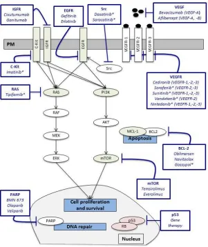
1-2; Figure 1). Angiogenesis plays an important role in tumour growth, invasion and
metastasis [47]. Vascular endothelial growth factors (VEGFs) stimulate cancer cell
migration, invasion, vascular permeability and vessel formation, and have been
shown to increase tumour growth and angiogenesis in pre-clinical models [48].
Increased levels of VEGF-A and high vascular counts are associated with a poor
prognosis in SCLC [48-50]. Moreover, aberrations within the VEGF pathway have
been identified in SCLC, the majority of which occur downstream of the VEGF:VEGFR
interaction (Table 3; Figure 1) [5-7]. These pre-clinical studies suggest that targeting
angiogenesis is an attractive strategy for the treatment of SCLC. Disappointingly,
nearly all trials of targeted agents inhibiting angiogenesis have demonstrated little or
no clinical benefit in SCLC (Tables 1-2; Figure 1) [13, 19-22, 25-28, 30, 31, 33-35, 37,
41, 45].
An area of potential promise is sunitinib, a small molecule inhibitor of multiple
1; Table 1-2) [51]. Ready and colleagues investigated maintenance sunitinib after
platinum and etoposide chemotherapy in untreated ED SCLC. Progression free
survival (PFS) and overall survival (OS) of patients receiving maintenance sunitinib
were 3.7 and 9.0 months compared to 2.1 and 6.9 months, respectively, in those
patients receiving placebo. The improvement in PFS was significant (P=0.02). The
most common grade 3-4 toxicities were fatigue (19%) and neutropenia (14%) [22].
Spigel and colleagues demonstrated the anti-tumour potential (PFS 7.6 months; OS
at 1 year was 54%) of sunitinib as maintenance therapy in patients with ED SCLC with
no evidence of progressive disease after first line chemotherapy with irinotecan and
carboplatin [26].
Sunitinib as monotherapy in patients with relapsed SCLC has not been well studied.
Sunitinib was poorly tolerated (63% grade 3-4 thrombocytopenia; 25% grade 3-4
neutropenia) in a phase II study, with little activity in patients who
relapsed/progressed after chemotherapy [35]. Recently, Abdelraouf and colleagues
reported anecdotal responses, comprising a RECIST partial response lasting 10
months and a second patient with stable disease that lasted 20 months, out of a
small cohort of nine patients (7 chemosensitive and 2 chemotherapy-naïve) treated
with sunitinib monotherapy [52]. Grade 3-4 thrombocytopenia and neutropenia
were observed in 33% of patients treated with sunitinib. The activity of sunitinib in
SCLC may thus warrant further investigation. Future studies should incorporate the
exploration of predictive biomarkers of response to identify patients with SCLC who
are most likely to benefit from treatment with sunitinib.
Aberrations in EGFR, c-KIT, PI3K/AKT/mTOR, IGFR1 and hedgehog signalling
pathways have been identified in SCLC (Table 3; Figure 1) [5-12, 53]. Preclinical
studies have reported overexpression of c-kit and IGFR1, and activation of Hedgehog
signalling in SCLC [38, 53-55]. Ross and colleagues performed targeted exome
sequencing of 236 cancer-related genes in ED SCLC and identified molecular
aberrations in a number of cell-signaling networks including rapamycin-insensitive
7%), PI3K catalytic subunit alpha (PIK3CA; 6%), epidermal growth factor receptor
(EGFR; 5%), phosphatase and tensin homolog (PTEN; 5%) and KRAS (5%) (Table 3)
[6]. Similarly, Umemura and colleagues identified high prevalence of genetic
mutations including PIK3CA (6% of cases), PTEN (4%), RICTOR (9%) and mTOR (4%)
(Table 3) [11]. Therapies targeting EGFR, c-KIT, PI3K/AKT/mTOR, IGFR1 and
hedgehog pathway inhibitors have disappointingly demonstrated minimal efficacy in
first-line, maintenance and relapsed SCLC (Tables 1-2; Figure 1) [14, 15, 18, 24, 29,
38, 40, 43].
Therapeutic strategies that induce apoptosis in SCLC have also been explored in
preclinical and clinical studies. The anti-apoptotic protein BCL-2 is expressed in SCLC
and correlates with poor prognosis [56]. In addition, inhibition of the proteasome by
bortezomib is associated with a reduction in BCL-2 levels and induction of apoptosis
in SCLC cell lines [57]. Similarly, HDAC inhibition reduces the growth of SCLC cell lines
and xenografts [58]. Targeted therapies that inhibit BCL-2 family proteins, HDACs
and the proteasome have demonstrated no benefit in patients with SCLC (Tables
1-2; Figure 1) [16, 23, 32, 36, 39, 42].
Overall, despite both widespread comprehensive genome analysis and pre-clinical
studies implicating angiogenesis, cell signalling and apoptosis in the pathogenesis of
SCLC, therapies that target these pathways have demonstrated little or no efficacy in
clinical trials (Tables 1-3; Figure 1) [5-7, 11, 13-16, 18-43, 45, 47-50, 53-55, 57, 58].
These results are likely multifactorial. The aberrations identified in recent genome
analyses may be present, but are probably not critical for the pathogenesis of SCLC.
Therefore, translational studies to understand the consequence of these aberrations
are urgently required. In addition, if such abnormalities are critical for the
progression and development of SCLC, then only those patients harboring such
aberrations are likely to benefit from treatment and therefore, clinical trials of
targeted therapies in small-unselected populations are unlikely to show clinical
benefit. Finally, most of those agents discussed have been developed for other
targets and develop targeted therapies based on the underlying biological drivers of
SCLC.
Targeting DNA repair pathways
Transcriptional regulation and DNA repair pathways have been implicated in SCLC
development. Sequencing of SCLC surgical samples has identified a high prevalence
of inactivating mutations in TP53 and RB1, amplification of MYC family members and mutations of histone modifiers (CREBBP and EP300) (Table 3) [6, 9-11]. Similarly,
SCLC has a very high level of poly(ADP-ribose) polymerase 1 (PARP1) expression
compared to other solid tumours implicating a potential role in SCLC [59]. PARP1 is a
highly abundant nuclear protein that is activated in response to DNA damage [60].
The PARP1 enzyme has a critical function in DNA damage recognition and
subsequent repair through base excision repair and has a role in homologous
recombinant repair [17, 60]. Loss of PARP1 activity in pre-clinical models leads to the
accumulation of DNA strand breaks along with sensitization to radiotherapy and
chemotherapy [17, 60-62]. Sensitization seems to be a result of increased DNA
damage and delayed DNA damage repair [17]. Platinum-based chemotherapy
induces high response rates in SCLC, and the use of PARP inhibitors to prevent the
ability of cancer cells to repair DNA damage induced by cytotoxic agents is thus an
attractive therapeutic strategy for the treatment of SCLC (Figure 1).
The safety of the PARP inhibitor veliparib in combination with cisplatin and
etoposide in first-line treatment of ED SCLC has been evaluated in the clinic (Table 1)
[63]. The addition of veliparib to cisplatin and etoposide did not improve the
response rate beyond that expected for chemotherapy alone [63]. PARP inhibitors
may demonstrate clinical benefit as maintenance treatment in patients with
platinum sensitive disease through the prolonged inhibition of DNA repair pathways.
Olaparib is currently being evaluated in a randomised placebo-controlled phase II
study as maintenance therapy in SCLC after RECIST response to first-line
chemotherapy (ISRCTN 73164486) (Table 1). Wainberg and colleagues evaluated the
PARP inhibitor BMN-673 in ED SCLC patients that progressed after platinum-based
3-4 toxicities) and two of 11 evaluable patients (18%) with SCLC demonstrated
RECIST partial responses [44].
Multiple putative predictive biomarkers of platinum sensitivity and clinical outcomes
in SCLC have been investigated [3, 64]. Karachaliou and colleagues identified a gene
expression signature from 184 patients with SCLC treated with cisplatin and
etoposide [3]. This favorable expression signature (low levels of excision repair cross
complementation group 1, pyruvate kinase isoform M2, DNA topoisomerase I and
DNA topoisomerase II mRNA) correlated with improved PFS and OS in patients with
both limited disease (LD) (P<0.001 and P=0.007, respectively) and ED (P=0.007 and
P=0.011, respectively) SCLC [3]. BMN-673 demonstrates anti-tumour activity in SCLC cell lines and xenografts [65]. The elevated expression of 17 DNA repair proteins as well as higher levels of expression of a novel “DNA repair protein score” predicted anti-tumour responses to BMN-673. In addition, PI3K activation was associated with resistance to BMN-673 [65]. These findings need to be validated in patients with SCLC as they may identify biomarkers that may predict for response and resistance to BMN-673. Germline BRCA1 and BRCA2 mutations in SCLC are low. However, the numerous somatic aberrations seen within DNA repair proteins mean SCLCs possess a BRCAness phenotype with potential associated sensitivity to platinum therapy and novel targeted agents such as PARP and ataxia telangiectasia and Rad3-related protein (ATR) inhibitors [66]. Therapies that target DNA repair defects in SCLC in an appropriately selected population have the potential to transform the treatment of this aggressive disease. The proposed BRCAness phenotype of SCLC, molecular alterations in transcriptional regulation, and observed early anti-tumour responses
to BMN-673 in relapsed SCLC warrant further investigation with the exploration of
biomarker-defined patient subgroups.
Targeting the immune system
Immune modulation provides an attractive method to harness a patients immune
system to achieve tumour control, stabilisation and potential eradication of disease
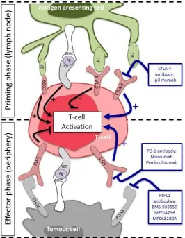
[67]. T-cells play a central role in cell-mediated immunity (Figure 2). Briefly, T-cell
independent co-regulatory signals. This leads to the activation and mobilisation of
T-cells (Figure 2) [68, 69]. Cytotoxic T-lymphocyte-associated protein 4 (CTLA-4) is
expressed on activated T-cells and down regulates the T-cell response [70]. Activated
T-cells mobilise to the tumour microenvironment, where if they recognise antigens
expressed on the tumour cell, they proliferate, release cytolytic enzymes and secrete
cytokines that attract other members of the immune system and eliminate tumour
cells (Figure 2). In comparison, programmed cell death protein-1 (PD-1) is expressed
on activated T-cells, which binds to its ligand PD-L1, causing T-cell inhibition. Tumour
cells hijack this pathway by expressing PD-L1 on their cell surface and directly
suppressing anti-tumour cytotoxic T-cell activity and thus evading the immune
system (Figure 2). Immune therapies that overcome inhibition of the immune
system by targeting CTLA-4, PD-1 and PD-L1 have shown great promise as novel
anti-cancer therapies in multiple malignancies [67].
Ipilimumab (CTLA-4 monoclonal antibody) has been evaluated in combination with
carboplatin and paclitaxel chemotherapy in first line treatment of patients with ED
SCLC (Table 1) [71]. In a pivotal 3-arm phase II trial, patients received either 6 cycles
of carboplatin and paclitaxel with or without ipilimumab or with ipilimumab in a
phased schedule [71]. Phased ipilimumab was associated with improved immune
related PFS when compared to the control arm (P=0.03) [71]. There was a trend
towards improved OS in the phased ipilimumab arm (P=0.13). The incidence of grade
3-4 toxicities was greater with ipilimumab treatment (phased 50%, concurrent 43%)
than without (30%). This study did not identify any potential biomarkers that may
predict for response to ipilimumab treatment in patients with SCLC. Further clinical
trials are now ongoing to evaluate ipilimumab with and without chemotherapy in LD
and ED SCLC (NCT01331525, NCT02046733 and NCT01450761).
Nivolumab and pembrolizumab are novel high-affinity, humanized monoclonal
antibodies that block PD-1. Nivolumab is currently being investigated as
monotherapy or in combination with ipilimumab in advanced tumours, including
SCLC (NCT01928394). Monoclonal antibodies that block PD-L1 (BMS 936559,
advanced cancers. Pembrolizumab demonstrated promising antitumor activity in the
KEYNOTE-028 trial involving patients with heavily pretreated, PD-L1–positive,
extensive-stage SCLC (Table 2) [72]. Patients received 10 mg/kg of intravenous
pembrolizumab once every 2 weeks. Response was assessed every 8 weeks for the
first 6 months, then every 12 weeks thereafter. Overall, the safety and toxicity
profile was consistent with previous studies with pembrolizumab in other tumor
types. Treatment-related adverse events affecting two or more patients occurred in
14 patients; two patients had grade 3-5 adverse events, including one case of colitis
that resulted in the patient’s death. One case of grade 2 autoimmune thyroiditis
resulted in treatment interruption [72]. The overall response rate to pembrolizumab
in antitumor activity was 35% (95% CI [15%, 59%]), and the responses appeared
durable, with six of seven ongoing at data cutoff. The median time to response was
8.6 weeks (range: 7.7-16.1 weeks), while the median duration of response was 29.1
weeks (range: 0.1-29.1 weeks). Five of seven patients who had a disease response
had reduction in tumor size from baseline of 50% or greater [72]. Interestingly, six of
seven responses occurred after 8 weeks of initiating treatment, and one patient who
had stable disease at 8 weeks then had a partial response at 16 weeks.
The distinct roles of CTLA-4 and PD-1/PD-L1 in regulating T-cell activation suggests
there is merit in exploring dual blockade (Figure 2). Combined blockade with CTLA-4,
PD-1 and PD-L1 antibodies inhibit growth in pre-clinical melanoma models [73].
CTLA-4 inhibition has been shown to upregulate PD-1 on tumour infiltrating cells
while PD-1 inhibition has been shown to upregulate CTLA-4 on tumour infiltrating
cells [73]. This potentially provides a mechanism of resistance to immune
monotherapy and therapeutic strategies that block CTLA-4 and PD-1 may
demonstrate synergistic benefit in patients. The phase I/II CheckMate 032 study of
nivolumab with or without ipilimumab for patients with recurrent SCLC
demonstrated that both nivolumab monotherapy and the nivolumab/ipilimumab
combination therapy showed activity and durable responses in patients with SCLC
(Table 2) [74]. The study comprised four arms, and included 128 patients with
progressive disease after one or more lines of therapy, including a first-line
2 weeks (40 patients), and two arms received 1 mg/kg of intravenous nivolumab in
combination with either 1 mg/kg or 3 mg/kg of intravenous ipilimumab every 3
weeks for four cycles (3 patients and 47 patients, respectively). Data have not been
reported from the fourth arm, which included 3 mg/kg of intravenous nivolumab
plus 1 mg/kg of intravenous ipilimumab every 3 weeks for four cycles [74].
Fifteen percent of patients in the arm treated with nivolumab monotherapy had
grade 3-4 adverse events; 34% had grade 3-4 adverse events in the arm treated with
nivolumab plus the higher dose (3 mg/kg) of ipilimumab. One patient died from
treatment-related causes (myasthenia gravis) in the combination arm receiving the
higher dose of ipilimumab. The ORR was 18% with nivolumab monotherapy and 17%
with nivolumab/ipilimumab. The disease control rate was 38% with monotherapy
and 54% with combination therapy. The median OS was 4.4 months with
monotherapy (95% CI [2.9, 9.4]) and 8.2 months with combination therapy (95% CI
[3.7, not reached]). Overall, the response rate and frequency of tumor reduction
suggested increased effects with the combination, activity was observed in both
platinum-sensitive and platinum-resistant/refractory disease. Interestingly,
responses occurred regardless of PD-L1 expression.
Only a subset of patients treated with immune therapies experience durable and
long-term disease control [67, 72, 74]. It will therefore be important to identify
predictive biomarkers of response to identify those patients that will benefit most
from CTLA-4 and/or PD-1/PD-L1 targeting. For example, PD-L1 expression is
currently the closest we have to a predictive biomarker at this point of time for
PD-1/PD-L1 inhibitors, but is clearly imperfect [67]. Variations in laboratory assays,
tumour type and previous treatments may contribute to observed heterogeneity in
PD-L1 expression [67]. Making treatment decisions on such a biomarker requires
careful consideration since many who are biomarker negative may still benefit. It is
likely that the selection of biomarkers for PD-L1/PD-1 inhibitors will involve
additional factors, such as non-synonymous mutational load. Candidate
neo-antigens have been identified that bind with high affinity to MHC class I receptors,
[75]. Patients with NSCLC harbouring tumors with high candidate neoantigen burden
had improved outcomes compared to controls when treated with pembrolizumab
(median PFS 14.5 months versus 3.5 months p=0.002) [76]. The application of
neoantigen burden as a predictive biomarker of response to different
immunotherapies is promising and should be explored in future clinical trials.
Antibody-drug conjugates (ADCs)
A number of antibodies that target tumor surface antigens have provided effective
anti-cancer therapies [77]. However, many unmodified antibodies lack therapeutic
activity and are instead being exploited to deliver potent cytotoxic drugs in the form
of ADCs. Neural cell adhesion molecule (also called CD56) is expressed on the cell
surface of SCLC (approximately 74%) [78, 79]. Lorvotuzumab mertansine (LM) is an
antibody drug conjugate (ADC) containing a CD56 binding antibody (N901) bound to
the microtubulin disruptive agent DM-1. LM has demonstrated activity in pre-clinical
models of SCLC both as monotherapy and in combination with chemotherapy [79,
80]. In two Phase I studies involving CD56-positive solid tumors, the CBR (partial
responses plus stable disease for ≥ 75 days) was 25% (17/68 patients) in patients
with small cell lung cancer (SCLC) from among 113 patients treated with LM [81].
Despite initial promise, a phase II study of LM in combination with carboplatin and
etoposide was stopped early by the independent data monitoring committee due to
concerns with efficacy of the combination and LM has not been developed further in
SCLC.
Rovalpituzumab tesirine, an ADC comprised of a humanized anti-delta-like 3 (DLL3)
monoclonal antibody conjugated to the DNA-damaging pyrrolobenzodiazepine (PDB)
dimer toxin, has shown promising activity in SCLC [82]. DLL3 expression by
immunohistochemistry is elevated in SCLC (72 to 85%), compared to
adenocarcinoma (3.7%), squamous cell carcinoma (0%) and normal lung tissue (0%)
[82]. DDL3 is overexpressed in primary SCLC and patient derived xenografts
compared to normal tissue [82]. Treatment with rovalpituzumab tesirine induced
durable responses in SCLC patient derived xenograft tumour models and eliminated
to be responsible for the rapid disease relapse seen clinically in SCLC [82]. The
efficacy of rovalpituzumab tesirine is a consequence of targeted delivery of PDB
dimer toxin either as naked DLL3 antibody or free PDB dimer toxin had
anti-tumour activity [82]. These encouraging pre-clinical data are supported by a recent
first-in-human phase I trial of rovalpituzumab tesirine in patients with recurrent
SCLC. In all evaluable patients (n=32), the overall response rate was 22% (n=7 partial
response), together with disease stabilization in 53% (n=17) of patients [83]. Sixteen patients had confirmed DLL3 positivity (H-Score ≥ 120) and of these, 7 patients (44%)
had partial response and 8 patients (50%) achieved stable disease [83]. Grade 3-4 toxicities were capillary leak syndrome (14%) and thrombocytopenia (6%) [83].
Rovalpituzumab tesirine in DLL3 positive patients is a promising treatment strategy, and the first in this class of drugs to show activity in SCLC. If these results can be reproduced in the planned follow-on trials, then rovalpituzumab tesirine has the potential to transform the treatment of relapsed SCLC.
Developing targeted therapies in SCLC: how can we improve?
Over the past 10 years, molecularly targeted agents have altered the management
of different malignancies, with significant patient benefit observed in biomarker
selected groups of patients. Despite the successes observed in multiple solid
tumours, there are still no targeted therapies approved for use in SCLC and there are
a number of reasons for these short fallings. Firstly, most agents explored in SCLC
have showed promise in other tumour types and have not been discovered or
developed specifically against the underlying biology of SCLC. Secondly, despite our
increase in the understanding of the molecular characteristics of SCLC, the
development of SCLC targeted treatments has continued in small underpowered
phase II studies involving unselected patients. We have learnt little from such
studies, and they have failed to demonstrate clinical benefit in selected groups of
patients with SCLC, resulting in a paucity of phase III studies involving targeted
agents in this disease.
Translational research that identifies molecular aberrations from extensive whole
Correlation of these findings with clinical endpoints is of paramount importance and
these studies have the potential to identify predictive biomarkers that determine
patient cohorts most likely to respond to molecularly targeted therapies and
immunotherapies. One constraint to such translational studies is the paucity of
tissue for molecular studies since SCLC is rarely resected and serial biopsies are a
major challenge. Therefore, alternative sources of tissue including circulating tumour
cells (CTCs) and circulating plasma DNA (cpDNA) needs to be identified for such
translational studies. Moreover, translational studies that can identify molecular
mechanisms of resistance in patients progressing on treatment with targeted
therapies and immunotherapies will be key to developing combination approaches
for the treatment of SCLC. These strategies have the potential to overcome and
reverse resistance to treatments, so as to provide further clinical benefit for patients
with SCLC.
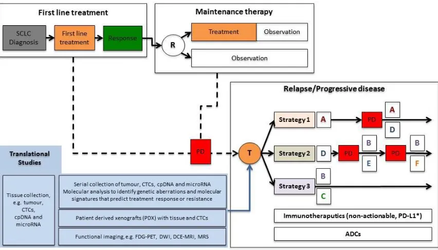
Taking all this together, we therefore propose a novel paradigm for SCLC that will
incorporate the upfront and sequential molecular analyses of patient tissue and
functional imaging studies to optimally select and monitor patients who may benefit
from such treatments (Figure 3). With this treatment pathway, patients with SCLC
should undergo a tumour biopsy at diagnosis to obtain suitable tissue for molecular
analysis alongside the collection of blood specimens for the isolation of CTCs, cpDNA
and microRNA (miRNA) (Figure 3). Such translational studies may identify genetic
aberrations and molecular signatures that predict treatment response and resistance
to therapies. In addition, co-clinical studies with patient tumour and/or CTC derived
xenografts (PDX) may potentially be used to guide therapeutic strategies for
individual patients. These models have already shown promise with CTC derived
xenografts from patients with SCLC [84]. One limitation currently is the time taken to
grow PDXs as clinically SCLC progresses rapidly and treatment decisions may be
required prior to knowledge being obtained from such model systems. However, as
technologies develop, these PDXs may have a critical role in guiding therapeutic
strategies to overcome drug resistance and therefore play an important role in
delivering precision medicine in SCLC. Overall, these hypothesis-testing,
respond to targeted therapies, immunotherapies and ADCs, and to provide greater
and more durable clinical benefit to patients with SCLC. Furthermore, serial
collection of tissue, CTCs, cpDNA and miRNA, with the development of PDX models,
will enable ‘real-time’ monitoring with sequential molecular analyses of tumour
biology while on targeted treatment and at progression.
Conclusions
SCLC has lagged behind other tumour types in terms of a canvas for novel
anti-cancer drug discovery and biomarker development. In the future, it is likely that with
our increased understanding of the underlying biology of SCLC and the identification
and validation of molecular signatures associated with this disease, we may finally
begin to see a new era of effective targeted agents, immunotherapeutics and ADCs
developed for the treatment of SCLC.
Acknowledgements
The authors acknowledge NHS funding to the NIHR Biomedical Research Centre at
The Royal Marsden and the ICR. AS is supported by the NIHR and the Medical
Research Council. TAY is the recipient of grants from the Academy of Medical
REFERENCES
1. Jemal, A., et al., Cancer statistics, 2008. CA Cancer J Clin, 2008. 58(2): p. 71-96.
2. Cancer Research UK. Cancer mortality for common cancers. 2014; Available
from: http://www.cancerresearchuk.org/cancer-info/cancerstats/mortality/cancerdeaths/.
3. Karachaliou, N., et al., Predictive value of BRCA1, ERCC1, ATP7B, PKM2, TOPOI, TOPOmicron-IIA, TOPOIIB and C-MYC genes in patients with small cell lung cancer (SCLC) who received first line therapy with cisplatin and
etoposide. PLoS One, 2013. 8(9): p. e74611.
4. Govindan, R., et al., Changing epidemiology of small-cell lung cancer in the United States over the last 30 years: analysis of the surveillance,
epidemiologic, and end results database. J Clin Oncol, 2006. 24(28): p.
4539-44.
5. Bordi, P., et al., Gene mutations in small-cell lung cancer (SCLC): results of a
panel of 6 genes in a cohort of Italian patients. Lung Cancer, 2014. 86(3): p.
324-8.
6. Ross, J.S., et al., Next-generation sequencing reveals frequent consistent
genomic alterations in small cell undifferentiated lung cancer. J Clin Pathol,
2014. 67(9): p. 772-6.
7. Wakuda, K., et al., Molecular profiling of small cell lung cancer in a Japanese
cohort. Lung Cancer, 2014. 84(2): p. 139-44.
8. Metro, G., et al., Emerging drugs for small cell lung cancer--an update.
Expert Opin Emerg Drugs, 2012. 17(1): p. 31-6.
9. Peifer, M., et al., Integrative genome analyses identify key somatic driver
mutations of small-cell lung cancer. Nat Genet, 2012. 44(10): p. 1104-10.
10. Rudin, C.M., et al., Comprehensive genomic analysis identifies SOX2 as a
frequently amplified gene in small-cell lung cancer. Nat Genet, 2012.
44(10): p. 1111-6.
11. Umemura, S., et al., Therapeutic priority of the PI3K/AKT/mTOR pathway in small cell lung cancers as revealed by a comprehensive genomic analysis.
J Thorac Oncol, 2014. 9(9): p. 1324-31.
12. Umemura, S., K. Tsuchihara, and K. Goto, Genomic profiling of small-cell
lung cancer: the era of targeted therapies. Jpn J Clin Oncol, 2015.
13. Arnold, A.M., et al., Phase II study of vandetanib or placebo in small-cell lung cancer patients after complete or partial response to induction
chemotherapy with or without radiation therapy: National Cancer Institute
of Canada Clinical Trials Group Study BR.20. J Clin Oncol, 2007. 25(27): p.
4278-84.
14. Belani, C., et al., Three-arm randomized phase II study of cisplatin and etoposide (CE) versus CE with either vismodegib (V) or cixutumumab (Cx) for patients with extensive stage-small cell lung cancer (ES-SCLC) (ECOG
1508). Journal of Clinical Oncology, 2013. 31.
15. Johnson, B.E., et al., Phase II study of imatinib in patients with small cell
lung cancer. Clin Cancer Res, 2003. 9(16 Pt 1): p. 5880-7.
16. Langer, C.J., et al., Randomized phase II study of carboplatin and etoposide with or without obatoclax mesylate in extensive-stage small cell lung
17. Owonikoko, T.K., et al., Poly (ADP) ribose polymerase enzyme inhibitor, veliparib, potentiates chemotherapy and radiation in vitro and in vivo in
small cell lung cancer. Cancer Med, 2014.
18. Pandya, K.J., et al., A randomized, phase II trial of two dose levels of temsirolimus (CCI-779) in patients with extensive-stage small-cell lung cancer who have responding or stable disease after induction
chemotherapy: a trial of the Eastern Cooperative Oncology Group (E1500). J
Thorac Oncol, 2007. 2(11): p. 1036-41.
19. Patton, D., et al., Irinotecan (I), carboplatin (C), and radiotherapy (RT) followed by maintenance bevacizumab (B) in the treatment (tx) of limited-stage small cell lung cancer (LS-SCLC): Update of a phase II trial of the
Minnie Pearl Cancer Research Network. Journal of Clinical Oncology, 2006.
24(18S): p. 7085.
20. Pujol, J.-L., et al., Randomized phase II-III study of bevacizumab in combination with chemotherapy in previously untreated extensive
small-cell lung cancer: Results from the IFCT-0802 trial. Journal of Clinical
Oncology, 2014. 32(5s).
21. Ready, N.E., et al., Cisplatin, irinotecan, and bevacizumab for untreated
extensive-stage small-cell lung cancer: CALGB 30306, a phase II study. J Clin
Oncol, 2011. 29(33): p. 4436-41.
22. Ready, N.E., et al., Chemotherapy With or Without Maintenance Sunitinib for Untreated Extensive-Stage Small-Cell Lung Cancer: A Randomized,
Double-Blind, Placebo-Controlled Phase II Study. Journal of Clinical
Oncology, 2015. Published on line before print.
23. Rudin, C.M., et al., Randomized phase II Study of carboplatin and etoposide with or without the bcl-2 antisense oligonucleotide oblimersen for
extensive-stage small-cell lung cancer: CALGB 30103. J Clin Oncol, 2008.
26(6): p. 870-6.
24. Schneider, B.J., et al., Phase II trial of imatinib maintenance therapy after irinotecan and cisplatin in patients with c-Kit-positive, extensive-stage
small-cell lung cancer. Clin Lung Cancer, 2010. 11(4): p. 223-7.
25. Sharma, N., et al., Phase II trial of sorafenib in conjunction with
chemotherapy and as maintenance therapy in extensive-stage small cell
lung cancer. Invest New Drugs, 2014. 32(2): p. 362-8.
26. Spigel, D.R., et al., Phase II study of maintenance sunitinib following irinotecan and carboplatin as first-line treatment for patients with
extensive-stage small-cell lung cancer. Lung Cancer, 2012. 77(2): p.
359-64.
27. Spigel, D.R., et al., Phase II trial of irinotecan, carboplatin, and bevacizumab
in the treatment of patients with extensive-stage small-cell lung cancer. J
Thorac Oncol, 2009. 4(12): p. 1555-60.
28. Spigel, D.R., et al., Tracheoesophageal (TE) fistula development in a phase II trial of concurrent chemoradiation (CRT) and bevacizumab (B) in
limited-stage small-cell lung cancer (LS-SCLC). Journal of Clinical Oncology, 2008.
26(15).
29. Spigel, D.R., et al., Irinotecan, carboplatin, and imatinib in untreated extensive-stage small-cell lung cancer: a phase II trial of the Minnie Pearl
30. Spigel, D.R., et al., Randomized phase II study of bevacizumab in
combination with chemotherapy in previously untreated extensive-stage
small-cell lung cancer: results from the SALUTE trial. J Clin Oncol, 2011.
29(16): p. 2215-22.
31. Allen, J.W., et al., Southwest Oncology Group S0802: a randomized, phase II trial of weekly topotecan with and without ziv-aflibercept in patients with
platinum-treated small-cell lung cancer. J Clin Oncol, 2014. 32(23): p.
2463-70.
32. de Marinis, F., et al., A phase II study of the histone deacetylase inhibitor
panobinostat (LBH589) in pretreated patients with small-cell lung cancer. J
Thorac Oncol, 2013. 8(8): p. 1091-4.
33. Gandhi, L., et al., A phase II trial of pazopanib in relapsed/refractory small
cell lung cancer (SCLC). Journal of Clinical Oncology, 2012. 30.
34. Gitlitz, B.J., et al., Sorafenib in platinum-treated patients with extensive stage small cell lung cancer: a Southwest Oncology Group (SWOG 0435)
phase II trial. J Thorac Oncol, 2010. 5(11): p. 1835-40.
35. Han, J.Y., et al., A phase II study of sunitinib in patients with relapsed or
refractory small cell lung cancer. Lung Cancer, 2013. 79(2): p. 137-42.
36. Heist, R.S., et al., Phase I/II study of AT-101 with topotecan in relapsed and
refractory small cell lung cancer. J Thorac Oncol, 2010. 5(10): p. 1637-43.
37. Jalal, S., et al., Paclitaxel plus bevacizumab in patients with chemosensitive relapsed small cell lung cancer: a safety, feasibility, and efficacy study from
the Hoosier Oncology Group. J Thorac Oncol, 2010. 5(12): p. 2008-11.
38. Krug, L.M., et al., Imatinib mesylate lacks activity in small cell lung
carcinoma expressing c-kit protein: a phase II clinical trial. Cancer, 2005.
103(10): p. 2128-31.
39. Lara, P.N., Jr., et al., Bortezomib (PS-341) in relapsed or refractory extensive stage small cell lung cancer: a Southwest Oncology Group phase II trial
(S0327). J Thorac Oncol, 2006. 1(9): p. 996-1001.
40. Moore, A.M., et al., Gefitinib in patients with sensitive and chemo-refractory relapsed small cell cancers: a Hoosier Oncology Group phase II
trial. Lung Cancer, 2006. 52(1): p. 93-7.
41. Mountzios, G., et al., Paclitaxel plus bevacizumab in patients with chemoresistant relapsed small cell lung cancer as salvage treatment: a
phase II multicenter study of the Hellenic Oncology Research Group. Lung
Cancer, 2012. 77(1): p. 146-50.
42. Rudin, C.M., et al., Phase II study of single-agent navitoclax (ABT-263) and
biomarker correlates in patients with relapsed small cell lung cancer. Clin
Cancer Res, 2012. 18(11): p. 3163-9.
43. Tarhini, A., et al., Phase II study of everolimus (RAD001) in previously
treated small cell lung cancer. Clin Cancer Res, 2010. 16(23): p. 5900-7.
44. Wainberg, Z., et al., Safety and antitumor activity of the PARP inhibitor BMN673 in a phase 1 trial recruiting metastatic small-cell lung cancer
(SCLC) and germline BRCA-mutation carrier cancer patients. Journal of
Clinical Oncology, 2014. 32: p. 5S.
45. Waterhouse, D.M., et al., Phase II study of oral topotecan plus bevacizumab (topo-bev) for second-line treatment of small cell lung cancer (SCLC).
46. Sculier, J.P., et al., Maintenance chemotherapy for small cell lung cancer: a
critical review of the literature. Lung Cancer, 1998. 19(2): p. 141-51.
47. Abidin, A.Z., et al., Targeted therapies in small cell lung cancer: a review.
Ther Adv Med Oncol, 2010. 2(1): p. 25-37.
48. Salven, P., et al., High pre-treatment serum level of vascular endothelial growth factor (VEGF) is associated with poor outcome in small-cell lung
cancer. Int J Cancer, 1998. 79(2): p. 144-6.
49. Fontanini, G., et al., A high vascular count and overexpression of vascular endothelial growth factor are associated with unfavourable prognosis in
operated small cell lung carcinoma. Br J Cancer, 2002. 86(4): p. 558-63.
50. Blackhall, F.H. and F.A. Shepherd, Angiogenesis inhibitors in the treatment
of small cell and non-small cell lung cancer. Hematol Oncol Clin North Am,
2004. 18(5): p. 1121-41, ix.
51. Zhang, Y. and J. He, The development of targeted therapy in small cell lung
cancer. J Thorac Dis, 2013. 5(4): p. 538-48.
52. Abdelraouf, F., et al., Sunitinib (SU11248) in patients with chemo-naïve extensive small cell lung cancer or who have a "chemosensitive" relapse: A
Single Arm Phase II study (EORTC-08061). Eur J Cancer (In-Press), 2015.
53. Badzio, A., et al., Increased insulin-like growth factor 1 receptor protein
expression and gene copy number in small cell lung cancer. J Thorac Oncol,
2010. 5(12): p. 1905-11.
54. Potti, A., et al., CD117 (c-KIT) overexpression in patients with
extensive-stage small-cell lung carcinoma. Ann Oncol, 2003. 14(6): p. 894-7.
55. Park, K.S., et al., A crucial requirement for Hedgehog signaling in small cell
lung cancer. Nat Med, 2011. 17(11): p. 1504-8.
56. Lawson, M.H., et al., Bcl-2 and beta1-integrin predict survival in a tissue
microarray of small cell lung cancer. Br J Cancer, 2010. 103(11): p.
1710-5.
57. Mortenson, M.M., et al., Reduction in BCL-2 levels by 26S proteasome
inhibition with bortezomib is associated with induction of apoptosis in small
cell lung cancer. Lung Cancer, 2005. 49(2): p. 163-70.
58. Crisanti, M.C., et al., The HDAC inhibitor panobinostat (LBH589) inhibits mesothelioma and lung cancer cells in vitro and in vivo with particular
efficacy for small cell lung cancer. Mol Cancer Ther, 2009. 8(8): p. 2221-31.
59. Byers, L.A., et al., Proteomic profiling identifies dysregulated pathways in small cell lung cancer and novel therapeutic targets including PARP1.
Cancer Discov, 2012. 2(9): p. 798-811.
60. Rouleau, M., et al., PARP inhibition: PARP1 and beyond. Nat Rev Cancer, 2010. 10(4): p. 293-301.
61. Virag, L. and C. Szabo, The therapeutic potential of poly(ADP-ribose)
polymerase inhibitors. Pharmacol Rev, 2002. 54(3): p. 375-429.
62. Nguewa, P.A., et al., Poly(ADP-ribose) polymerases: homology, structural
domains and functions. Novel therapeutical applications. Prog Biophys Mol
Biol, 2005. 88(1): p. 143-72.
63. Owonikoko, T.F., et al., A phase 1 study of veliparib, a poly (ADP-ribose) polymerase (PARP) inhibitor, in combination with cisplatin and etoposide in extensive-stage small cell lung cancer (SCLC) patients: An Eastern
Cooperative Oncology Group study (E2511). Journal of Clinical Oncology,
64. Sereno, M., et al., ERCC1 and topoisomerase I expression in small cell lung
cancer: prognostic and predictive implications. Int J Oncol, 2012. 40(6): p.
2104-10.
65. Cardnell, R.J., et al., Proteomic markers of DNA repair and PI3K pathway activation predict response to the PARP inhibitor BMN 673 in small cell
lung cancer. Clin Cancer Res, 2013. 19(22): p. 6322-8.
66. Turner, N., A. Tutt, and A. Ashworth, Hallmarks of 'BRCAness' in sporadic
cancers. Nat Rev Cancer, 2004. 4(10): p. 814-9.
67. Naidoo, J., D.B. Page, and J.D. Wolchok, Immune modulation for cancer
therapy. Br J Cancer, 2014. 111(12): p. 2214-9.
68. Mostafa, A.A. and D.G. Morris, Immunotherapy for Lung Cancer: Has it
Finally Arrived? Front Oncol, 2014. 4: p. 288.
69. Yoon, S.H., Immunotherapy for non-small cell lung cancer. Tuberc Respir Dis (Seoul), 2014. 77(3): p. 111-5.
70. Collins, A.V., et al., The interaction properties of costimulatory molecules
revisited. Immunity, 2002. 17(2): p. 201-10.
71. Reck, M., et al., Ipilimumab in combination with paclitaxel and carboplatin as first-line therapy in extensive-disease-small-cell lung cancer: results from
a randomized, double-blind, multicenter phase 2 trial. Ann Oncol, 2013.
24(1): p. 75-83.
72. Ott, P.A., et al., Pembrolizumab (MK-3475) in patients (pts) with extensive-stage small cell lung cancer (SCLC): Preliminary safety and efficacy results
from KEYNOTE-028. J Clin Oncol, 2015. 33.
73. Ott, P.A., F.S. Hodi, and C. Robert, CTLA-4 and PD-1/PD-L1 blockade: new immunotherapeutic modalities with durable clinical benefit in melanoma
patients. Clin Cancer Res, 2013. 19(19): p. 5300-9.
74. Antonia, S.J., et al., Phase I/II study of nivolumab with or without ipilimumab for treatment of recurrent small cell lung cancer (SCLC):
CA209-032. J Clin Oncol, 2015. 33.
75. Snyder, A., et al., Genetic basis for clinical response to CTLA-4 blockade in
melanoma. N Engl J Med, 2014. 371(23): p. 2189-99.
76. Rizvi, N.A., et al., Cancer immunology. Mutational landscape determines
sensitivity to PD-1 blockade in non-small cell lung cancer. Science, 2015.
348(6230): p. 124-8.
77. Panowksi, S., et al., Site-specific antibody drug conjugates for cancer
therapy. MAbs, 2014. 6(1): p. 34-45.
78. Kontogianni, K., et al., CD56: a useful tool for the diagnosis of small cell lung
carcinomas on biopsies with extensive crush artefact. J Clin Pathol, 2005.
58(9): p. 978-80.
79. Whiteman, K.R., et al., Lorvotuzumab mertansine, a CD56-targeting antibody-drug conjugate with potent antitumor activity against small cell
lung cancer in human xenograft models. MAbs, 2014. 6(2): p. 556-66.
80. Roy, D.C., et al., Elimination of neuroblastoma and small-cell lung cancer
cells with an anti-neural cell adhesion molecule immunotoxin. J Natl Cancer
Inst, 1996. 88(16): p. 1136-45.
82. Saunders, L.R., et al., A DLL3-targeted antibody-drug conjugate eradicates
high-grade pulmonary neuroendocrine tumor-initiating cells in vivo. Sci
Transl Med, 2015. 7(302): p. 302ra136.
83. Rudin, C.M., et al. A DLL3-Targeted ADC, Rovalpituzumab Tesirine, Demonstrates Substantial Activity in a Phase I Study in Relapsed and
Refractory SCLC. in 16th World Conference on Lung Cancer. 2015. Denver,
Colorado, USA.
84. Hodgkinson, C.L., et al., Tumorigenicity and genetic profiling of circulating
FIGURES
Figure 1: Molecular targets in small cell lung cancer
Figure 2: Targeting the immune system in small cell lung cancer
Initial T-cell activation occurs within lymph nodes (priming phase). Tumour antigen (Ag) is presented by the major histocompatibility complex (MHC) of the antigen presenting cells (APC) to the T-cell receptor (TCR) of the T-cell. This is followed by antigen independent coregulatory signals in which CD28 on the T-cell binds B7 on the APC leading to T-cell activation and mobilisation. Cytotoxic
T-lymphocyte-associated protein 4 (CTLA-4) that is expressed on the activated T-cell competes with CD28 for binding to B7 and down regulates the T-cell response. Activated T-cells mobilise to the tumour microenvironment and if they recognise antigens expressed on the MHC of the tumour cell they proliferate, release cytolytic enzymes and secrete cytokines that attract other members of the immune system and eliminate the tumour (effector phase). Programmed cell death protein-1 (PD-1) is found on the surface of activated T-cells where it binds to its ligands PD-L1, causing T-cell
inhibition and down regulation of the acquired T-cell response. Tumour cells may hijack this pathway by expressing PD-L1 on their cell surface and directly suppressing anti-tumour cytotoxic T-cell activity and evading the immune system.
Figure 3: Future treatment paradigm for small cell lung cancer
TABLES
Table 1: Targeted therapies in first line treatment of small cell lung cancer
Putative Target
Agent Author Phase Therapy Setting Outcome
Targeting angiogenesis
VEGF-A Bevacizumab Pujol2015 Patton2006 Spigel2008 Ready2011 Spigel2009 Spigel2011 II/III II II II II II Combination Monotherapy Combination Combination Combination Combination
F (M) R M F (M) F F (M) F (M) R
Negative 15m OS Stopped 7m PFS, 11.6m OS 9.1m TTP, 12.1m Negative
RAF-1, VEGFR-2, VEGFR-3 and PDGFRβ
Sorafenib Sharma2014 II Combination F (M) 7.4m OS
VEGFR-1, VEGFR-2, VEGFR-3, PDGFR, c-KIT, FLT-3 and RET
Sunitinib Spigel2012 Ready2015 II II Monotherapy Monotherapy M M R 7.6m PFS
Improved PFS not OS
VEGFR-2, EGFR and RET
Vandetanib Arnold2007 II Monotherapy M R Negative
Targeting cell signaling BCR-Abl,
c-KIT and PDGFR
Imatinib Johnson2003 Spigel2007 Schneider2010 II II II Monotherapy Monotherapy Monotherapy F M F (M) 0.8m TTP 5.4m PFS, 8.4m OS 4.3m PFS, 7.8m OS
mTOR Temsirolimus Pandya2007 II Monotherapy M 2.5m PFS
IGF1R Cixutumumab Belani2013 II Combination F R Negative
Smoothened Vismodegib Belani2013 II Combination F R Negative Targeting apoptosis
BCL-2 Oblimersen Rudin2008 II Combination F Negative
BCL-2, MCL-1, BCL-W, BCL-XL
Obatoclax Langer2014 II Combination F Negative
Targeting DNA repair defects PARP Veliparib
Olaparib Owonikoko2014 Ongoing I II Combination Monotherapy F M R Negative ISRCTN73164486 Targeting the immune system
CTLA-4 Ipilimumab Reck2013 Ongoing Ongoing Ongoing II II II III Combination Combination Combination Combination F F F (M) F Improved iRPFS NCT01331525 NCT02046733 NCT01450761
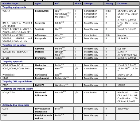
Table 2: Targeted therapies in treatment of relapsed small cell lung cancer
Putative Target Agent Ref Phase Therapy Setting Outcome Targeting angiogenesis
VEGF-A Bevacizumab Jalal2010 Waterhouse2010 Mountzios2012 II II II Combination Combination Combination R (M) R R
14.7w PFS, 30w OS 17.4w PFS, 31.6w OS
2.7m PFS, 6.3m OS RAF-1, VEGFR-2, VEGFR-3
and PDGFRβ
Sorafenib Gitlitz2010 II Monotherapy R 6.7m OSPS, 5.3m OSPR
VEGFR-1, VEGFR-2, VEGFR-3, PDGFR, c-KIT, FLT-3 and RET
Sunitinib Han2013 II Monotherapy R 1.4m PFS, 5.6m OS
VEGFR-1 and VEGFR-2 Aflibercept Allen2014 II Combination R Ra Negative VEGFR-1, VEGFR-2 and
VEGFR-3, PDGF and c-Kit
Pazopanib Gandhi2012 II Monotherapy R 14.1w PFS
Targeting cell signaling
EGFR Gefitinib Moore2006 II Monotherapy R 50d TTP
BCR-Abl, c-KIT and PDGFR Imatinib Johnson2003
Krug2005 II II Monotherapy Monotherapy R R 1.2m TTP All PD by 4w (n=12) mTOR Everolimus Tarhini2010 II Monotherapy R 1.3m PFS, 6.7m OS Targeting apoptosis
BCL-2, BCL-W, BCL-XL Navitoclax Rudin2012 II Monotherapy R 1.5m PFS, 3.2m OS BCL-2, BCL-XL, BCL-W, MCL-1 Gossypol Heist2010 I/II Combination R 17.4w PFSPS, 11.7w
PR
Proteasome Bortezomib Lara2006 II Monotherapy R 1m PFS, 3m OS HDAC Panobinostat De Marinis2013 II Monotherapy R Negative Targeting DNA repair defects
PARP BMN673 Wainberg2014 I Monotherapy R 18% RR
Targeting the immune system
PD-1/CTLA-4 Nivolumab Ipilimumab
Antonia2015 I/II Combination R Nivolumab 18% ORR and 4.4m OS, Combination 17% ORR and 8.2m OS Antibody-drug conjugates
CD56 Lorvotuzumab
mertansine
Beck2012 I Monotherapy R 25% PR/SD
DLL3 Rovalpituzumab
tesirine
Rudin2015 I Monotherapy R
Table 3: Molecular basis of small cell lung cancer
Ref Bordi et al 2014
Ross et al 2014*
Wakuda et al 2014 Umemura et al 2014*
Rudin et al 2012* Peifer et al 2012*
Patients 113 98 60 47 36 99
Technique DNA from FFPE
DNA from FFPE
DNA from FFPE (50), FF (8) and pleural effusions (7)
DNA from FFPE (43) and
MFPE (12).
DNA from FF DNA from FF
Coverage 6 gene panel
236 cancer genes
9 gene panel Whole exome and copy number analysis Whole exome, transcriptome and copy number analysis Whole exome sequencing and transcriptome analysis Aberrations Mutation Mut and amp Mut and amp Mut and amp Mut and amp Mut and amp
EGFR 1.8% 5% 1.7% NR Mutation hotspots
TP53 RB1 PIK3CA CDKN2A PTEN EP300 MLL2 Mutation clustering PIK3CA AKT1-3 MTOR NOTCH1-3 SMO SOX 3-6,9,11,14,17 Amplification SOX2 MYC KIT Mutations TP53 RB1 CREBBP EP300 MLL PTEN SLIT2 EPHA7 Amplification MYC family FGFR1
BRAF 0% NR 0% 2.1%
c-KIT 0% 7% NR
c-MET 4.4% NR 1.7% NR
KRAS 0% 5% 1.7% 2.1%
NRAS NR 0% NR
PDGFR 0% NR NR
TP53 86% 78.7%
RB1 54% 44.7%
MLL2 17% NR
RICTOR 10% 8.5%
MYCL1 8% 8.5%
FGF10 8% NR
LRP1B 7% NR
PIK3CA 6% 15% 6.4%
PTEN 5% 4.3%
MYST3 5% NR
MEK1 NR 0% 4.3%
AKT NR 1.7% 12.8%
HER2 NR 0% NR
CREBBP 3% 4.3%