P1
Development of a Nanolantern™ Assay for Rapid Detection of Enterobacter Cloacae
Angelo J. Cambio,1Jennifer G. Rothschild,1Hsin-P Peng,2Robert D. Mayer,1
Benjamin L. Miller2
1University of Rochester Medical Center, Rochester, NY, USA; 2University of Rochester, Rochester, NY, USA
Background: Urinary tract infections (UTI) are among the most common
bacterial infections in the United States. The gold standard for diagnosis is urine culture. This provides bacteria identification and sensitivity, but is labor intensive and time-consuming. As such, many UTIs are treated empir-ically contributing to resistance and cost. There is an urgent need for a diag-nostic tool that can rapidly identify uropathogens and their sensitivities. We present the development of a PCR-based assay for Enterobacter cloa-cae, and evidence for its further use in the context of NanoLantern™ tech-nology. The NanoLantern™ is a chip sensor that utilizes DNA hairpins immobilized onto a gold surface. In the presence of complementary (tar-get) DNA, the hairpin stem is opened, exposing a quantifiable fluorophore.
Methods: A synthetic DNA hairpin, unique to Enterobacter cloacae, was
immobilized onto 9 gold coated chips. Baseline fluorescence intensity was recorded. The chip was exposed to synthetic target and then imaged with a fluorescence microscope. Fluorescence intensity was recorded and compared to baseline. PCR, asymmetric PCR and linear after the expo-nential PCR were used to amplify the target DNA from the bacteria. The amplicon was exposed to the NanoLantern™ chip and fluorescence inten-sity was quantified.
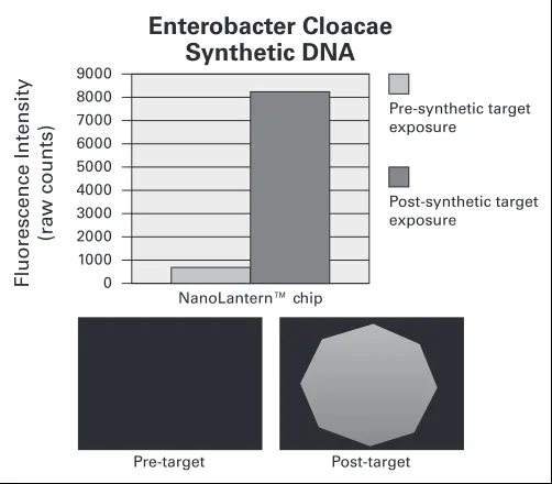
Results: Pre-synthetic target fluorescence imaging of 9 chips exposed to
synthetic DNA resulted in a mean raw count of 665 (SD ± 244). Post-synthetic target fluorescence imaging of 9 chips resulted in a mean raw
count of 8197 (SD ± 648), a greater than 12-fold increase in intensity. See Fig. 1.
Conclusions: We have demonstrated the feasibility of a novel DNA
hair-pin probe. Work is ongoing to develop a PCR assay and to begin testing the NanoLantern™ chip with bacteria derived amplicon and urine sam-ples. Ultimately, these integrated assays will provide rapid analysis, as part of an automated system, to be used in the clinical setting.
P2
WITHDRAWN
P3
Nox4 Inhibitor as a Potential Therapeutic Agent for Renal Cell Carcinoma
Li Chen, Guimin Chang, Jodi K. Maranchie
University of Pittsburgh Medical Center, Pittsburgh, PA, USA
Background: Nox4 is a member of the NADP(H) oxidase enzyme family
that transports electrons across biological membranes to produce reactive oxygen species (ROS). Loss of the von Hippel-Lindau tumor suppressor (VHL) leads to activation of hypoxia-inducible factor-2α(HIF-2α) with increased VEGF, Glut-1, TGF-αexpression. We have shown that Nox4 is essential for HIF-2αtranscriptional activity and that inhibition of Nox4 abro-gates HIF transactivation in VHL-deficient renal cell cancer (RCC) cells. Therefore, Nox4 may serve as a therapeutic target to for RCC. To test this, we screened novel compounds with activity against Nox4 in a human VHL-deficiency renal cell carcinoma xenograft tumor model.
Methods: One million 786-O cells in 100 μl HBSS were injected subcu-taneously in the left flank of 6-week-old female SCID Beige mice. Tumors were measured in two dimensions and estimated tumor weights were cal-culated using the formula: V (mm3) = (length × width2)/2. When tumors reached 100-150 mm3, mice were stratified into 6 equal groups of 10 mice each. Compounds, GKT137892 (100 mg/kg), GKT137928 (100 mg/Kg), GKT136901 (100 mg/Kg) or sutent (40 mg/kg) were administrated by daily gavage for 34 days. Controls arms received no gavage or gavage with vehicle only. Mice were sacrificed at 24h after last gavage, and tumors were harvested and weighed. Significance was determined by ANOVA and Student T tests.
Results: Sutent inhibition of xenograft tumors was observed by day 1.
GKT136901 treatment inhibited tumor growth by day 15. The average volume of the tumors in GKT136901-treated group was 208.36 mm3 on day 22 of treatment vs 279.26 mm3 and 275.24 mm3for the two control groups, respectively, representing a 26% growth inhibition (p = 0.012). For compounds, GKT137892 and GKT137928, no growth inhibition was seen. No toxicity was observed for any of the drugs.
Conclusions: GKT136901, an inhibitor of Nox4, inhibits 786-O cell xenograft
tumor growth, providing proof of principle for Nox4 as a therapeutic tar-get for RCC. Efficacy may be further enhanced by structural alterations that improve tumor penetration.
Moderated Poster Session I: Basic Science
Thursday, September 23: 4:00-5:00 p.m.
9000
F
lu
o
re
s
c
e
n
c
e
I
n
te
n
s
it
y
(r
a
w
c
o
u
n
ts
)
Enterobacter Cloacae
Synthetic DNA
Post-synthetic target exposure
Pre-target Post-target 8000
7000 6000 5000 4000 3000 2000 1000 0
Pre-synthetic target exposure
NanoLantern™ chip
P4
A Dual Adenoviral Amplification System Based on PSA-promoter to Increase Sensitivity of Detection of Prostate Cancer Cells In Vivo By Non Invasive Imaging Frederic Pouliot,1Makoto
Sato,2Lily Wu2
1Université Laval, Québec, QC, Canada; 2University of California, Los Angeles, Los Angeles, CA, USA
Introduction and Objectives: When PSA recurs after prostatectomy, localization of prostate can-cer cells by imaging is important to decide which
patients should receive salvage radiation therapy (RT) as opposed to androgen deprivation therapies. However, the threshold at which prostate cancers cells must be detected for RT to be efficient is <1 ng/mL of serum PSA necessitating sensitive imaging tools. We have developed a dual transcriptional amplification system (DTSTA) that allows sensitive detection of prostate cells by non invasive imaging and tested it in a local recurrence prostate cancer model.
Methods: Adenovirus (Ad) expressing firefly luciferase (fl) under the
con-trol of a modified PSA promoter and the Two-Step-Amplification-Transcription system (Ad-PSATSTAfl) was constructed as previously described (Sato et al. 2008). We have also constructed an oncolytic adenovirus expressing viral early genes E1A and E1B under TSTA (Ad-PSATSTAE1AE1B). When the two viruses are co-administered, we named the combination DTSTA for dual-TSTA system. LAPC-9 cells expressing Renilla luciferase were implanted subcutaneoulsy or in the peritoneum (i.p., as our local recurrence model) of scid/beige mice and tumor growth was monitored in vivo by bioluminescence. Intratumoral (i.t.) reporter activity was assessed after i.p. or i.t. injections of the viruses alone or in combination.
Results: In vitro, DTSTA infection of CWR-22Rv1 prostate cancer cells
increased FL activity by 4.3-fold after 3 days when compared to Ad-PSATSTAfl alone. In vivo, i.t. injection of DTSTA in LAPC-9 tumors resulted in increased Ad-PSAPTSTAfl reporter activity by up to 25-fold (Fig. 1). Real-time PCR on tumor DNA confirmed viral genome replica-tion in tumors infected with DTSTA. Finally, using an i.p. local recur-rence after prostatectomy model, we show that DTSTAfl can detect and localize specifically prostate cancer cells.
Conclusion: We describe a new adenoviral based amplification system,
named DTSTA, which can specifically detect prostate cancer cells by non-invasive imaging in vivo. If translated to the clinic, this nanotech-nology could help treatment decision making when there is PSA recur-rence after prostatectomy.
P5
Increased Cancer Cell Proliferation in Prostate Cancer Patients with High Levels of Serum Folate
Jeffrey J. Tomaszewski, Jessica L. Cummings, Anil Parwani, Rajiv Dhir, Joel B. Mason, Joel B. Nelson, Dean J. Bacich, Denise S. O’Keefe University of Pittsburgh School of Medicine, Pittsburgh, PA, USA
Background: A recent clinical trial revealed that folic acid
supplemen-tation is associated with an increased incidence of prostate cancer. Given mandatory folic acid fortification of cereal grains in the United States, the potential link between prostate cancer and increased folic acid intake warrants further investigation
Objective: Determine the relationship between patient folate status and
the proliferative capacity of Gleason 7 tumors in men with prostate cancer.
Methods: Serum and prostate samples from 86 patients undergoing
sur-gery for prostate cancer and from 25 cancer-free organ donors were uti-lized to measure serum and/or prostate tissue folate concentrations, and assess Ki67 expression. Patients were assessed for genetic polymorphisms in methyltetrahydrofolate reductase and dihydrofolate reductase genes.
Results: Mean serum and tissue folate levels were significantly higher
in men with prostate cancer compared to cancer-free organ donors (p < 0.002 and p < 0.02, respectively) (Fig. 1). Fasting serum folate lev-els were positively correlated with prostate cancer tissue folate content (n = 15; Spearman Correlation r = 0.577, p < 0.03). Fasting serum folate was significantly higher in users of folic acid supplements (p < 0.05). When divided into quartiles, there were no significant differences in serum folate levels between users and non-users of supplements. Among patients with Gleason 7 disease, the mean proliferation index was 6.17 ± 3.2% and 0.86 ± 0.92% in patients in the highest (117 ± 15nM) and lowest (18 ± 9nM) quintiles for serum folate, respectively (p < 0.0001) (Fig. 2).
Conclusions: This is the first report of a positive correlation between
serum and prostate tumor folate. Increased cancer cell proliferation in men with higher serum folate concentrations is consistent with an increase in prostate cancer incidence observed with folate supplementation. Unexpectedly, more than 25% of our patients had serum folate levels greater than 6-fold adequate. Only half of these men reported supple-ment use, suggesting altered folate metabolism and/or consumption of folic acid from fortified foods.
Fig. 1.P4.
0 20 40 50 60 100 120 140 160
S
e
ru
m
F
o
la
te
(
n
M
)
Prostate Cancer Patients Cancer-Free Organ Donors p<0.002
Fig. 1.P5. Fasting serum folate concentrations (nM) in patients with prostate cancer vs. cancer-free organ donors.
P6
Nitric Oxide Signalling Mediates Hypoxic Upregulation of Macrophage Inhibitory Factor in Prostate Cancer
D. Robert Siemens
Queen’s University, Kingston, ON, Canada
Background: Macrophage inhibitory factor (MIF) is an important chemokine
influencing progression of prostate cancer. We have demonstrated that tumor hypoxia mediates many factors leading to a malignant phenotype in prostate cancer, including invasion, metastases and drug-resistance. Such hypoxia-induced phenotypes can be attenuated by manipulating nitric oxide (NO) signaling through classic, cGMP mediated pathways. The aim of this study was to determine the role of NO signalling in hypoxia-induced upregulation of MIF in prostate carcinoma cells.
Methods: MIF production by DU-145 prostate cancer cells (as well as
the MDA breast cancer cell line) was determined by ELISA in different oxygen culture conditions (0.5-20% O2). The role of NO signalling in
hypoxia-mediated upregulation of MIF was determined by pharmaco-logic inhibitors and mimics of classic NO signalling with 10 nM GTN, 100 μM L-NMMA as well as non-hydrolysable analogue of cGMP, 8-bromo-cGMP (10 nM).
Results: These studies demonstrate that exposure of DU145 (as well as
the MDA cell lines) to low oxygen tension for 24 hours consistently increased the secretion of MIF into the supernatant (1720 ± 245 ng/mL vs. 240 ± 46 ng/mL, p < 0.05). Incubation of the cell lines with the inhibitor of nitric oxide synthase L-NMMA in 20% oxygen resulted in a similar increase in MIF secretion (832 ± 66 ng/mL vs. 220 ± 33 ng/mL, p< 0.029). Restoring classical NO signalling in these cells with low concentrations of GTN or 8-bromo-cGMP was also able to significantly (p < 0.05) reverse the hypoxia-mediated increase in MIF.
Conclusions: These results contribute to our understanding of MIF
regu-lation, an important chemokine linked to cancer progression in numer-ous cancer sites. It appears that decreased NO signalling, as a result of microenvironmental hypoxia, is at least partially responsible for increased MIF secretion by cancer cells. These results justify further in vivo inves-tigations of the role of nitric oxide signalling and MIF action and may represent a novel target for pharmacologic therapy for prostate cancer.
P7
Reconstruction of a Human Vesical Equivalent Using Adipose-Derived Stem Cells
Alexandre Rousseau, Jonathan Cloutier, Geneviève Bernard, Guillaume Marceau Fortier, Robert Gauvin, Sara Bouhout, Julie Fradette, Stéphane Bolduc
LOEX, Centre de recherche FRSQ du CHA Universitaire de Québec, Université Laval, Québec, QC, Canada
Background: For several years, fibroblast cells have been primarily used
for tissue engineering but adipose-derived stem/stromal cells (ASCs) show promising potential due to their facility to obtain, their capacity to differentiate and their ability to secrete mediators. Our group previ-ously reported on the production of a bioengineered vesical equivalent using dermal fibroblasts without exogenous matrix. The aim of this study was therefore to evaluate the possibility of engineering an autologous vesical equivalent with human ASCs in order to validate if our model can benefit from the attributes of the ASCs.
Methods: ASCs were obtained from lipoaspirated adipose tissue and
fibroblasts were extracted from a dermal biopsy. These human cells were cultured with serum and ascorbic acid to stimulate the formation of extracellular matrix and obtain cell sheets. Cells were cultured with constant media motion (GyrotwisterTM, Woodbridge, NJ) during three weeks and then three cell sheets of ASCs or fibroblasts were superim-posed. After 4 days of maturation allowing cell sheet fusion, human urothelial cells were seeded on top of the construction and matured at the air/liquid interface. The vesical equivalents were characterized by histology, immunofluorescence as well as mechanical and suture resist-ance tests.
Results: Complete vesical equivalents were obtained with ASCs or
fibrob-lasts. The histology clearly showed that cell sheets forming the ASC
vesi-cal equivalents featured a strong cohesion between cell sheets and were 1.8 fold thicker than the fibroblast vesical equivalents. Immunolabelings of the mature constructions showed the presence of cytokeratin 8\18, a differentiation marker for urothelial cell; and collagen 1 and 3, which are the major components of the extracellular matrix. The ASC vesical equivalents were easy to manipulate resistant enough to suture, there-fore allowing the 3D reconstruction of a bladder shaped tissue engi-neered substitute.
Conclusions: Human vesical equivalents were successfully produced
using either dermal fibroblasts or ASCs, without the use of exogenous scaffolding components. The ASC vesical equivalents could sustain sutur-ing without tearsutur-ing. Considersutur-ing their accessibility, abundance and increased matrix production ACSs therefore represent a great cell source to further optimize our innovative model for vesical reconstruction.
P8
Stray Electrical Currents in Laparoscopic Instruments Used in DaVinci Robotic Surgery
Carlos E. Mendez Probst,1George Vilos,1Paul Borg,2David Galloway,2
Stephen E. Pautler1
1Schulich School of Medicine and Dentistry, The University of Western Ontario, London, ON, Canada; 2St Joseph’s Health Care London, London, ON, Canada
Background: The use of the DaVinci robotic surgical system in
laparo-scopic procedures has gained wide acceptance and popularity across all surgical disciplines. This system however requires the utilization of monopolar electrosurgery and a finite reuse of electrosurgical instru-ments both of which provide opportunities for stray electrical currents from capacitive coupling and/or insulation failure. We report the preva-lence and magnitude of such stray currents measured in DaVinci instru-ments that had reached the end of their duty cycle.
Methods: We tested 30 such instruments, 6 monopolar scissors, 1 Maryland
bipolar forceps, 1 monopolar hook, 5 plasma kinetic dissecting forceps, 7 prograsp forceps, 10 large needle drivers using a Valleylab Force 2 ESU at pure coag and cut waveforms in open circuit at 4 different set-tings (open air) at 40 w, and sequentially gel coated instruments at 40 w, 80 w and maximum ESU output (coag 120 w, cut 300 w). The mag-nitude of stray currents was measured by an electrosurgical analyzer (454A Dynatec, Nevada). Visual inspection did not identify insulation defects in any of the instruments.
Results: At coag waveform in open air, 86% of instruments leaked a
mean of 0.4 w (0-0.7 w). In the presence of gel coated instruments, stray currents were detected in all instruments with means and (range) of 4.2 w (1.5-7.7), 5 w (1.8-9.7), and 5 w (1.9-10.5) at 40 w, 80 w and 120 w, respectively. At cut waveform in open air, none of the instruments leaked current, while gel coated instruments leaked a mean of 2.7 w (0.6-4.3), 2.7 w (0.8-8.2) and 4 w (1.6-8.2) at 40 w, 80 w and 300 w, respectively. Compared by instrument group, the highest leakage was in PKDF (mean 4.1 w, one >8 w), followed by LND (3.3 w), PF (2.8 w), MBF (2.4 w), MCS (2.3 w), and MH (1.1 w).
Conclusions: At the end of their life cycle, all tested instruments showed
energy leakage with one over 8 w, at >80 w of ESU power. Stray cur-rents were higher during coag waveforms and the magnitude was not always proportionally related to ESU settings. Such stray currents may cause electrical burns to patients and/or operating room personnel.
P9
Vitamin D3Therapy Increases Cryoablation Efficacy: A Novel
Strategy for the Treatment of Prostate Cancer
John M. Baust,1Daniel Klossner,2Anthony Robilotto,1Robert Van Buskirk,1
Andrew Gage,1Vladimir Mouraviev,3Thomas Polascik,3John G. Baust2
1CPSI Biotech, Owego, NY, USA; 2Binghamton University, Binghamton, NY, USA; 3Duke University Medical Center, Durham, NC, USA
Introduction and Objective: Adjuvant therapies contribute to the
shown to exhibit cytotoxic properties preferentially inducing apoptosis in a variety of human cancer cells. Human prostate cancer cells are known to be resistant to many cytodestructive agents, including cryoab-lation and VD3. Here, we evaluated the efficacy of VD3combined with
cryoablation on androgen insensitive human prostate cancer (PC-3 and LNCaP-HP) cell death.
Methods: Freezing and VD3exposure to PC-3 and LNCaP-HP cells were
performed using both 2D and 3D culture systems and efficacy was deter-mined by cell viability assays. Resultant cell death and specific signal-ing components were determined ussignal-ing apoptotic inhibitors, fluores-cence microscopy, protease activity assays and immunoblotting.
Results: Exposure of LNCaP-HP cells to freezing (-15°C) or VD3(50 uM)
results in minimal cell death (15% and 10% respectively 3 days post treatment), while a complete loss of viability was observed with the combination. The synergistic effect was found to be due to a marked increase in apoptosis. Western blot analysis revealed a decrease in pro-caspase-9 and -3 between 6 and 18 hours post-exposure. Caspase acti-vation assays confirmed the reduction in pro-caspase levels was a result of caspase activation. Protease inhibitors were incorporated into the combination protocol to determine the overall contribution of caspase activity in cell death. Inhibition of caspase-9 significantly blocked the combination induced cell death compared to cells that did not receive the inhibitor (55% vs. 21% viable, respectively). The addition of the caspase-8 inhibitor resulted in only minimal protection, indicating a specific mitochondrial-mediated event. Importantly, the combination was not effective when applied to normal prostate cells.
Conclusions: VD3 sensitizes CaP cells to cryoablation. The significant
increase in cell death was attributed to the activation of apoptosis, specif-ically through mitochondrial -mediated events. The results describe a novel therapeutic model for the treatment of prostate cancer and pro-vide support for future in vivo studies.
P10
Rapid Induction of Apoptosis at Ultra Low Temperatures Enhances the Efficacy of Prostate Cancer Cryoablation Anthony Robilotto,1John M. Baust,1Robert VanBuskirk,1Andrew Gage,1
John G. Baust2
1CPSI Biotech, Owego, NY, USA; 2Binghamton University, Binghamton, NY, USA
Introduction: Investigations into the molecular-based responses of prostate
cancer following cold exposure have led to the discovery of delayed-onset, apoptotic cell death within the periphery of cryolesions. The apoptotic pathway typically attributed to this delayed death is the intrin-sic/mitochondrial-mediated pathway characterized by a loss of mito-chondrial potential, release of cytochrome c, and activation of caspase-9. Recent studies, however, have shown that at lower temperatures within the core of the cryogenic lesion (< -20°C) a rapid programmed cell death response occurs. Using an engineered, 3-dimensional prostate tumor model, we investigated these events to determine the signaling pathway(s) responsible for the cell death as a means of developing improved molec-ular based approaches for the cryoablation of prostate cancer.
Methods: Human prostate cancer cells (PC3) were cultured in the 3D
matrices for 7 days prior to experimentation. The tumor models were then frozen to -30 or -15°C and analyzed at various times post-thaw using fluorescence microscopy, flow cytometry, and Western blots.
Results: Results demonstrated that the activation of apoptotic cell death
occurred within 30 min of thawing at ultra low temperatures. At -30°C, ~25% of cells were apoptotic at 30 min and by 6 hr levels had dropped near those of controls. At elevated temperatures (-15°C), the activation and progression of apoptosis was considerably delayed, peaking at ~20% by 6 to 24 hr post-thaw. Additionally, it was determined that early onset apoptosis was regulated through a unique, caspase dependent process compared to that seen within the freeze margins. This induction was found to progress through a membrane mediated pathway associated with more severe thermal stressors as indicated by the activation of cas-pase-8 at low (-30°C) but not mild (-15°C) temperatures.
Conclusion: These data suggest that an apoptotic continuum exists
through-out the cryolesion whereby the more severe the cryogenic stress, the faster programmed cell death is manifested. The identification of this rapid-onset apoptosis within the core of the ablative zone represents a novel finding in a region previously thought to be only necrotic. Ultimately, it is our aim to decipher the signaling pathways involved in triggering rapid-onset apoptosis such that these events can be manipulated to enhance cell death, thus improving the overall efficacy of cryosurgical procedures.
P11
The VHL Tumor Suppressor Promotes Renal Cancer Cell Apoptosis by Inhibiting Casein Kinase 2 Activity and Impairing NF-κB Signaling
Guan Wu, Xiangrong He, Edward M. Messing University of Rochester, Rochester, NY, USA
Background: Renal cell carcinoma (RCC) responds notoriously poor to
cytotoxic chemotherapy, indicating aberrant anti-apoptotic signal acti-vation. Meanwhile, pVHL inactivation is implicated in most RCC cases, suggesting a potential link between pVHL and RCC apoptosis. In this report, we investigate abnormal anti-apoptotic behavior in pVHL-defi-cient RCC cells. Our objectives are to identify the molecular mechanism involved in RCC resistance to cytotoxicity-induced apoptotic signaling and to provide information that may lead to better RCC treatment design.
Methods: HEK293, Hela, and RCC cell lines, including 786-O, RCC4,
cell lines derived from these two cell lines were used in our experiments. The GST pull-down and co-IP assays were conducted to confirm protein interactions. Western blotting was performed for evaluating protein expres-sion/activation. Kinase activity was measured by in vitro kinase assay. NF-κB activation was dedected by EMSA and luciferase reporter gene assay. RNAi techniques were applied for transient knockdown of spe-cific protein expressions and for the construction of RNAi stable RCC cell lines. Apoptosis was assessed via FACS or fluorescence microscope.
Results: In this study, we found that pVHL inhibits NF-κB activity by interacting with casein kinase 2α(CK2α), an I B-independent pathway. The CK2αprotein level was not regulated by pVHL or hypoxia. Upon TNFαtreatment, pVHL-deficient RCC cells exhibit much stronger
NF-κB activation and dramatically elevated resistance to apoptosis. pVHL-deficient RCC cells also showed increased NF-κB p65 serine529 phos-phorylation and enhanced association between p65 and CBP/p300. Knockdown of CK2αin pVHL-deficient RCC cells inhibited p65 ser529 phosphorylation and abnormal NF-κB activation and blunted NF-κB downstream anti-apoptotic gene expressions, which lead to restored sensitivity to TNFα-induced apoptosis. Moreover, a chemical inhibitor of CK2, 5,6-dichlororibifuranosylbenzimidazole (DRB) also showed robust inhibition of abnormal NF-κB activation in pVHL-deficient RCC cells.
Conclusions: Our findings suggest that pVHL regulate NF-κB anti-apop-totic pathway by suppressing CK2 kinase activity and p65 serine529 phosphorylation - a process that culminates in decreased p65/p300/CBP interaction and enhanced RCC apoptosis. This represents a novel mech-anism of how pVHL inactivation reduces RCC cells susceptibility to cyto-toxicity-induced apoptosis by promoting CK2 and NF-κB activations.
P12
The Effect of Dietary Folate on Prostate Carcinogenesis in an in vivoModel of Tumorigenesis
Jeffrey J. Tomaszewski, Jessica L. Cummings, Anil Parwani, Dean J. Bacich, Denise S. O’Keefe
University of Pittsburgh School of Medicine, Pittsburgh, PA, USA
Introduction: Recent studies demonstrate that folic acid
supplementa-tion is associated with an increased incidence of prostate cancer. We previously reported a positive correlation between serum and prostate tumor folate, and increased cancer cell proliferation in men with higher serum folate concentrations.
Objective: To determine the effect of dietary folate manipulation on
Methods: Utilizing the subrenal prostatic recapitulation model for
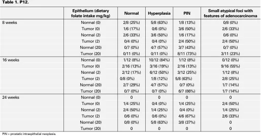
tis-sue recombination, rat urogenital mesenchyme (rUGM) was microdis-sected from rat embryos and combined with 10-20 mg slivers of prostate tumor or normal prostate tissue from patients with prostate cancer. Tissue recombinants were grafted beneath the renal capsule of male SCID mice. Mice were treated hormonally at the time of grafting with testosterone propionate and 17β-estradiol. Mice were randomly assigned to receive amino-acid defined diets which were deficient (0 mg/kg), folate-sufficient (2 mg/kg), or excessively folate-fortified (20 mg/kg). Tissue recombinants were serially grafted and harvested after 8, 16, and 24 weeks under the renal capsule. Histological and morphological features were assessed based upon 20 fields per recombinant, with the worst histological phenotype assigned to that recombinant. Immunohistochemical analysis to detect basal cells (p63), proliferation (Ki67), DNA methyla-tion (5-methylcytosine), and tumor aggressiveness (EZH2) was performed.
Results: Recombinants from normal and cancerous prostate tissue formed
normal, hyperplastic, focal PIN like glandular structures, and adenocar-cinoma. The incidence of normal glands, hyperplasia, PIN, and adeno-carcinoma in human prostate cancer tissue recombinants was 6.5%, 6.5%, 50% and 37%, respectively; incidence in normal prostate tissue recombinants was 17%, 62.5%, 18.7%, and 4.7% (Table 1). Among normal prostate tissue recombinants, excess dietary folate intake increased the incidence of PIN and adenocarcinoma when compared to recombi-nants grown in mice receiving a folate deficient diet (31.3% vs. 7%, respectively; p < 0.05). An increased incidence of PIN and adenocarci-noma was also observed among prostate cancer recombinants in mice with adequate or excess dietary folate intake when compared to folate deficient mice (97.6% vs. 75.3%, respectively; p < 0.05).
Conclusions: Adequate to excess levels of dietary folate promote prostate
carcinogenesis compared to folate deficiency in an in vivo human prostate tissue recombinant model of tumorigenesis.
P13
Nox4 is Required for Nuclear Translocation of HIF-2αin Renal Cancer Cells
Guimin Chang, Li Chen, Jodi K. Maranchie
University of Pittsburgh Medical Center, Pittsburgh, PA, USA
Background: Renal cell carcinoma (RCC) causes nearly 12,000 deaths
each year in America. Nox4 belongs to the NADPH oxidase family that generates reactive oxygen species (ROS). The kidney is the site of great-est abundance of the Nox4. We previously showed that Nox4 is critical to activation of HIF-2αand necessary to support the tumor phenotype of RCC cells. However, the mechanism of Nox4 induction of HIF-2αis unknown. Inactive HIF-2αis located in the cytoplasm and activation requires translocation to the nucleus where it dimerizes with ARNT and binds DNA. To determine if Nox4 is required for nuclear translocation, we localized exogenous HIF-2αby immunofluorescence in RCC cells in the presence or absence of Nox4. We further mutated key HIF-2α
hydroxylation sites to measure their impact on translocation.
Methods: 786-0 human kidney cancer cells with stable expression of
Nox4 shRNA (KD) or a non-targeting shRNA (NS) were transiently trans-fected with pSNAP- HIF-2αto express wild type HIF-2αtagged at the N-terminus with SNAP. Cells were cultured 48 hr under 21% or 1% oxygen conditions and then fixed and bound to an anti-SNAP primary antibody. Nuclei were counterstained with DAPI. pSNAP- HIF-2α-PA was cloned by site directed mutagenesis of Pro531 to Ala, and pSNAP-HIF-2α-NA, was derived by mutation of Asp851 to Ala.
Results: 786-0 NS cells showed moderate nuclear HIF-2αlocalization at 21% O2with a shift to 100% nuclear expression under hypoxia. With
Nox4 silencing, the 786-0 KD cells demonstrated only cytoplasmic local-ization regardless of oxygen conditions, suggesting that Nox4 is critical to HIF-2αnuclear translocation in normoxia or hypoxia. HIF-2α-PA mutation did not alter the localization pattern at 21% or 1% O2.
HIF-2α-NA mutants, however, demonstrated cytoplasmic localization under all conditions. HIF-2αstaining intensity was significantly lower in KD than NS. Addition of the proteasome inhibitor, MG132 (0.1 M) increased staining intensity to levels seen in 786-0 NS.
Table 1. P12.
Epithelium (dietary
folate intake mg/kg) Normal Hyperplasia PIN
Small atypical foci with features of adenocarcinoma
8 weeks Normal (0) 2/8 (25%) 5/8 (63%) 1/8 (13%) 0/8 (0%) Tumor (0) 1/6 (17%) 0/6 (0%) 3/6 (50%) 2/6 (33%) Normal (2) 2/6 (33%) 3/6 (50%) 1/6 (17%) 0/6 (0%)
Tumor (2) 0/4 (0%) 0/4 (0%) 2/4 (50%) 2/4 (50%) Normal (20) 0/7 (0%) 4/7 (57%) 3/7 (43%) 0/7 (0%)
Tumor (20) 0/11 (0%) 0/11 (0%) 8/11 (73%) 3/11 (23%) 16 weeks Normal (0) 1/12 (8%) 10/12 (84%) 1/12 (8%) 0/12 (0%)
Tumor (0) 2/16 (13%) 3/16 (19%) 2/16 (13%) 9/16 (55%)
Normal (2) 2/12 (17%) 6/12 (50%) 3/12 (25%) 1/12 (8%) Tumor (2) 0/8 (0%) 1/8 (12%) 5/8 (63%) 2/8 (25%) Normal (20) 2/7 (29%) 4/7 (57%) 0/7 (0%) 1/7 (14%)
Tumor (20) 0/7 (0%) 0/7 (0%) 6/7 (86%) 1/7 (14%)
24 weeks Normal (0) 0 0 0 0
Tumor (0) 1/4 (25%) 0/4 (0%) 1/4 (25%) 2/4 (50%)
Normal (2) 2/4 (50%) 1/4 (25%) 0/4 (0%) 1/4 (25%) Tumor (2) 0/6 (0%) 0/6 (0%) 4/6 (67%) 2/6 (33%)
Normal (20) 0/8 (0%) 5/8 (63%) 3/8 (37%) 0
Tumor (20) 0 0 0 0
Conclusions: We show for the first time that Nox4 expression is required
for HIF-2αnuclear translocation in RCC cells under both hypoxic and non-hypoxic conditions. Asp851 appears to be required for Nox4-medi-ated translocation. We speculate that Nox4-derived ROS may inhibit FIH, the enzyme responsible for Asp851 hydroxylation, thereby trigger-ing this “hypoxic switch” in the absence of hypoxia. Furthermore, stabi-lization of HIF-2αby proteasome inhibitors suggests that Nox4 protects HIF-2αfrom proteasomal degradation via a pVHL-independent path-way. Ongoing investigations aim to further elucidate these pathways.
Funding: ACS RSG-09-023-01-CNE
P14
Temporal RF-Ultrasound Augmentation of Prostate (TRAP): Enhancing Prostate Cancer Detection Utilizing Temporal Ultrasound Radio Frequency Signals
D. Robert Siemens, Purang Abolmaesumi, Parvin Mousavi, Mehdi Moradi, Eric Sauerbrei, Sandy Boag
Queen’s University, Kingston, ON, Canada
Background: Ultrasound echo signals are affected by the geometrical
deregulation of cellular architecture in neoplastic tissue and can be exploited to differentiate normal from various grades of cancerous tis-sue, information that is not visible in post-processed B-scan ultrasound images. We have proposed to apply an innovative approach to process raw transrectal ultrasound (TRUS) radio-frequency (RF) signals for the early detection of prostate cancer.
Methods: Ex vivo experiments have involved evaluation of a time series,
through fractal and frequency analyses, of raw temporal RF ultrasound signals to differentiate tissue types in 35 human prostates obtained after radical prostatectomy. Previously described ultrasound texture features for tissue typing were included in these studies for comparison. Detailed whole mount sectioning of the prostate specimens were regarded as the gold standard and compared to probability maps created by analysis of the temporal RF signals.
Results: Comparing the generated probability maps of prostates scanned
ex vivo to the detailed pathology reports have demonstrated sensitivity and specificity values of 90% and 85% respectively, in characterizing cancerous tissue. The area under receiver operating characteristic curve for the subset of RF time series evaluation was 0.87, which increased to 0.95 when combined with other ultrasound texture features. Validation utilizing leave one patient out resulted in an area under the curve of 0.82.
Conclusions: These results suggest that the temporal ultrasound echo
signals can be employed to differentiate different tissues in the prostate and subsequently improve cancer detection. Furthermore, we propose to train classifiers in order to visualize this information as color-coded probability maps on real-time US images and have initiated an in vivo study for men undergoing TRUS guided biopsy.
P15
Urinary MicroRNA As An Accurate Urinary Diagnostic Marker For Urothelial Cancer
D. Robert Siemens, Jaime Snowden, Jason Izard, Sandy Boag, Harriet Feilotter
Queen’s University, Kingston, ON, Canada
Background: MicroRNAs (miRNAs) are a class of small RNAs that are
important regulatory molecules, involved in several cell processes such as developmental timing, stem cell division, and apoptosis. Dysregulated miRNAs have been identified in several human malignancies, includ-ing bladder cancer tissue samples, and may confer a “tumor signature” that can be exploited for diagnostic purposes. We report on a prospec-tive pilot study investigating the diagnostic capability of miRNAs in the urine of patients with urothelial cancer.
Methods: Voided urine samples were collected from 8 patients with
urothelial carcinoma just prior to bladder tumor resection as well as 5 age-matched healthy control patients. Pathology demonstrated both low grade and high-grade cancer. Total RNA was isolated and quantitative reverse transcriptase-polymerase chain reaction was performed on the
RNA extracts using primers for 4 miRNAs shown previously to be dys-regulated in solid urothelial carcinomas with RNU6B as the endoge-nous control. Standard urine cytology was performed on all samples in a blinded fashion.
Results: Two miRNAs were found to be significantly dysregulated in the
urine from cancer patients with miR-A showing an average 10.42-fold decrease (p < 0.05) and miR-B showing an average 2.70-fold increase (p > 0.05) in the cancer samples compared to the normal controls. Using these 2 miRNAs, a decision-tree prediction model was generated yield-ing a specificity of 100% and a sensitivity of 87.5%. The sensitivity and specificity of the cytology on the same urine samples was 50% and 80% respectively.
Discussion: MiRNA expression levels are altered in bladder cancer and
may have diagnostic and prognostic value. This preliminary study of candidate urinary miRNA in patients with both low grade and high-grade urothelial cancer demonstrated a significantly improved diagnos-tic accuracy over cytology. These results provide rationale for further studies on discovery and validation of candidate miRNAs in voided urine and may potentially lead to the development of a non-invasive and sensitive test for bladder cancer diagnosis and prognosis.
P16
Development of a Realtime Intraoperative Electrical Impedance Tomography Sensor for Cavernous Nerve Mapping for Radical Prostatectomy
Henry H. Tran
University of British Columbia, New Westminster, BC, Canada
Introduction and Objective: Radical prostatectomy is a proven
effica-cious treatment locally confined prostate cancer, however rates of post operative impotency continue to range between 10-30% in contempo-rary series. Thus, in addition to satisfactory cancer control, accurate localization of the cavernous nerves and the contributing plexus during resection is exceedingly important during radical prostatectomy. We have developed a realtime, intraoperative tissue impedance sensor for nerve localization based on electrical impedance tomography (EIT) tech-nology, which will serve to reduce post prostatectomy impotence
Methods: The prototype constructed consists of a probe with a needle
array to interface with the tissue, and a signal generator and analysis system. The device cycles through each set of electrodes on the probe, and injects current while simultaneously computing the voltage change. Reconstruction algorithms determine nerve location based on tissue impedance properties. The system was functionally validated in an in vitro model system and following further refinement, rat sciatic nerve identification. For each trial, the probe was placed on the tissue and the device sequentially injected 5mA sinusoidal current across each of its electrodes with the resulting voltages recorded.
Results: Each trial demonstrated the ability of the device to detect changes
in impedance of different tissues. Electrodes closest to the wire in the in vitro model had the smallest voltage change, corresponding to a lower impedance value. V=0.135V for the electrodes near the wire, compared to V=0.256V for other electrodes. Localization of rat sciatic nerve was also successful demonstrating V=0.294V for the electrodes near the nerve, compared to V=0.467V for the remaining electrodes.
Conclusions: We have developed a realtime intraoperative tissue
P17
Mapping the Cytokine Profile of Interstitial Cystitis in Human Bladder and Urine Specimens: A Pilot Study
Anthony T. Corcoran,1Vikas Tyagi,1Masa Kita,1Pradeep Tyagi,2Wendy
W. Leng,1Naoki Yoshimura1
1University of Pittsburgh Medical Center, Pittsburgh, PA, USA, 2William Beaumont Hospital, Royal Oak, MI, USA
Introduction: Evidence suggests that increases in cytokine activity may
be involved in the pathogenesis of painful bladder syndrome/interstitial cystitis (PBS/IC). However, data on cytokine activity in human PBS/IC patients are lacking. This pilot study investigated the cytokine profile in human bladder tissue and urine of PBS/IC patients.
Methods: Ten PBS/IC patients (ICS definition) were enrolled in this pilot
study. Human bladder (cold cup biopsy) and urine specimens were col-lected intraoperatively before hydrodistention (HD) under general anes-thesia. Follow-up urine was also collected post-HD (mean 27 days). Specimens were compared to a control group of banked human blad-der tissue and urine specimens (n = 10) from non-PBS/IC patients. A comparison of 22 cytokines was performed using multiplex analysis with a multiple antigen bead assay (Luminex 100 IS). Statistical analysis was performed using two-tailed t-tests (p≤ 0.5).
Results: Compared to control bladder tissue specimens; IL-16, IL-18,
CTACK, ICAM-1, MCP-3, SCGFb, TRAIL and VCAM1 were significantly elevated in PBS/IC bladder tissue. When comparing the cytokine profile of control and pre-HD PBS/IC urine specimens, no significant differ-ences were noted. However, when comparing pre-HD and post-HD urine specimens within the PBS/IC patients; MCP-3 and TRAIL were noted to be significantly decreased after hydrodistension. Standardized measures of clinical symptoms (pain, urgency and frequency (PUF) over-all score [mean 25.8 ± 5.5 vs. 20.3 ± 7, p = 0.04] and PUF symptom score [mean 18.2 ± 3.2 vs. 12.2 ± 5.9; p = 0.009]) showed improve-ment in the pre- and post-HD comparison.
Conclusions: These results indicate that several cytokines are