1556-6811/06/$08.00⫹0 doi:10.1128/CVI.00112-06
Copyright © 2006, American Society for Microbiology. All Rights Reserved.
Development and Validation of a Fourfold Multiplexed Opsonization
Assay (MOPA4) for Pneumococcal Antibodies
Robert L. Burton
1and Moon H. Nahm
1,2*
Departments of Pathology1and Microbiology,2University of Alabama at Birmingham, Birmingham, Alabama
Received 23 March 2006/Returned for modification 31 May 2006/Accepted 12 July 2006
Opsonophagocytic killing assays (OPAs) are essential for developing and improving pneumococcal vaccines. There is a need for a high-throughput, reliable, standardized, and fully characterized OPA for pneumococcal antibodies. To meet the need, we have developed and characterized a fourfold multiplexed OPA (MOPA4) against 13 serotypes (1, 3, 4, 5, 6A, 6B, 7F, 9V, 14, 18C, 19A, 19F, and 23F) of pneumococci. Thirteen target bacteria were made resistant to only one of the following antibiotics: optochin, streptomycin, spectinomycin, and trimethoprim. Following optimization of assay conditions, accuracy of MOPA4 was determined by testing 30 sera from old adults in the MOPA4 and the single-serotype assays. The opsonization titers obtained with
both assays agreed well (r2
> 0.95). Although 22 (out of 390;⬃6%) results differed more than twofold, the
differences were not reproducible. The assay was specific: preabsorbing test sera with homologous
polysac-charide (PS) completely abrogated opsonic activity, but a pool of unrelated PS (5g/ml of each) had no effect.
Intra- and interassay coefficients of variation were 10 and 22%, respectively. MOPA4 results were unaffected by having different target pneumococcal serotypes in each assay group. Also, HL60 cell-to-bacteria ratios could be varied twofold without affecting the results. We conclude that MOPA4 is sensitive, accurate, specific, precise, and robust enough for large-scale clinical studies. Furthermore, MOPA4 should allow evaluation of multiva-lent pneumococcal vaccines with the limited volume of serum typically available from young children.
Streptococcus pneumoniaeis a significant human pathogen, pri-marily for young children and elderly adults (4). A pneumococcal vaccine effective for these two target populations is therefore highly desirable. To obtain such a vaccine, there are active efforts to improve currently available vaccines, which are designed to elicit antibodies to pneumococcal capsular polysaccharide (PS). A 23-valent pneumococcal PS vaccine (PPV23) contains capsular PS of 23 different serotypes and is widely used for the elderly (15). Because PPV23 is not useful for young children, a 7-valent pneu-mococcal conjugate vaccine (PCV7) was developed (14, 21), and its use has significantly reduced the incidence of invasive pneu-mococcal infections (20, 23). To extend the serotypic coverage of PCV7, 10- and 13-valent conjugate vaccines are now in late stages of development.
The development or improvement of pneumococcal vac-cines requires measuring their immunogenicity, determined primarily by measuring anticapsular PS antibody levels with the use of enzyme-linked immunosorbent assay (ELISA). Use of the pneumococcal antibody ELISA has established that an antibody level of 0.35g/ml is associated with protection from invasive pneumococcal infections in children (7). However, old adults generally have more than 0.35g/ml of pneumococcal antibodies and yet are susceptible to pneumococcal infections (16, 19). Thus, old adults may have nonfunctional pneumococ-cal antibodies (18), and ELISA for pneumococpneumococ-cal antibodies may be inadequate to accurately measure immunogenicity of pneumococcal vaccines in the elderly. It is preferable to
mea-sure the protective capacity of pneumococcal antibodies di-rectly.
Since antibodies to pneumococcal capsular PS protect the host by opsonizing pneumococci for phagocytes, the opsono-phagocytic killing assay (OPA) has been widely accepted as the reference method for measuring the protective capacity of pneumococcal antibodies (16). Because pneumococcal vac-cines contain many (7 to 23) serotypes, a comprehensive eval-uation requires many OPAs to be performed. Since many vac-cine studies involve infants, only a small amount of serum is available for such analyses. To overcome these limitations, we have demonstrated the feasibility of a multiplexed OPA with antibiotic-resistant pneumococci as target bacteria (9, 11). To help meet the need for a practical assay useful for large-scale pneumococcal vaccine evaluations, we have now developed, optimized, and validated a fourfold multiplexed OPA (MOPA4) for 13 serotypes.
MATERIALS AND METHODS
Bacteria.Thirteen different antibiotic-resistant strains used for the assay and their wild-type origins are described in Table 1. Table 1 also shows the concen-trations of the four antibiotics used in the assay (all purchased from Sigma, St. Louis, MO). Wild-type strains DBL5, L82006, BG25-9, and EF6796 came from D. Briles at the University of Alabama at Birmingham. EMC23F (strain 1212458) and EMC9V (derived from strain 1081748) were provided by P. Her-mans in The Netherlands (2). Wu2 was previously described (3). All other wild-type strains came from G. Carlone at the Centers for Disease Control and Prevention (Atlanta, GA).
EMC23F was a clinical isolate naturally resistant to trimethoprim only, but the remaining 12 strains listed in Table 1 were derived from the wild-type strains by selecting natural variants resistant to increasing concentrations of antibiotics as described before (2, 11). In addition to the 13 strains listed in Table 1, OREP19F and STREP23F, which were described previously (9), were used to evaluate washing of the target bacteria for OPA.
Phagocytes.HL60 cells were originally obtained from ATCC (catalog no. CCL-240, lot no. 3346359; Manassas, VA) and were cryopreserved (5). An
* Corresponding author. Mailing address: University of Alabama, Department of Pathology, 845 19th Street South (BBRB 614), Bir-mingham, AL 35294. Phone: (205) 934-0163. Fax: (205) 975-2149. E-mail: [email protected].
1004
on August 17, 2020 by guest
http://cvi.asm.org/
aliquot of HL60 cells was thawed and propagated up to 6 months in culture medium (CM), consisting of RPMI 1640 with 10% fetal bovine serum (FBS)
(Fetalclone I; HyClone, Logan, UT) and 1%L-glutamine. To obtain phagocytes
for the OPA, HL60 cells were differentiated for 5 to 6 days in CM with 0.8%
dimethyl formamide at a starting density of 4⫻105cells/ml.
Serum samples.Pool 18 was prepared by mixing sera from three adult donors who were vaccinated with a 23-valent PS vaccine. Pool 20 was prepared from equal volumes of 180 elderly individuals who had been vaccinated with either a PPV23 (Merck, West Point, PA) or 9-valent conjugate vaccine (Wyeth Vaccines, Rochester, NY). Pool 22 was made by pooling two outdated plasma samples from a blood bank and converting the pool to serum using thromboplastin. Thirty single-donor serum samples used throughout this study were obtained from old adults (more than 65 years old). Five of these sera were obtained prevaccination, and the remainders were collected 4 months after vaccination with a PPV23. All serum samples and pools contained no antibiotics, as judged by their inability to inhibit growth of a rough strain of pneumococcus (R36A). The serum samples were collected and used following the guidelines approved by the institutional review board.
Rabbit complement.Several lots of complement from 3- to 4-week-old rabbits (PelFreeze Biologicals, Rogers, Arkansas) were tested for opsonophagocytic killing of the 13 strains in the absence of human sera (see Results) using the MOPA4 procedure described below. Lots demonstrating less than 30% nonspe-cific killing were selected.
OPA.All serum samples were incubated at 56°C for 30 min before serial
dilutions. Serially diluted serum samples (20l/well) were tested in duplicate in
round-bottom 96-well plates (Corning Inc., Corning, NY). Eleven 2.3-fold serial dilutions were used for interassay variability experiments, and eight threefold dilutions were used for all other experiments. Frozen aliquots of target pneu-mococci were thawed, washed twice (unless otherwise indicated) with opsoniza-tion buffer B (Hanks’ balanced salt soluopsoniza-tion [HBSS] with Mg and Ca, 0.1%
gelatin, and 10% FBS) by centrifugation (12,000⫻g, 2 min), and diluted to the
proper bacterial density (⬃105
CFU/ml for single-serotype assays and⬃2⫻105
CFU/ml of each serotype for multiplexed assays). For MOPA4, equal volumes of four bacterial suspensions in one assay group were pooled. Ten microliters of bacterial suspension was added to each well.
After 30 min of incubation at room temperature, 10l of complement and 40
l of HL60 cells were added to each well. HL60 cells were washed twice before
use with HBSS by centrifugation (350⫻g, 5 min), and 4⫻105
cells were added to each well (unless otherwise indicated). Plates were incubated in a tissue
culture incubator (37°C, 5% CO2) with shaking (mini orbital shaker; Bellco
Biotechnology, Vineland, NJ) at 700 rpm. After a 45-min incubation, plates were
placed on ice for 10 to 15 min and an aliquot of the final reaction mixture (5l
for single-serotype assays, 10l for all other assays) was spotted onto four
different Todd-Hewitt broth–yeast extract (0.5%) agar plates for MOPA4 (9). After application of an overlay agar containing one of the four antibiotics to each Todd-Hewitt broth–yeast extract (0.5%) agar plate and overnight incubation at 37°C, the number of bacterial colonies in the agar plates was enumerated (9). Opsonization titers (OT) were defined as the serum dilution that kills 50% of bacteria (9). A detailed protocol is posted at a website (www.vaccine.uab.edu).
RESULTS
(i) Development of target bacterial strains. The 13
pneu-mococcal isolates that were selected as target bacteria (sum-marized in Table 1) were made resistant to one of the four antibiotics (optochin, spectinomycin, streptomycin, or tri-methoprim). All strains were resistant to the antibiotic used for
the selection, even at twofold-higher antibiotic concentrations than those used in the assay, but they were sensitive to the other three antibiotics, even at half the concentration used in the assay. All strains were sensitive to many other antibiotics. Commercially available antibiotic disks (Sensi-disc; Becton Dickinson and Company, Sparks, MD) containing erythromy-cin, cefotaxime, clindamyerythromy-cin, tetracycline, vancomyerythromy-cin, or ox-acillin produced a large zone of growth inhibition in blood agar plates. OREP18C consistently displayed a mixture of transpar-ent and opaque colonies, whereas all other strains were pre-dominately (⬎98%) opaque.
(ii) Assay optimization. To develop a reliable OPA, many
analytical parameters were investigated and optimized.
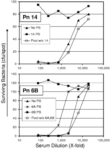
Vari-FIG. 1. Number of surviving bacteria (yaxis) at various dilutions of serum (xaxis). Serotype of pneumococci (Pn) was serotype 14 (top panel) or serotype 6B (bottom panel). For the top panel, the serum sample was absorbed with nothing (solid triangle), 5g/ml of serotype 14 PS (solid square), or a mixture of heterologous PS (open square). For the bottom panel, the serum sample was absorbed with nothing (solid triangle), 5g/ml of serotype 6B PS (solid square), 5g/ml of serotype 6A PS (symbol “X”), or a mixture of heterologous PS (open square). The heterologous PS pool had PS (5g/ml each) of all the unrelated serotypes, but PS of cross-reactive serotypes was omitted for serotypes 6A, 6B, 19A, and 19F. The cross-reactive PS was tested separately (also at 5g/ml). w/o, without.
TABLE 1. Bacteria strain composition of opsonization assay groups and their antibiotic resistance
Assay group
Assay group strain no.a
1 2 3 4
A OREP4 (DS2382-94) SPEC6B (BG25-9) STREP14 (DS2214-94) TREP19A (DS3519-97)
B OREP18C (GP116) SPEC19F (DS2217-94) EMC9V (1081748) EMC23F (1212458)
C OREP7F (DS2617-97) SPEC1 (L82006) STREP5 (DBL5) TREP6A (EF6796)
D OREP3 (Wu2) None None None
a
The names of the wild-type strains are included in parentheses. The antibiotics (and concentrations in overlay) used were the following: strain 1, optochin (2g/ml);
strain 2, spectinomycin (150g/ml); strain 3, streptomycin (250g/ml); strain 4, trimethoprim (25g/ml).
on August 17, 2020 by guest
http://cvi.asm.org/
ous amounts of free capsular PS are present in target bacterial preparations, and the PS can function as a competitive inhib-itor even at a very low concentration (⬍1g/ml) (6). We found that the washing of target bacteria reduced variability in assay sensitivity associated with using different preparations of target bacteria (data not shown). Hu et al. reported that assay plates for OPA required a unique shaking rate for each serotype (6). Since MOPA includes multiple serotypes simultaneously, one cannot employ serotype-specific shaking speeds. After investi-gating different shakers, we found that a shaker with a small shaking radius could produce comparable opsonization titers for all serotypes at shaking speeds from 650 to 800 rpm (data not shown). The shaker and 700 rpm were chosen for MOPA4. A major difficulty in OPA is in nonspecific killing of pneu-mococci by rabbit serum that is used as a source of comple-ment. For some serotypes, especially 6A, 6B, and 14, many lots of rabbit serum had a high level (ⱖ50%) of nonspecific killing (data not shown). Nonspecific killing was defined as 100⫻[1⫺ (CFU in wells containing rabbit serum and HL60)/(CFU in
wells with bacteria alone)]. All CFU were determined at the end of the assay due to bacterial growth during the assay. Nonspecific killing was considerably reduced when the phago-cytosis phase of the assay was performed in air containing 5% CO2(data not shown). For all subsequent assays, the
phago-cytosis for phase was performed in air with 5% CO2.
(iii) Assay validation. (a) Specificity.Once the assay
condi-tions were optimized, we investigated assay specificity by neu-tralizing a serum pool with homologous PS (5g/ml), a pool of heterologous capsular PS (5 g/ml of each), or with no PS before being tested in OPA (Fig. 1). Heterologous PS pools contained PS from all 13 serotypes, except for the homologous PS and PS from the same serogroup. For instance, the heter-ologous pool for 6B did not contain 6B and 6A PS but con-tained the remaining 11 PSs; for serotype 14, the heterologous PS pool contained 12 other PSs (all except for serotype 14). Evaluation was performed for all 13 serotypes using MOPA4, and two representative results are shown in Fig. 1. For serotype 14, the heterologous pool did not inhibit the opsonic capacity
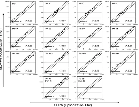
FIG. 2. Comparison of opsonization titers of 30 serum samples determined with MOPA4 (yaxis) and with single-serotype OPA (SOPA) (xaxis). Each panel contains an identity line (dotted line) and two lines indicating twofold deviation from identity (solid lines). The serotypes and ther2values are indicated in each panel. In the bottom row of the panels, two panels show comparisons for serotype 7F. In run 1, six samples (open
circles) showed a more-than-twofold deviation, but their results deviated less than twofold in run 2. In most panels, several samples had opsonization titers below the detection limit, which was 4. These samples were assigned an opsonization titer of 2, and the number of such serum samples is indicated in each panel. Pn, pneumococcal serotype.
on August 17, 2020 by guest
http://cvi.asm.org/
of the serum pool, but serotype 14 PS completely abrogated the opsonic capacity (upper panel). In the case of serotype 6B, opsonic activity was completely abrogated by 6B PS, reduced (⬃3-fold) by 6A PS, and unaffected by the heterologous PS mixture (lower panel). These data indicate that the multi-plexed OPA is serotype specific.
(b) Accuracy.A primary concern with the multiplexed OPA
was its ability to produce results comparable to those with the single-serotype OPA. To address this issue, 30 serum samples (5 prevaccination, 25 postvaccination) were tested in the sin-gle-serotype OPA format and the MOPA4 format (Fig. 2). The data for all 13 serotypes are shown in Fig. 2 along with the line of identity (dotted line) and lines showing twofold deviation (solid lines) from the identity for each serotype. The data obtained from each format agreed very well, and only 22 data points out of 390 data points (13 serotypes/sample⫻30 sam-ples) deviated more than twofold (i.e., outside of the two solid lines). Since six outlier data points were found in 7F serotype (run 1), we investigated potential difficulties in the 7F assay by retesting the six samples. Upon retesting (run 2), the six sam-ples produced comparable results (Fig. 2, bottom two panels). Thus, we believe the outliers were not due to assay inaccuracy but rather random errors.
(c) Precision.Intra-assay variation for the 13 serotypes was
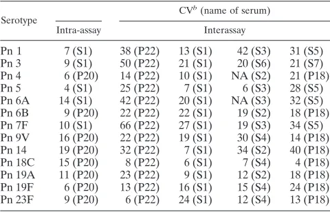
assessed by testing a serum pool 10 times in a single assay run. As shown in Table 2, the average coefficient of variation (CV) was about 10%, ranging from 4% to 19%. Interassay variation was estimated for the 13 serotypes by testing several sera in six independent experiments over a 2-week interval (Table 2). For this study, the same lot of target bacteria and lot of comple-ment were used for each assay. Under these conditions, the overall average CV of the serum samples for 13 serotypes was 22%. No specific serotypes could be associated with high CV. There was a slight trend for high CVs for samples with high OT, suggesting that serial dilution errors of these samples may
slightly increase the imprecision. Two cases with high CVs (ⱖ50%) were observed with one sample (pool 22) but not with other samples. Pool 22 was made by converting plasma to serum, whereas all other samples were collected as serum. Thus, the high CVs may be associated with the sample (pool 22) itself rather than the assay.
(iv) Effect of different E:T ratios.As a part of assessing the
robustness of MOPA4, we determined the effect of different numbers of HL60 cells in each well (8⫻105, 4⫻105, 2⫻105,
or 1⫻105cells/well) by performing OPA for all 13 serotypes
(serotype 3 in single-serotype OPA format, other serotypes in MOPA4 format) with one serum sample (serum 1) with a constant number of bacteria (500 CFU/well of each strain) per well (Fig. 3). The OT for most serotypes tended to decrease as effector-to-target (E:T) ratios were reduced from 400:1 to 100:1, but the reduction was modest. When an E:T ratio of 200:1 was compared to 400:1, the maximum OT reduction for all serotypes was less than 20%, except serotypes 6B (25%) and 9V (44%). When an E:T ratio of 100:1 was compared to 200:1, the maximal reduction in OT was 29% for 23F and 21% for 9V but remained less than 20% for all other serotypes. When the E:T ratio was reduced to 50:1, however, the reduction in OT was more pronounced and occurred in multiple serotypes. The reduction, however, was not consistently reproduced (data not shown). For the 13 serotypes, the average OT at 200:1 was 86% of that obtained at 400:1, and the average OT at 100:1 was 92% of that at 200:1. These data show that the E:T ratio of 200:1 used in the MOPA4 produces results comparable to the con-ventional ratio of 400:1 and has an adequate margin of safety (twofold).
DISCUSSION
To accommodate large demands for OPA, we have devel-oped and optimized a fourfold multiplexed OPA (MOPA4) for pneumococcal antibodies against 13 serotypes. Once opti-mized, MOPA4 was found to be robust: assay conditions could be varied slightly without significant changes in assay results. Above all, E:T ratios can be changed twofold with little
vari-FIG. 3. Opsonization titers of one serum sample for 13 serotypes with the E:T ratio of 400:1 (hatched bars), 200:1 (open bars), 100:1 (gray bars), or 50:1 (black bars). Because serotype 3 is an odd member, it was tested in the single-serotype OPA format. All other serotypes were tested in MOPA4 format. Since opsonization titers for serotypes 6A, 9V, and 23F displayed a marked trend of decreasing opsonization titers with decreasing E:T ratios, the experiment was repeated for these three serotypes. This trend was not reproducible (data not shown).
TABLE 2. Intra- and interassay precision of MOPA4a
Serotype
CVb(name of serum)
Intra-assay Interassay
Pn 1 7 (S1) 38 (P22) 13 (S1) 42 (S3) 31 (S5) Pn 3 9 (S1) 50 (P22) 21 (S1) 20 (S6) 21 (S7) Pn 4 6 (P20) 14 (P22) 10 (S1) NA (S2) 21 (P18) Pn 5 4 (S1) 25 (P22) 7 (S1) 6 (S3) 28 (S5) Pn 6A 14 (S1) 42 (P22) 20 (S1) NA (S3) 32 (S5) Pn 6B 9 (P20) 22 (P22) 22 (S1) 19 (S2) 18 (P18) Pn 7F 10 (S1) 66 (P22) 27 (S1) 19 (S3) 34 (S5) Pn 9V 16 (P20) 22 (P22) 19 (S1) 30 (S4) 14 (P18) Pn 14 19 (P20) 32 (P22) 7 (S1) 34 (S2) 40 (P18) Pn 18C 15 (P20) 8 (P22) 6 (S1) 7 (S4) 4 (P18) Pn 19A 11 (P20) 23 (P22) 9 (S1) 12 (S2) 18 (P18) Pn 19F 6 (P20) 13 (P22) 16 (S1) 15 (S4) 24 (P18) Pn 23F 9 (P20) 6 (P22) 24 (S1) 12 (S4) 13 (P18)
aCV is expressed in percentages. NA (not applicable) was used when the
sample had results below the detection limit for the serotype. The name of the serum sample is listed in parenthesis. P18, P20, and P22 indicate pool 18, pool 20, and pool 22, respectively. S1 through S7 indicate single-donor serum 1 through serum 7, respectively. These sera were postvaccination sera.
bIntra-assay average CV, 10%; interassay average CV, 22%. When the
interassay CV was determined with the samples diluted only once, the inter-assay CV could be reduced to 15%. The average intra-inter-assay CV was deter-mined from 10 experiments; the average interassay CV was deterdeter-mined from 6 experiments.
on August 17, 2020 by guest
http://cvi.asm.org/
ation in results. In addition, we found that opsonization titers obtained by MOPA4 did not change when the assay was per-formed with a different combination of pneumococcal sero-types (r2⬎0.98) (data not shown). Also, the degree of
multi-plexing was not significant, and we could obtain comparable results with the twofold, threefold, and fourfold MOPA (data not shown).
MOPA4 was validated by investigating the assay’s precision, accuracy, and specificity. Not surprisingly, the assay was found to be highly specific. Since no OPA standards with assigned values are currently available, we assessed the assay’s accuracy by comparing MOPA4 results with those obtained with a sin-gle-serotype OPA. Both assays were found to yield similar results, and deviations from the identity were less than twofold in most samples in our comparison studies. Although MOPA4 gave slightly higher results than the single-serotype OPA in serotype 7F for several samples with low titers, the difference was not reproducible. In the future, we plan to compare our MOPA4 with other single-serotype OPAs by studying samples from vaccinated children and exchanging samples with other laboratories that perform single-serotype OPAs.
Under our assay conditions, we found the MOPA4 to be precise, with an interassay CV of about 20%. Indeed, this level of CV is comparable to those observed for ELISA results (12). Since this is short-term interassay precision, assay precision would be reduced as the assay is performed for a longer period using different batches of bacteria and complement. Thus, we are investigating various approaches to improve precision fur-ther, such as the use of a standard serum for each assay and a data analysis program that would utilize data obtained at all serum dilutions. With these improvements, we believe that MOPA4 would become as precise as ELISA, even though MOPA4 is a bioassay.
It is difficult to demonstrate assay sensitivity, because there are no established OPA standards. Since most serum samples obtained after a vaccination have measurable opsonization ti-ters, our MOPA4 appears to have adequate assay sensitivity. However, assay sensitivity may depend on target bacteria, since we observed up to 5- to 10-fold variation in the magnitude of opsonization titers when different target bacteria were used (unpublished observation). Interstrain variation may occur be-cause strains produce different amounts of capsule, express different phases (8), or differ in the expression of molecules affecting complement deposition on the bacterial surface (13). Due to these considerations, a set of target strains of pneumo-cocci should be adopted as the standard. We plan to make our antibiotic-resistant bacteria available to qualified investigators. Since pneumococci are pathogens, the safety of using anti-biotic-resistant pneumococci should be a concern. However, MOPA4 uses antibiotics that are not relevant to managing pneumococcal infections. Also, we used natural antibiotic re-sistance by pneumococci and did not insert “antibiotic resis-tance” genes which could be transferred to other bacteria. In addition, we used an overlay method which seals the pneumo-cocci within the agar plate and prevents exposure of the patho-gen to the air. In addition to reducing biohazard, we found the overlay kept the colony size of type 3 pneumococci small. In conventional OPA, because of their large colony size, type 3 pneumococci required large agar surfaces, and this caused significant practical problems. Although the antibiotics we
used do not pose serious safety concerns, one may develop target bacterial strains that depend on specific nutrient factors. In this way, the multiplex assay can be performed without using antibiotics.
Because of its importance in pneumococcal vaccine devel-opment, various multiplexed opsonization assay formats have been developed (2, 9–11). While we have described here a killing-type multiplexed opsonization assay, Martinez et al. de-veloped a flow-cytometric multiplexed opsonization assay (10). This approach uses, in place of target pneumococci, a mixture of latex particles that have distinct fluorescent characteristics and are coated with different capsular PSs. The particles are opsonized with antisera and complement, and their uptake into phagocytes is then determined with a flow cytometer. The flow-cytometric assay uses artificial targets instead of bacteria, measures phagocytosis but not actual killing, requires an ex-pensive instrument, and requires more hands-on effort than the opsonophagocytic killing assay employing automated col-ony counting. Also, a killing-type multiplexed opsonization assay is similar to the conventional opsonization assay used as the reference assay in the past (17). Thus, multiplexed opsonophagocytic killing assays may be easier to adopt and validate than the multiplexed phagocytosis assay. Nevertheless, these multiplexed opsonization assays may be robust and rapid enough to replace pneumococcal antibody ELISA, which may not be specific for functional antibodies (1, 18, 22).
ACKNOWLEDGMENT
The work was supported by an NIH contract (AI-30021) from NIH/ NIAID/DMID.
REFERENCES
1.Bhushan, R., B. F. Anthony, and C. E. Frasch.1998. Estimation of group B streptococcus type III polysaccharide-specific antibody concentrations in
hu-man sera is antigen dependent. Infect. Immun.66:5848–5853.
2.Bogaert, D., M. Sluijter, R. De Groot, and P. W. Hermans.2004. Multiplex opsonophagocytosis assay (MOPA): a useful tool for the monitoring of the
7-valent pneumococcal conjugate vaccine. Vaccine22:4014–4020.
3.Briles, D. E., M. Nahm, K. Schroer, J. Davie, P. Baker, J. Kearney, and R. Barletta.1981. Antiphosphocholine antibodies found in normal mouse
se-rum are protective against intravenous infection with type 3Streptococcus
pneumoniae. J. Exp. Med.153:694–705.
4.Fedson, D. S., and D. M. Musher.1994. Pneumococcal vaccine, p. 517–564.
InS. A. Plotkin and E. A. Mortimer (ed.), Vaccines, 2nd ed. W. B. Saunders
Co., Philadelphia, Pa.
5.Fleck, R. A., S. Romero-Steiner, and M. H. Nahm.2005. Use of HL-60 cell line to measure opsonic capacity of pneumococcal antibodies. Clin. Diagn.
Lab. Immunol.12:19–27.
6.Hu, B. T., X. Yu, T. R. Jones, C. Kirch, S. Harris, S. W. Hildreth, D. V. Madore, and S. A. Quataert.2005. Approach to validating an
opsonophago-cytic assay forStreptococcus pneumoniae. Clin. Diagn. Lab. Immunol.12:
287–295.
7.Jodar, L., J. C. Butler, G. Carlone, R. Dagan, C. E. Frasch, D. Goldblatt, H. Ka¨yhty, K. Klugman, B. D. Plikaytis, G. Siber, R. Kohberger, I. Chang, and T. Cherian.2003. Serological criteria for evaluation and licensure of
pneu-mococcal conjugate vaccine formultions for use in infants. Vaccine21:3265–
3272.
8.Kim, J. O., S. Romero-Steiner, U. B. Sorensen, J. Blom, M. Carvalho, S. Barnard, G. Carlone, and J. N. Weiser.1999. Relationship between cell surface carbohydrates and intrastrain variation on opsonophagocytosis of
Streptococcus pneumoniae. Infect. Immun.67:2327–2333.
9.Kim, K. H., J. Yu, and M. H. Nahm.2003. Efficiency of a pneumococcal opsonophagocytic killing assay improved by multiplexing and by coloring
colonies. Clin. Diagn. Lab. Immunol.10:616–621.
10.Martinez, J. E., E. A. Clutterbuck, H. Li, S. Romero-Steiner, and G. M. Carlone.2006. Evaluation of multiplex flow cytometric opsonophagocytic
assays for determination of functional anticapsular antibodies to
Streptococ-cus pneumoniae. Clin. Vaccine Immunol.13:459–466.
11.Nahm, M. H., D. E. Briles, and X. Yu.2000. Development of a
multi-specificity opsonophagocytic killing assay. Vaccine18:2768–2771.
on August 17, 2020 by guest
http://cvi.asm.org/
12.Plikaytis, B. D., D. Goldblatt, C. E. Frasch, C. Blondeau, M. J. Bybel, G. S. Giebink, I. Jonsdottir, H. Kayhty, H. B. Konradsen, D. V. Madore, M. H. Nahm, C. A. Schulman, P. F. Holder, T. Lezhava, C. M. Elie, and G. M. Carlone.2000. An analytical model applied to a multicenter pneumococcal
enzyme-linked immunosorbent assay study. J. Clin. Microbiol.38:2043–2050.
13.Ren, B., A. J. Szalai, S. K. Hollingshead, and D. E. Briles.2004. Effects of PspA and antibodies to PspA on activation and deposition of complement on
the pneumococcal surface. Infect. Immun.72:114–122.
14.Rennels, M. B., K. M. Edwards, H. L. Keyserling, K. S. Reisinger, D. A. Hogerman, D. V. Madore, I. Chang, P. R. Paradiso, F. J. Malinoski, and A. Kimura. 1998. Safety and immunogenicity of heptavalent pneumococcal
vaccine conjugated to CRM197 in United States infants. Pediatrics101:604–
611.
15.Robbins, J. B., R. Austrian, C. J. Lee, S. C. Rastogi, G. Schiffman, J. Henrichsen, P. H. Makela, C. V. Broome, R. R. Facklam, R. H. Tiesjema, and J. C. Parke, Jr.1983. Considerations for formulating the second-gen-eration pneumococcal capsular polysaccharide vaccine with emphasis on the
cross-reactive types within groups. J. Infect. Dis.148:1136–1159.
16.Romero-Steiner, S., C. E. Frasch, G. Carlone, R. A. Fleck, D. Goldblatt, and M. H. Nahm.2006. Use of opsonophagocytosis for the serological evaluation
of pneumococcal vaccines. Clin. Vaccine Immunol.13:165–169.
17.Romero-Steiner, S., D. Libutti, L. B. Pais, J. Dykes, P. Anderson, J. C. Whitin, H. L. Keyserling, and G. M. Carlone.1997. Standardization of an opsonophagocytic assay for the measurement of functional antibody activity
againstStreptococcus pneumoniaeusing differentiated HL-60 cells. Clin.
Diagn. Lab. Immunol.4:415–422.
18.Romero-Steiner, S., D. M. Musher, M. S. Cetron, L. B. Pais, J. E. Groover, A. F. Fiore, B. D. Plikaytis, and G. M. Carlone.1999. Reduction in functional
antibody activity against Streptococcus pneumoniaein vaccinated elderly
individuals highly correlates with decreased IgG antibody avidity. Clin.
In-fect. Dis.29:281–288.
19.Rubins, J. B., M. Alter, J. Loch, and E. N. Janoff.1999. Determination of antibody responses of elderly adults to all 23 capsular polysaccharides after
pneumococcal vaccination. Infect. Immun.67:5979–5984.
20.Shinefield, H. R., and S. Black.2000. Efficacy of pneumococcal conjugate
vaccines in large scale field trials. Pediatr. Infect. Dis. J.19:394–397.
21.Shinefield, H. R., S. Black, P. Ray, I. Chang, N. Lewis, B. Fireman, J. Hackell, P. R. Paradiso, G. Siber, R. Kohberger, D. V. Madore, F. J. Malinowski, A. Kimura, C. Le, I. Landaw, J. Aguilar, and J. Hansen.1999. Safety and immunogenicity of heptavalent pneumococcal CRM197
conju-gate vaccine in infants and toddlers. Pediatr. Infect. Dis. J.18:757–763.
22.Wernette, C. M., C. E. Frasch, D. Madore, G. Carlone, D. Goldblatt, B. Plikaytis, W. Benjamin, S. A. Quataert, S. Hildreth, D. J. Sikkema, H. Kayhty, I. Jonsdottir, and M. H. Nahm.2003. Enzyme-linked immuno-sorbent assay for quantitation of human antibodies to pneumococcal
poly-saccharides. Clin. Diagn. Lab. Immunol.10:514–519.
23.Whitney, C. G., M. M. Farley, J. Hadler, L. H. Harrison, N. M. Bennett, R. Lynfield, A. Reingold, P. R. Cieslak, T. Pilishvili, D. Jackson, R. R. Facklam, J. H. Jorgensen, and A. Schuchat.2003. Decline in invasive pneumococcal disease after the introduction of protein-polysaccharide conjugate vaccine.
N. Engl. J. Med.348:1737–1746.