N A N O E X P R E S S
Open Access
Capillary-driven surface-enhanced Raman
scattering (SERS)-based microfluidic chip for abrin
detection
Hao Yang
1†, Min Deng
2†, Shan Ga
1†, Shouhui Chen
2, Lin Kang
1, Junhong Wang
1, Wenwen Xin
1, Tao Zhang
1,
Zherong You
1, Yuan An
1, Jinglin Wang
1*and Daxiang Cui
2*Abstract
Herein, we firstly demonstrate the design and the proof-of-concept use of a capillary-driven surface-enhanced Raman scattering (SERS)-based microfluidic chip for abrin detection. The micropillar array substrate was etched and coated with a gold film by microelectromechanical systems (MEMS) process to integrate into a lateral flow test strip. The detection of abrin solutions of various concentrations was performed by the as-prepared microfluidic chip. It was shown that the correlation between the abrin concentration and SERS signal was found to be linear within the range of 0.1 ng/mL to 1μg/mL with a limit of detection of 0.1 ng/mL. Our microfluidic chip design enhanced the operability of SERS-based immunodiagnostic techniques, significantly reducing the complication and cost of preparation as compared to previous SERS-based works. Meanwhile, this design proved the superiority to conventional lateral flow test strips in respect of both sensitivity and quantitation and showed great potential in the diagnosis and treatment for abrin poisoning as well as on-site screening of abrin-spiked materials.
Keywords:Capillary force; Microfluidic chip; SERS; Abrin; Phytotoxin
Background
Recently, ricin has caught the public's attention by the toxin-tainted letters sent to US President Barack Obama, Mississippi Senator Roger Wicker, and a Mississippi just-ice official, while abrin, its 70 times more toxic analogue, is less known to the general public. Abrin and ricin are toxic proteins with similar structure and properties, both of which are classified as category B select agents by the US Health and Human Services [1]. Compared with ricin, abrin is much more poisonous with an estimated human fatal dose of 0.1~1.0 μg/kg [2]. Although there are re-ported deaths on account of intentional poisoning, most cases occur in children by unintentional ingestion [3].
After ingestion, the major symptoms of abrin poisoning may occur in less than 6 h, and the deaths in children dying of ingestion of one or more abrin seeds have been documented in literature [4]. Therefore, a fast, readily available confirmatory testing will greatly facilitate the timely diagnosis and treatment for abrin poisoning.
Surface-enhanced Raman scattering (SERS) is a surface-sensitive technique that provides a highly enhanced Raman signal from Raman-active molecules that have been adsorbed onto rough metal surfaces. The reported surface enhancement factor ranges from 103to 1015, which means that the technique may detect proper analytes at a single molecule level [5-8]. There are two effects, chemical and electromagnetic, attributing to the enhancement. The former involves the formation of a charge-transfer state be-tween the metal surface and adsorbate, contributing 1 to 2 orders of magnitude to the overall enhancement, while the latter is the dominant effect, arising from the collective os-cillation of conduction electrons due to the irradiation of a metal by light [8]. Besides high sensitivity, the Raman scat-ter possesses 10~100 times narrower bands than those of fluorescence and excellent anti-photobleaching properties,
* Correspondence:[email protected];[email protected]
†Equal contributors
1State Key Laboratory of Pathogen and Biosecurity, Beijing Institute of
Microbiology and Epidemiology, No. 20 Dongda StreetFengtai District, Beijing 100071, People's Republic of China
2
Institute of Nano Biomedicine and Engineering, Key Laboratory for Thin Film and Microfabrication Technology of the Ministry of Education, Research Institute of Micro/Nano Science and Technology, Shanghai Jiao Tong University, Dongchuan Road 800, 200240 Shanghai, People's Republic of China
which avail to reduce undesirable spectral overlap and pro-vide long and stable signal readout [9]. So far, there have been many different SERS-based analytical techniques that have been developed for cancer markers, infectious dis-eases, pH sensing, etc. [8-15]. These techniques unleash tremendous potential for ultrasensitive biomedical analysis. However, it still remains a great challenge to reduce the overall cost while maintaining the advantages of sensitivity, because most SERS-based detection systems are strongly dependent on the relatively expensive process of micro-electromechanical systems (MEMS), especially sputtering of a noble metal layer.
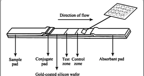
Herein, we introduce a proof-of-concept use of the capillary-driven SERS-based microfluidic chip for abrin detection (Figure 1). A micropillar array was fabricated by MEMS process on silicon wafer and sputtered with noble metal. After proper hydrophilic modification, anti-abrin polyclonal antibodies and secondary antibodies were immobilized on different places of the micropillar array as the detection zone and control zone. The sam-ple liquid dissolved the external anti-abrin SERS probes in the conjugate pad and reacted with them and then was driven through the whole micropillar array by capil-lary action. The detection signal was provided by the external SERS probes captured on the detection and control zones. This proof-of-concept design combined the advantages of SERS-based detection and previous ca-pillary action-driven chip, providing a novel and feasible solution for the application of SERS-based point-of-care test (POCT).
Methods
All animal experiments (No. SYXK2007-0025) were approved by the Institutional Animal Care and Use Committee of Shanghai Jiao Tong University.
Extraction of natural abrin
Natural abrin was extracted according to the previous method with slight modifications [16]. Briefly, the decor-ticated seeds ofAbrus precatorius(approximately 100 g)
were soaked in 200 mL of 0.01 M phosphate buffer solu-tion (PBS) at pH 7.4 and 4°C for 24 h. After thorough homogenization, the puree was centrifuged at 10,000g for 30 min. Then, the aqueous layer was saturated with ammonium sulfate (95%w/v) and centrifuged at 10,000g for 30 min. The precipitate was dissolved in 100 mL of 0.01 M PBS and applied to a 1.5 × 10 cm Gal-agarose column (EY Laboratories Inc., San Mateo, CA, USA). The bound abrin was eluted with 0.125 M D-galactose solution. The collected fractions were dialyzed and applied to a Sephacryl S-100 prepacked column (GE Healthcare Bio-Sciences Corp, Piscataway, NJ, USA) equilibrated in PBS. The as-prepared abrin was analyzed by 15% sodium dodecyl sulfate polyacrylamide gel elec-trophoresis (SDS-PAGE).
Preparation of anti-abrin polyclonal antibodies
The purified abrin was inactivated by formalin and used to hyperimmunize a rabbit, and 0.5 mL of abrin toxoid (80 mg/mL) was mixed with an equal volume of Freund's complete adjuvant and injected subcutaneously to the rabbit. Seven days later, immunization was carried out four times including one booster immunization with the mixture of the abrin toxoid and Freund's incomplete adju-vant as well as three injections with the toxoid at weekly intervals. Ten days after the final injection, the immunized blood was collected by jugular puncture, and the serum was separated for subsequent purification of anti-abrin polyclonal antibodies with rProtein A Sepharose Fast Flow (GE Healthcare Bio-Sciences Corp., Piscataway, NJ, USA). The antibody titers were evaluated by enzyme-linked immunosorbent assay (ELISA).
Preparation of external SERS probes
[image:2.595.56.291.580.704.2]The external SERS probes were prepared according to a published method [6]. DTNB (5,5′-dithiobis (2-nitroben-zoic acid), Sigma-Aldrich Co. LLC, St. Louis, MO, USA) was used as the Raman-active tag. One milliliter of puri-fied anti-abrin polyclonal antibodies (approximately 75 mg/mL in 0.01 M PBS) was dropwise added to 1 mL of 20-nm colloidal gold solution (Sigma-Aldrich Co. LLC) under stirring. After 1 h of incubation at 4°C, the antibody-coated colloidal gold was separated by centrifu-gation at 12,000g for 1 h. Bovine serum albumin (BSA) was used to block the unmodified colloidal gold at a final concentration of 0.5% (w/v). The labeled colloidal gold was centrifuged at 12,000gfor 1 h and resuspended in 1 mL 0.01 M PBS solution. Twenty microliters of DTNB solution (1 mM in 0.01 M PBS) was added to the gold solution and incubated at 4°C for 1 h. The resultant SERS probes were centrifuged again at 12,000g for 1 h and then resuspended in 0.01 M PBS for later use. Figure 1The schematic view of capillary-driven SERS microfluidic
Fabrication and surface modification of gold-coated silicon wafer
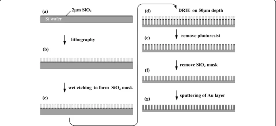
The gold-coated silicon wafer was fabricated by MEMS technique. The process was shown in Figure 2. Firstly, a 2-μm-thick layer of SiO2was grown onto a 3-in. Si wafer (Mouser Ltd., Hefei, China) using wet oxidation in a thermal furnace (TS-6304, Tempres Ltd., Vaasen, The Netherlands). Then, a photoresist (AZ 4562, Micro Chemicals Ltd., Japan) was spin-coated at 3,000 rpm to a thickness of approximately 20 μm and soft-baked for 90 min at 80°C. The layer was patterned subsequently by photolithography. The buffered hydrofluoric acid (BHF, composition of BHF solution for SiO2etching: HF 84 mL, NH4F 339 g, H2O5 10 mL; etching condition: 45°C, pH 3) was used to etch SiO2 uncovered by the photoresist. Afterwards, the microchips were etched into Si to a depth of 50 μm using deep reactive ion etching (DRIE, AMS-200SE, Annecy Cedex Ltd., France), and the micro silicon cylindrical array formed. The photo-resist and SiO2 mask were removed by acetone (Great Fortune, Zibo Ltd, China) and DRIE, respectively. The as-prepared chips were cut into strips (1 × 4 mm) using a laser scribing apparatus (WL-9030, Titan Ltd., USA). After being cleaned with the reactive ion etcher (Nex-tral-100, Alcatel Ltd., France) at 30 W and 1.2 Torr for 45 s, the chips were then incubated in a solution of acetone for 20 min, rinsed with deionized water, and dried under an N2 stream. The deposition of gold film (200 nm) on the chip was carried out with the sputtering system (ZT-550, L-H Ltd., Germany). After being soaked in piranha solution (H2SO4/30% H2O2= 3:1) for 10 min,
the gold-coated chips were cleaned with deionized water and rinsed in 1 mM of HS-C2H4-CONH-PEG-C3H6 -COOH (Rapp Polymere GmbH Ltd., Tuebingen, Germany) solution for 4 h. Finally, the chips were cleaned with deion-ized water and dried at room temperature. Scanning elec-tron microscopy (SEM) (JEOL Ltd. Tokyo, Japan) was used to explore the surface ultrastructure of the as-prepared chip. PBS was used to evaluate the flow rate of the sample solution on the chip.
Assembly of capillary-driven chip
Anti-abrin polyclonal antibodies and goat anti-rabbit secondary antibodies (1 mg/mL) were dispensed on the gold-coated wafer with a Biodot XYZ3000 dispenser (Biodot Inc., Irvine, CA, USA) as test zone and control zone, respectively. After drying for 30 min, the wafer was blocked with PBS containing 1% BSA (w/v). The SERS probes were printed on a glass fiber filter as conju-gate pad and dried at room temperature. The absorbent pad, conjugate pad, and sample pad were cut into strips of 1 mm in width with a guillotine cutter and overlapped on the laminating card with the dried wafer as shown in Figure 1.
SERS signal measurement
[image:3.595.58.539.474.694.2]The purified abrin was diluted with a series of concen-trations from 0.1 ng/mL to 10 mg/mL in 0.01 M PBS solution. Fifty microliters of the diluted toxin solution was added to the sample pad, and the SERS signal was read out with i-Raman-785S (B&W TEK Inc., Newark, DE, USA)
after 5 min. The intensity of the peak at approximately 1,330 cm−1was used to quantify the abrin in PBS solution.
Results and discussion
Characterization of natural abrin and anti-abrin antibody Abrin consists of two subunits which are linked by a di-sulfide bond between Cys247 of the A subunit and Cys8 of the B subunit [2]. Their molecular weights are ap-proximately 30 and 35 kDa, respectively. The A subunit inactivates ribosomes and inhibits protein synthesis, while the B subunit shows the activity of lectin that facilitates the entrance into the cell. The extraction of natural abrin with high purity is the key in production of polyclonal antibody, which determines the quality of in-duced antibody. However, the process of the purification of abrin from seeds of A. precatorius was complicated due to the existence of abundant agglutinin that pos-sesses nearly identical galactose-binding properties as abrin. Given their differences in galactose-binding avidity and molecular mass between the abrin and agglutinin, a two-step purification was exploited to separate abrin from raw extracts (Figure 3). As shown in Figure 2, the purified abrin in the final step could be broken into two subunits under reducing condition, and the sizes of bands were in accordance with their theoretical molecu-lar weight. In addition, the purity was over 95% by Quan-tity One software analysis (Bio-Rad Laboratories Inc., Hercules, CA, USA). After being inactivated with forma-lin, the abrin toxoid was used to produce polyclonal
antibody. In this experiment, the as-prepared antibody could yield a positive result by ELISA under 100,000-fold dilution, which reflected the good immunogenicity of the abrin toxoid and good affinity of the antibodies.
Characterization of microfluidic chip
The assembled microchip is shown in Figure 4. From the appearance, it resembled a traditional lateral flow (LF) test strip except for its width (1 mm) and gold-coated substrate. The SEM image showed the micropil-lar array on the chip. The micropilmicropil-lars were about 50
[image:4.595.58.541.458.704.2]μm high and had a diameter of 35 μm and a center-to-center distance of 90 μm. The flow rate of PBS was about 4 mm/s on the chip. In this experiment, the design of microchip referred to the microstructure of micropost array of 4castchip® developed by Åmic AB [17,18]. It is important to note that the LF strip is one of the most successful commercial POCT products. So far, there was no available commercial POCT product that overmatches the lateral flow test strip in cost and uni-versality of application. However, the main weaknesses of the colloidal gold or latex-based traditional LF test trip are sensitivity and quantitation as a result of the in-trinsic property of the cellulose membrane [19-22]. Par-ticularly, it is only the superficial colorimetric signal that could be used for quantitation, while the deep signal in the membrane is lost. The planar structure of 4castchip® addressed the problem well and retained the capability of capillary-driven force. However, it is obvious that the
cost for sputtering noble metal will be high if this struc-ture is wholly introduced into the SERS-based chip. In fact, except for cellulose membrane, other components of the traditional LF strip function well and cost little in practical application. Therefore, only the cellulose mem-brane was replaced by the gold-coated micropillar array substrate in our design strategy. This strategy has several advantages as follows: First of all, it economizes the consumption of gold-coated substrate and facilitates the homogeneous batch processing. Second, it is a mature technique in practice to process the sample pad, conju-gate pad, and absorbent pad, which works well and does not need further optimization. Third, wide center-to-center distance guarantees a good passing ability, avoid-ing the blockavoid-ing of possible residual coarse materials passing through sample pad in samples. Last but not the least, this design, which decreased the width of flow path in conventional LF test strips from 4 to 1 mm, facilitates the enriching of analytes on the surface of the capture zone, improving the sensitivity under the condition of high flow rate. In other words, this strategy reduced not only the complication of fabrication, but also the overall cost.
Analysis of abrin-spiked sample
Figure 5 shows the SERS spectra of the abrin-spiked sample at various concentrations. The intensity of the peak at approximate 1,330 cm−1was proportional to the concentration of abrin in the PBS solution. The concen-tration of abrin ranged from 0.1 ng/mL to 1μg/mL. The characteristic peak under 0.1 ng/mL became difficult to distinguish from that of the blank sample, indicating the limit of detection (LOD). Because of the absence of the washing step, some SERS probes remained on the gold-coated substrate, resulting in a weak nonspecific binding peak at approximate 1,330 cm−1. Figure 6 shows the dose-response curve calculated by averaging the readout
at three different locations of each concentration from 0.1 to 100 ng/mL. The linear regression equation was y=1,430.7x−2,312.5 and the correlation coefficient (R2) was 0.9902. The LOD of this capillary-driven SERS-based microfluidic chip was 0.1 ng/mL.
[image:5.595.58.293.86.273.2]As previously mentioned, SERS-based techniques showed many potential advantages including high sensitivity, nar-row bandwidths, and photobleaching resistance. It still remains a challenge to develop a SERS-based immunodiag-nostic technique of both low cost and good operability. Some pioneering researchers have published their works focusing on the ultrasensitivity from the level of picograms per milliliter to femtograms per milliliter [6,8,9,11,14,23-28]. Compared with their work, our de-sign strategy emphasized the operability of SERS-based technique. In other words, this strategy is aimed at not just a comparative LOD, but a balanced solution Figure 4Characterization of capillary-driven SERS
[image:5.595.307.539.88.255.2]microfluidic chip.
Figure 5SERS spectra of the abrin-spiked sample at different concentrations.
[image:5.595.305.539.520.704.2]concentrations
between the complication of new techniques and the universality of traditional ones.
Conclusions
We have designed and demonstrated the proof-of-concept use of our capillary-driven SERS-based microfluidic chip for abrin detection. The microfluidic chip was fabricated by MEMS process. The combination of the traditional LF test strip with capillary-driven gold-coated substrate results in the enhancement of sensitivity as well as the reduction of cost for SERS-based immunodiagnostic tech-niques. In this work, a calibration curve was obtained to detect the concentration of abrin in the range from 0.1 ng/mL to 1μg/mL, which is superior to the traditional LF test strip for the same purpose in respect of both sensitiv-ity and quantitation [21]. What is critically important is the operability of our design strategy, that is, the perform-ance of traditional LF test strips is improved without ex-cessive increase in complication and cost of fabrication. In addition, this SERS-based microfluidic chip can be further developed and applied to other on-demand and point-of-care detection for a substance of interest.
Competing interests
The authors have declared that no competing interest exists.
Authors’contributions
HY carried out antibody preparation and SERS experiments. MD finished the microfabrication of the micropillary chip. SG finished the surface modification of the micropillary chip. SHC finished the antibody conjugation with the surface of the chip. LK and JW finished the characterization of the chip. WX, TZ, and ZY finished the result analysis. HY and YA finished the draft. JW and DC finished the experiment design and manuscript revision. All authors of this paper have read and approved the final manuscript.
Acknowledgements
This work is supported by the National Key Basic Research Program (973 Project) (No. 2011CB933101), National Natural Scientific Fund (Nos. 81225010, 81327002, and 31100717), 863 project of China (2012AA022703), Shanghai Science and Technology Fund (Nos. 13NM1401500 and 11nm0504200), and Shanghai Jiao Tong University Innovation Fund for Postgraduates (No. AE340011).
Received: 19 February 2014 Accepted: 11 March 2014 Published: 24 March 2014
References
1. Felder E, Mossbrugger I, Lange M, Wolfel R:Simultaneous detection of ricin and abrin DNA by real-time PCR (qPCR).Toxins2012,4:633–642. 2. Dickers KJ, Bradberry SM, Rice P, Griffiths GD, Vale JA:Abrin poisoning.
Toxicol Rev2003,22:137–142.
3. Jang DH, Hoffman RS, Nelson LS:Attempted suicide, by mail order: Abrus precatorius.J Med Toxicol2010,6:427–430.
4. Balali-Mood M, Moshiri M, Etemad L:Medical aspects of bio-terrorism.
Toxicon2013,69:131–142.
5. Yang D-P, Chen S, Huang P, Wang X, Jiang W, Pandoli O, Cui D: Bacteria-tem-plate synthesized silver microspheres with hollow and porous structures as excellent SERS substrate.Green Chem2010,12:2038–2042.
6. Lin CC, Yang YM, Chen YF, Yang TS, Chang HC:A new protein A assay based on Raman reporter labeled immunogold nanoparticles.Biosens Bioelectron2008,24:178–183.
7. Nie S, Emory SR:Probing single molecules and single nanoparticles by surface-enhanced Raman scattering.Science1997,275:1102–1106.
8. Porter MD, Lipert RJ, Siperko LM, Wang G, Narayanan R:SERS as a bioassay platform: fundamentals, design, and applications.Chem Soc Rev2008,
37:1001–1011.
9. Grubisha DS, Lipert RJ, Park HY, Driskell J, Porter MD:Femtomolar detection of prostate-specific antigen: an immunoassay based on surface-enhanced Raman scattering and immunogold labels.Anal Chem2003,
75:5936–5943.
10. Zong S, Wang Z, Chen H, Hu G, Liu M, Chen P, Cui Y:Colorimetry and SERS dual-mode detection of telomerase activity: combining rapid screening with high sensitivity.Nanoscale2014,6:1808–1816.
11. Wu P, Gao Y, Lu Y, Zhang H, Cai C:High specific detection and near-infrared photothermal therapy of lung cancer cells with high SERS active aptamer-silver-gold shell-core nanostructures.Analyst2013,138:6501–6510. 12. Zhang P, Zhang R, Gao M, Zhang X:Novel nitrocellulose membrane
substrate for efficient analysis of circulating tumor cells coupled with surface-enhanced Raman scattering imaging.ACS Appl Mater Interfaces 2014,6:370–376.
13. Yuen C, Liu Q:Optimization of Fe3O4@Ag nanoshells in magnetic field-enriched surface-enhanced resonance Raman scattering for malaria diagnosis.Analyst2013,138:6494–6500.
14. Lee S, Chon H, Lee J, Ko J, Chung BH, Lim DW, Choo J:Rapid and sensitive phenotypic marker detection on breast cancer cells using surface-enhanced Raman scattering (SERS) imaging.Biosens Bioelectron2014,
51:238–243.
15. Kong KV, Dinish US, Lau WK, Olivo M:Sensitive SERS-pH sensing in biological media using metal carbonyl functionalized planar substrates.
Biosens Bioelectron2014,54:135–140.
16. Tang J, Xie J, Shao N, Yan Y:The DNA aptamers that specifically recognize ricin toxin are selected by two in vitro selection methods.Electrophoresis 2006,27:1303–1311.
17. Jonsson C, Aronsson M, Rundstrom G, Pettersson C, Mendel-Hartvig I, Bakker J, Martinsson E, Liedberg B, MacCraith B, Ohman O, Melin J:Silane-dextran chemistry on lateral flow polymer chips for immunoassays.Lab Chip 2008,8:1191–1197.
18. Melin J, Rundstrom G, Peterson C, Bakker J, MacCraith BD, Read M, Ohman O, Jonsson C:A multiplexed point-of-care assay for C-reactive protein and N-terminal pro-brain natriuretic peptide.Anal Biochem2011,409:7–13. 19. Posthuma-Trumpie GA, Korf J, van Amerongen A:Lateral flow (immuno)
assay: its strengths, weaknesses, opportunities and threats. A literature survey.Anal Bioanal Chem2009,393:569–582.
20. Yang H, Li D, He R, Guo Q, Wang K, Zhang X, Huang P, Cui D:A novel quantum dots-based point of care test for syphilis.Nanoscale Res Lett 2010,5:875–881.
21. Gao S, Nie C, Wang J, Kang L, Zhou Y, Wang JL:Colloidal gold-based immunochromatographic test strip for rapid detection of abrin in food samples.J Food Protect2012,75:112–117.
22. Yang H, Guo Q, He R, Li D, Zhang X, Bao C, Hu H, Cui D:A quick and parallel analytical method based on quantum dots labeling for ToRCH-related antibodies.Nanoscale Res Lett2009,4:1469–1474.
23. Tian S, Zhou Q, Gu Z, Gu X, Zheng J:Fabrication of a bowl-shaped silver cavity substrate for SERS-based immunoassay.Analyst2013,138:2604–2612. 24. Chon H, Lee S, Son SW, Oh CH, Choo J:Highly sensitive immunoassay of lung cancer marker carcinoembryonic antigen using surface-enhanced Raman scattering of hollow gold nanospheres.Anal Chem2009,81:3029–3034. 25. Wu L, Wang Z, Zong S, Huang Z, Zhang P, Cui Y:A SERS-based
immuno-assay with highly increased sensitivity using gold/silver core-shell nanorods.Biosens Bioelectron2012,38:94–99.
26. Xu S, Ji X, Xu W, Zhao B, Dou X, Bai Y, Ozaki Y:Surface-enhanced Raman scattering studies on immunoassay.J Biomed Optic2005,10:031112. 27. Yoo JH, Han HS, Lee C, Yoo KP, Kang T:Surface-enhanced Raman
scattering-based detection of molecules in an aqueous solution via lipid-modified gold nanorods.J Nanosci Nanotechnol2013,13:7239–7244. 28. Pekdemir ME, Erturkan D, Kulah H, Boyaci IH, Ozgen C, Tamer U:
Ultrasensitive and selective homogeneous sandwich immunoassay detection by surface enhanced Raman scattering (SERS).Analyst2012,
137:4834–4840.
doi:10.1186/1556-276X-9-138