Soner Yalçınkaya
1, b, Esin Eren
2, a, b, Muzaffer Eroglu
1, b, Ozgur aydin
3, d, f,
Necat Yilmaz
3, 4, a, c–fDeficiency of Vitamin D and Elevated Aldosterone
in Prostate Hyperplasia
1 Urology clinic of the antalya Education and Research Hospital of the Ministry of Health, antalya, Turkey 2 antalya Public Health center of the Ministry of Health, antalya, Turkey
3 batman Maternity and children’s Hospital, batman, Turkey
4 central Laboratories of the antalya Education and Research Hospital of the Ministry of Health, antalya,
Turkey
A – research concept and design; B – collection and/or assembly of data; C – data analysis and interpretation;
D – writing the article; E – critical revision of the article; F – final approval of article; G – other
Abstract
Background. Epidemiological studies have confirmed the association between vitamin d deficiency and benign prostate hyperplasia (bPH). Lately, serum calcium and parathyroid hormones were shown to stimulate prostate growth, assuming an interplay between elements of the calcium metabolism rather than a sole role of any. finally, aldosterone actions were found to be affected by vitamin d.
Objectives. We have sufficient reason to believe that human disease, bPH in this case, is a dysfunction of a fine net-work rather than a failure of a particular substance. Unfortunately, previous studies include results of studies that fall short in combining the overall structure. This study aimed to investigate these four parameters in bPH patients.
Material and Methods. Twenty five patients with bPH (median age 62 years) and 30 volunteer healthy controls (median age 63.5 years) were enrolled. Serum total prostate specific antigen (PSa), intact parathormone (PTH), calcium, 25-hydroxy vitamin d (25-(OH) 2d), aldosterone and lipids were measured.
Results. We found serum aldosterone levels significantly higher in bPH patients (p = 0.04). bPH patients had signi-ficantly higher serum PSa levels (p < 0.0001). 25-(OH) 2d levels were lower in the bPH group (p = 0.05). Median serum 25-(OH) 2d levels in both groups were lower than the threshold reference limit (20 ng/mL).
Conclusions. The co-existence of vitamin d deficiency and elevated levels of aldosterone in bPH, presented for the first time in literature, strongly favors a link between the renin-angiotensin system (RaS), vitamin d and bPH pathogenesis. Our findings may influence studies with larger groups of subjects (Adv Clin Exp Med 2014, 23, 3, 441–446).
Key words: aLdO, aldosterone, bPH, prostate, vitamin d.
adv clin Exp Med 2014, 23, 3, 441–446 ISSN 1899–5276
ORIGINaL PaPERS
© copyright by Wroclaw Medical University
benign prostatic hyperplasia (bPH) is the non-malignant overgrowth of prostatic glandular and stromal tissue, leading to the enlargement of the gland that eventually blocks the flow of urine through the urethra. although a long-known dis-ease, the exact etiology of bPH is still obscure. In daily practice, bPH is not considered a life-threaten-ing condition, yet the symptoms it causes may dras-tically reduce the quality of the patient’s life [1].
Medical records present histologically-proven bPH in about 8% of men in their third decade of life. The prevalence increases with age reaching to about 90% by the ninth decade. bPH is apparently a chronic disease, indolent, but doggedly persistent
through years that span decades [1]. as any disease with a high frequency in the elderly, bPH is usually present with one or more comorbidities. The prev-alence of cardiovascular disease (cVd) and hyper-tension (HT) in aging men may also relate in part to a high incidence of type 2 diabetes and metabol-ic syndrome in this population [2, 3].
all in concordance with plasma sodium deficien-cies, stimulate adrenal gland aldosterone synthe-sis [4, 5].
RaS is present in the human prostate and may be activated in bPH, possibly contributing to the pathophysiology of this disorder by several patho-physiological effects like enhancing local sympa-thetic tone and cell growth. In addition, ang-II has been demonstrated to be a cytokine, especially act-ing as a growth factor. The functional role of RaS in the prostate, however, is not clear [6].
Large epidemiological studies warrant the association between vitamin d deficiency and bPH. The new focus of attention is the extent of involvement of vitamin d-related factors. a recent population-based study reported that serum calci-um and parathyroid hormones stimulate prostate growth [6].
as is well known, aldosteronism, or chronic el-evation in plasma aldosterone (aLdO; inappropri-ate for dietary Na+ intake), is accompanied by an adverse structural remodeling of the heart and vas-culature. aLdO is now considered a cardiovascu-lar risk factor (alongside obesity, diabetes, hyper-tension and hyperlipidemia). also, Rutledge et al. have reported prostate vascular resistance in pa-tients with bPH to have positive correlations with cardiovascular risk factors and prostate size [7]. apparently, the overall impact is more than in-dividual actions of the elements in concern, but a shift in the fair balance between.
a number of investigators have clarified that aldosterone collaborates in the remodeling of renal and cardiac tissue [8]. although, particular clues presently point to it, the association between aldos-teronism and prostatic tissue and prostate remod-eling has not been widely recognized. We inves-tigated the hypothesis that a relationship between chronic elevation in plasma aldosterone and lower levels o 25-hydroxy vitamin d(25-(OH)2d) in the prostate gland may be a feature of bPH.
Material and Methods
Our patient group was composed of 25 pa-tients, suffering lower urinary tract disease symp-toms suggesting benign prostate hyperplasia (me-dian age 62 years, IQR (59.9–73.5). Their physical examinations at the Urology Outpatient clinic were also suggestive of their prediagnosis. Thirty volunteer healthy controls among the laboratory stuff (median age 63.5 years, IQR (60–70) without prominent urinary symptoms or history of urinary operations composed our control group.
The patients and controls were evaluated by the same urologist. all our subjects underwent a full,
detailed physical examination. We asked them to complete a general questionnaire. as a rule they all gave an informed consent. The questions includ-ed: age, social-economic status, origin of ancestors, status of physical activity, smoking, alcohol con-sumption and detailed medical history. We used a manual sphygmomanometer to measure blood pressure. after measuring the weight and height of the subjects, body mass index was calculated as weight (in kilograms) divided by height (in meters squared). We defined hypertension as ≥ 140 mm Hg systolic blood pressure and ≥ 90 mm Hg diastolic blood pressure.
Those with a known past history of any ma-jor diseases like hypertension, diabetes, cardiac disease, renal, hepatic or endocrine disease were excluded. None of the participants in the present study were using drug medications including anti-hypertensive and lipid lowering agents, vitamins or antioxidant drugs. Smokers and alcohol users were also excluded. The subjects were locals with average family income, strongly favoring common dietary habits, and routine daily life. final diagnosis of each bPH patient was confirmed by a histopathological evaluation of prostate needle biopsies. This study was performed in accordance with the ethical stan-dards set by the Helsinki declaration and the local ethics committee approved it before onset.
Analytical Methods
Sample Collection
all samples were in an overnight fasting state at the time of blood sampling. Serum samples were immediately carried to the laboratory. clear se-rum was separated from the cells by centrifuga-tion (3000 rpm, 10 min). Serum total PSa, intact PTH, calcium and lipids (cholesterol, triglyceride, HdL and VLdL) levels were measured in fresh se-rum samples. We prepared multiple sese-rum por-tions, stored them at –80°c refrigerator. Only after all the subjects were completed were the portions used to analyze 25-(OH) 2d and aldosterone.
The PTH and PSa kits used in our laboratory were methodically immunoenzymatic sandwich assays (beckman coulter, USa). Serum calcium levels and the lipid panel were determined by us-ing commercially-available, ready-to-use assay kits (abbott diagnostics, USa). The LIaISON® 25 OH
Vitamin d assay (diaSorin) is a direct competitive chemiluminescence immunoassay.
aldosterone was measured by means of a com-petitive enzyme immunoassay test (dRG®
Statistical Analysis
We performed our statistical analyses using a computer program (Medcalc®, belgium). as
ad-vised, the normally distributed group results are presented with means and all others with medi-ans. We tested the significance of the differences between the two groups using a Student’s unpaired t-test when groups showed normal distributions, and by using the Mann–Whitney U-test when they showed abnormal distributions. Still consid-ering the distribution patterns, Pearson and Spear-man correlation coefficients were used to test the strength of any associations. a P value ≤ 0.05 was accepted as the statistical significance level.
Results
The demographic data from the questionnaire and clinical findings of patients and controls are summarized in Table 1. The patients and controls were similar in age, and bMI. VLdL levels were significantly lower in bPH patients.
We evaluated associations between total PSa, total serum calcium, serum intact PTH, serum 25-(OH) d, and serum aldosterone levels in patients with bPH, compared to a non-bPH control group. We found serum aldosterone levels significantly higher in bPH patients (p = 0.04). bPH patients had significantly higher serum PSa levels (p < 0.0001). 25-(OH) 2d levels were lower in the bPH group (p = 0.05). Me-dian 25-(OH) 2d levels in both groups were lower than the threshold reference limit (20 ng/mL). Serum PTH, and calcium levels were indifferent between the groups, and were within normal limits (Table 2).
Table 1. The patients and controls were similar in age and bMI. Lipid parameters showed no difference other than VLdL levels, which were significantly lower in bPH patients
Parameter (median-IQR) BPH Patients (n = 25) Controls (n = 30) p
age (years) 62 (59.9–73.5) 63.5 (60–70) 0.90 bMI (kg/m2) 27 (24.2–29.7) 27.7 (25.9–28.7) 0.51
Tc (mg/dL) 194 (167–237) 194 (177–221) 1.00 LdL-c (mg/dL) 127 (107–158) 119 (104–146) 0.38 HdL-c (mg/dL) 44 (36–54) 42 (35–47) 0.33 VLdL (mg/dL) 23 (16–36) 35 (23–42) 0.03
Table 2. Serum aldosterone and PSa levels were significantly higher, while 25-(OH)2d levels were lower in the bPH patient group compared to the control group
Parameter BPH (25) Controls (30) Normal range p
aldosterone (pg/mL) 279 (185–694) 221 (172–256) 25–315 0.04 25-(OH)2d (ng/mL) 13.2 (7.3–20.4) 17.3 (11.7–22.4) 20–60 0.05 PTH (pg/mL) 39.4 (35.2–59.4) 42.6 (34.2–54.9) 15–88 0.55 calcium (mg/dL) 9.7 (9.4–10.3) 9.7 (9.5–9.7) 8.2–10.9 0.49 PSa (ng/mL) 6.4 (5.4–8.9) 1.3 (0.9–1.7) 0.01–1 < 0.0001
Table 3. a complete list of optimal cut-off levels and associated diagnostics of 25-(OH)2d, PTH, aldosterone and calcium based on ROc analysis
Biomarker Cut-off Sensitivity (%) Specificity (%) AUC* +LR# –LR#
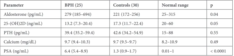
Table 3 shows the ROc analysis, presenting optimal cut-off levels and associated diagnostic performances. 25-(OH) 2d showed a sensitivity
and specificity of 36% and 96.7%, respectively, and the area under curve (aUc) was 0.644. The diag-nostic sensitivity and specificity of PTH were 92% and 23.3%, respectively, and the aUc was 0.546 (fig. 1). The sensitivity, specificity and aUc for aldosterone were 48%, 90%, and 0.643. The sensi-tivity, specificity and aUc for calcium were 40%, 88.7% and 0.553. PTH was superior in sensitivity whereas the other three were within close specifi-city percentages.
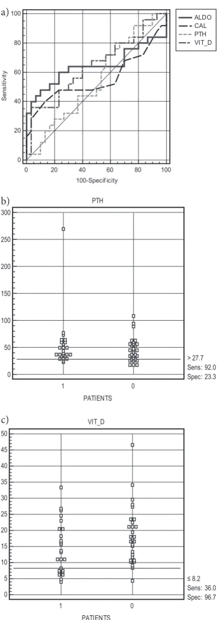
a positive correlation was seen between PTH and PSa levels in the control group (p = 0.006) (fig. 2).
Discussion
The most striking finding in this study was the higher aldosterone levels in patients with bPH. Our study may be the first to present the link between RaS and bPH. The vitamin d deficiency found in our bPH group was in line with previous studies. The co-existence of vitamin d deficiency and ele-vated levels of aldosterone composed the backbone of our assumptions [9].
Recent findings raise the possibility that aldo-sterone could influence a broad range of physio-logical and pathophysiophysio-logical processes, such as intestinal calcium absorption, vascular inflamma-tion and calcificainflamma-tion, and blood pressure [10]. Of concern, aldosterone makes some of these hap-pen through modifying the response of target tis-sues to 1,25-dihydroxy vitamin d(1,25(OH)2d) stimulation. Several 1,25(OH)2d target tissues, in-cluding intestinal epithelial cells and the cardio-vascular system, have also been identified as sites of non-genomic aldosterone regulation [4, 11]. Fig. 1. PTH was superior in sensitivity whereas the
other three were within close specificity percentages based on ROc analysis; a) ROc analysis of all 4 para-meters, b) PTH, and c) 25-(OH)2d
ALDO CAL PTH VIT_D
0 20 40 60 80 100
100-Specif icity 100
80
60
40
20
0
S
en
si
tiv
ity
PTH
PATIENTS 300
250
200
150
100
50
0
1 0
> 27.7 Sens: 92.0 Spec: 23.3
VIT_D
PATIENTS 50
45
40 35
30
25
20 15
10
5
0
1 0
≤ 8.2 Sens: 36.0 Spec: 96.7
Fig. 2. There was a positive correlation between PTH and PSa levels in the control group (p = 0.006)
0 20 40 60 80 100 120
CONT_PTH 4,0
3,5
3,0
2,5
2,0
1,5
1,0
0,5
0,0
C
O
N
T
_P
S
A
a)
b)
Vitamin d, as a versatile actor of the human metabolism, takes part in a wide variety of biologi-cal processes, including the immune response, in-sulin secretion, cardiovascular function and blood pressure, in addition to its very well-known role in calcium and phosphate homeostasis. To be an in-fluencing factor in our study 1,25(OH)2d has re-cently been identified as a regulator of the renin-angiotensin-aldosterone system (RaaS). It seems that aldosterone and 1,25(OH)2d cooperate in the regulation of cell function and this interaction is very likely to be the result of cross talk between non-genomic and genomic steroid hormone sig-naling pathways [12, 13].
How vitamin d deficiency in patients might cause an overproduction of aldosterone was pro-posed to be through the activation of the RaaS axis. The mechanism of the inverse relation-ship between serum vitamin d levels and plasma renin hormone activity remained elusive until the study by chopra et al. in which 1,25(OH)2d was found to be a negative regulator of genetic renin expression [14]. The profit of this valuable find-ing was achieved by vitamin d analogues used in combination with angiotensin inhibitors. The pa-tients taking combination therapy did not show the compensatory renin increase. The additive ef-fect clearly increased the potential of these drugs. Sigmund has hypothesized in his paper that the caMP signaling pathway was the target of 1,25-di-hydroxy vitamin d, and through this, the vitamin functioned as a regulator of other renin stimu-lating factors that prevent the overproduction of renin [15]. In turn, hypocalcemia promotes sec-ondary hyperparathyroidism with elevated circu-lating levels of PTH. Elevations in PTH lead to a rise in L-type ca2+ channel entry and intracellu-lar ca2+ overloading involving diverse tissues, cluding the prostate [16–18]. There follows the in-duction of oxidative stress and ultimately necrosis of parenchymal cells with consequent tissue repair. Lost parenchyma and the appearance of reparative fibrosis each compromise organ function [19].
collectively referred to as aldosteronism, this mineralocorticoid excess state is accompanied by aLdO-mediated classic responses that contribu-te to a heighcontribu-tened excretion of K+ and elevation
in arterial pressure, and frequently the retention of salt and water [7]. The putative risk factor for cVd, HT, shares the same characteristics of preva-lence with bPH [1]. although these two are separa-te disease processes, age-relasepara-ted increases in aLdO have been proposed to play a role in both of their pathophysiologies. McVary has extrapolated from statistical findings that if about 50% of men in the-ir sixties have bPH and 50% of them have hyper-tension, then approximately 25% of men ≥ 60 ye-ars have bPH with co-morbid hypertension [1].
aldosterone has also been linked to oxidative stress induction, which is the main path through multi-organ pathologies [19]. Recently, calo et al. have reported in a human model that increased al-dosterone production has effects on enzyme sys-tems related to oxidative stress, enhancing the systemic fibrogenic effects of aldosterone excess through TGf-β and PaI-1 expression [20]. Studies have also demonstrated that ang II induced oxida-tive stress, which was essential for vascular remod-eling. aldosterone was shown to induce fibrosis and remodeling through direct effect on some tis-sues [4, 5]. another study showed that the drug Salinomycin induces aldosterone levels, which are known to act as oxidative stress inducers in pros-tate cells [21].
Serum calcium and parathyroid hormones are two actors shown to stimulate prostate growth. although we could not determine an increase in PTH, the relationship between aldosterone, vita-min d and PTH should not be ignored. This cal-citropic hormone is very probably involved in any process leading to cell death through intracellu-lar ca2+ overloading in diverse tissues, including prostate and their mitochondria, with an induc-tion of oxidative stress [4, 5]. Yet one study showed that patients with histopathologically-proven bPH, high PTH, vitamin d, and calcium levels did not stimulate prostate growth [22, 23].
after all, the results of this study were derived from a small number of subjects, but still repre-sent an important hypothesis for further research in a larger number of cases to clarify the role of aldosterone overproduction and its clinical rele-vance in human disease, particularly of the pros-tate [24, 25].
References
[1] McVary KT: bPH: epidemiology and comorbidities. am J Manag care 2006, 12, 122–128.
[2] Nandeesha H: benign prostatic hyperplasia: dietary and metabolic risk factors. Int Urol Nephrol 2008, 40, 649–656.
[3] Parsons JK: Lifestyle factors, benign prostatic hyperplasia, and lower urinary tract symptoms, curr Opin Urol 2011, 21, 1–4.
[4] Bauman DR, Steckelbroeck S, Peehl DM, Penning TM: Transcript profiling of the androgen signal in normal prostate, benign prostatic hyperplasia, and prostate cancer. Endocrinology 2006, 147, 5806–5816.
[6] Skinner HG, Schwartz GG: The relation of serum parathyroid hormone and serum calcium to serum levels of prostate-specific antigen: a population-based study. cancer Epidemiol biomarkers Prev 2009, 18, 2869–2873.
[7] Rutledge MR, Farah V, Adeboye AA, Seawell MR, Bhattacharya SK, Weber KT: Parathyroid Hormone, a crucial Mediator of Pathologic cardiac Remodeling in aldosteronism. cardiovasc drugs Ther 2012 [Epub ahead of print].
[8] Zhao W, Ahokas RA, Weber KT, Sun Y: aNG II-induced cardiac molecular and cellular events: role of aldoste-rone. am J Physiol Heart circ Physiol 2006, 291, 336–343.
[9] Vaidya A, Pojoga L, Underwood PC, Forman JP, Hopkins PN, Williams GH, Williams JH: The association of plasma resistin with dietary sodium manipulation, the renin-angiotensin-aldosterone system, and 25-hydroxyvi-tamin d3 in human hypertension. clin Endocrinol (Oxf) 2011, 74, 294–299.
[10] Ullah MI, Uwaifo GI, Nicholas WC, Koch CA: does vitamin d deficiency cause hypertension? current evidence from clinical studies and potential mechanisms. Int J Endocrinol 2010, 579640. Epub 2009 Nov 10.
[11] Judd SE, Tangpricha V: Vitamin d deficiency and risk for cardiovascular disease. am J Med Sci 2009, 338, 40–44.
[12] Kota SK, Kota SK, Jammula S, Meher LK, Panda S, Tripathy P, Modi KD: Renin-angiotensin system activity in vitamin d deficient, obese individuals with hypertension: an urban Indian study. Indian J Endocrinol Metab 2011, 15 Suppl 4, 395–401.
[13] Forman JP, Williams JS, Fisher ND: Plasma 25-hydroxyvitamin d and regulation of the renin-angiotensin system in humans. Hypertension 2010, 55, 1283–1288.
[14] Chopra S, Cherian D, Jacob JJ: The thyroid hormone, parathyroid hormone and vitamin d associated hyperten-sion. Indian J Endocrinol Metab 2011, 15, Suppl 4, 354–360.
[15] Sigmund CD: Regulation of renin expression and blood pressure by vitamin d(3): J clin Invest 2002, 110, 155–156.
[16] Kamalov G, Deshmukh PA, Baburyan NY, Gandhi MS, Johnson PL, Ahokas RA, Bhattacharya SK, Sun Y, Gerling IC, Weber KT: coupled calcium and zinc dyshomeostasis and oxidative stress in cardiac myocytes and mitochondria of rats with chronic aldosteronism. J cardiovasc Pharmacol 2010, 55, 248–254.
[17] Donkena KV, Young CY: Vitamin d, sunlight and prostate cancer risk. adv Prev Med 2011, 2011, 281863. Epub 2011 Jun 8.
[18] Manchanda PK, Konwar R, Nayak VL, Singh V, Bid HK: association of genetic variants of the vitamin d recep-tor (VdR) gene (fok-I, Taq-I and bsm-I) with susceptibility of benign prostatic hyperplasia in a North Indian population. asian Pac J cancer Prev 2010, 11, 1005–1008.
[19] Sun Y, Ahokas RA, Bhattacharya SK, Gerling IC, Carbone LD, Weber KT: Oxidative stress in aldosteronism. cardiovasc Res 2006, 15, 71, 300–309.
[20] Calò LA, Pagnin E, Davis PA, Armanini D, Mormino P, Rossi GP, Pessina AC: Oxidative stress-related proteins in a conn’s adenoma tissue. Relevance for aldosterone’s prooxidative and proinflammatory activity. J Endocrinol Invest 2010, 33, 48–53.
[21] Ketola K, Hilvo M, Hyötyläinen T, Vuoristo A, Ruskeepää AL, Orešič M, Kallioniemi O, Iljin K: Salinomycin inhibits prostate cancer growth and migration via induction of oxidative stress. br J cancer 2012, 106, 99–106.
[22] Kim WT, Choi YD, Park C, Kim YW, Yun SJ, Kim IY, Kim WJ: Parathyroid hormone is not involved in prostate growth in patients with benign prostatic hyperplasia. Prostate 2011, 71, 1210–1215.
[23] Chaimuangraj S, Thammachoti R, Ongphiphadhanakul B, Thammavit W: Lack of association of VdR polymor-phisms with Thai prostate cancer as compared with benign prostate hyperplasia and controls. asian Pac J cancer Prev 2006, 7.
[24] Lupp A, Klenk C, Röcken C, Evert M, Mawrin C, Schulz S: Immunohistochemical identification of the PTHR1 parathyroid hormone receptor in normal and neoplastic human tissues. Eur J Endocrinol 2010, 162, 979–986.
[25] Schwartz GG, Whitlatch LW, Chen TC, Lokeshwar BL, Holick MF: Human prostate cells synthesize 1,25-dihy-droxyvitamin d3 from 25-hy1,25-dihy-droxyvitamin d3. cancer Epidemiol biomarkers Prev 1998, 7, 391–395.
Address for correspondence:
Necat Yilmaz
antalya Education and Research Hospital central Laboratory 07050 antalya
Turkey
Tel.: +90 505 357 83 05
E-mail: [email protected]
conflict of interest: None declared