original papers
andrzej Wojtowicz
1, a, B, D, e, artur Kamiński
2, a, D, e, ewa olender
2, a, B, D–F,
remigiusz Czerkies
3, B, Tomasz Kamiński
1, B, eClinical Aspects of Gene Expression for Bone
Morphogenetic Proteins 2, 4, 6 in Bone Augmentation*
Kliniczne aspekty ekspresji genów dla białek morfogenetycznych
kości 2, 4, 6 w augmentacji tkanki kostnej
1 Department of oral surgery, Warsaw Medical University, poland
2 Department of Transplantology and Central Tissue Bank, Warsaw Medical University, poland 3 aW Clinic, private practice, Warszawa, poland
A – koncepcja i projekt badania; B – gromadzenie i/lub zestawianie danych; C – opracowanie statystyczne;
D – interpretacja danych; E – przygotowanie tekstu; F – zebranie piśmiennictwa
Abstract
Background. Human jaw bone augmentation is a procedure applied as a part of dental implant therapy. Bone substitutes are very well known and described. The cascade of bone substitute remodeling process leads to bone substitute resorption and new bone formation, called creeping substitution. The most important aspect for this cascade promotion is the activation of stem cells by local or applied growth factors. BMps are a group of growth factors present in bone allograft (proteins synthesized by the donor) as well as in the newly formed bone (BMps synthesized by the recipient’s cells).
Objectives. The purpose of this study was to evaluate the clinical restoration of jaw bone defects using guided bone regeneration, and analyzed three isoforms, BMp-2, BMp-4 and BMp-6 in newly formed bone tissue, used real time pCr method on the mrna synthesis stage, i.e. synthesized in response to the graft application.
Material and Methods. 12 patients aged 20–56 years underwent therapy by implanting: 1) allogenic cortico-can-cellous bone granules with xenogeneic bovine mineral into bone defects (6 patients); 2) allogenic cortico-cancortico-can-cellous bone granules with synthetic beta-tricalcium phosphate (BTCp) into bone defects (6 patients).
Results. synthesis of BMp2 and 4 was observed after application of both: natural bovine mineral and synthetic BTCp, but expression of BMp6 was not found. synthesis of BMp2 was comparable in control tissue and newly formed tissue in the site of bovine mineral application. after BTCp application, the synthesis of BMp2 was signifi-cantly lower. synthesis of BMp-4 was two times higher in the control tissue.
Conclusions. natural bovine mineral seems to be more effective than synthetic BTCp in bone inducing through the synthesis of BMp2, the main osteogenic growth factor in the human jaw bone augmentation methodology. BMp isoform tests revealed a significant concentration of BMp-2 mrnas, a lower expression of BMp-4 and trace amounts or no presence of BMp6 mrna in all newly formed bone samples (Dent. Med. Probl. 2012, 49, 3, 337–344).
Key words: bone allograft, human bone augmentation, bovine mineral, BTCp, BMps.
Streszczenie
Wprowadzenie. augmentacja kości szczęk jest zabiegiem wspomagającym leczenie implantoprotetyczne. Znanych jest wiele materiałów kościopodobnych i substytutów kości. Kaskada procesu przebudowy, stopniowego zastępo-wania tych materiałów prowadzi do ich resorpcji i powstania nowej tkanki kostnej. istotne jest aktywowanie tej kaskady przez pobudzenie miejscowych komórek macierzystych przez miejscowe lub egzogenne czynniki
wzro-Dent. Med. probl. 2012, 49, 3, 337–344
issn 1644-387X © Copyright by Wroclaw Medical University and polish Dental society
* The project was supported by the grant no. 3F/36 by Ministry of science and High education and realized in Medical Warsaw University.
stowe. Białka morfogenetyczne kości są obecne w tkance kostnej przeszczepów allogenicznych (syntetyzowane w organizmie dawcy), a także w nowotworzonej tkance kostnej (syntetyzowane przez komórki biorcy).
Cel pracy. Kliniczna ocena regeneracji uszkodzeń tkanki kostnej szczęk po zastosowaniu metodologii sterowa-nej regeneracji kości i analiza trzech izoform białek morfogenetycznych kości: BMp-2, BMp-4, BMp-6 w nowo powstającej tkance kostnej, z wykorzystaniem metodologii real time-pCr i oceny syntezy mrna w odpowiedzi na zastosowanie implantu.
Materiał i metody. Badania prowadzono u 12 pacjentów (20–56 lat), którym wszczepiono: 1) kostną macierz allo-geniczną wraz z minerałem wołowym (6 pacjentów), 2) kostną macierz alloallo-geniczną wraz z syntetycznym beta-trój-fosforanem wapniowym (6 pacjentów). Kość allogeniczna miała postać granulatu (1–2 mm średnicy)
Wyniki. W badaniach 15 eksplantów tkanki kostnej pobranej trepanem o średnicy 3 mm i długości 10 mm z miejsc implantacji poddano analizie rt-pCr. Zaobserwowano syntezę BMp-2 i BMp-4 w przypadku zastosowania obu materiałów: naturalnego minerału wołowego i syntetycznego BTCp.nie stwierdzono syntezy BMp-6. instensywność syntezy BMp-2 była porównywalna w kości kontrolnej ektopowej i po zastosowaniu minerału wołowego. po zasto-sowaniu BTCp synteza BMp-2 była znacząco słabsza. synteza BMp-4 była dwa razy większa w kości kontrolnej.
Wnioski. naturalny minerał ksenogenny jest bardziej skuteczny niż syntetyczny BTCp w procesie kościotworzenia w sterowanej reakcji tkanki kostnej przez ekspresję BMp-2, który okazał się najbardziej skuteczną izoformą BMps. Mniejszą ekspresję wykazała izoforma BMp-4, nie stwierdzono natomiast obecności BMp-6 lub ekspresja była na poziomie dokładności metody. BMp-6 nie jest czynnikiem niezbędnym w procesie resorpcji, osteogenezy i prze-budowy tkanki kostnej, ale być może interferuje z procesem degradacji tkankowej (Dent. Med. Probl. 2012, 49, 3, 337–344).
Słowa kluczowe: przeszczep kostny, augmentacja kości u ludzi, minerał wołowy, BTCp, BMps.
advanced methods of bone augmentation us-ing growth factors and/or stem cells constitute one of the most rapidly developing areas of oral im-plantology, especially when bone quantity is not sufficient.
The crucial role of bone morphogenetic pro-teins (BMps) in craniomaxillofacial and neurolog-ical surgery were discussed [1, 2], but the efficacy of a different methodology is still under investi-gation.
a recent significant breakthrough in tissue engineering therapy was the application of isolat-ed CD34+ bone marrow stem cells [3]. Their total amount of approximately 1,000,000 per 1 ccm of transplant appeared to be too small to achieve de-tectable bone tissue regeneration, the more so that the proposed recombinant human growth factors BMp-2, BMp-4 and lately BMp-7 and egF-2 re-quire also, as mitogens, a significant local stem cell pool [4–6]. BMp-2 has been acknowledged as one of the most effective growth factors in dental thera-py and has a synergistic effect with BMp-4 [7]. Both of these factors in a recombinant form of rhBMp2 are already used in clinical conditions for bone tis-sue augmentation, e.g. in Usa, and so is rhpDgF BB in periodontal regeneration. However, the ac-tion of the above factors is known to be of a cas-cade nature, and there is feedback because pDgF is a mitogen, while BMp-2 and BMp-4 plays the role of a morphogen [8–11]. Bone morphogenetic pro-teins stimulate human stable cell lines, human mes-enchymal stem cells in vitro [12–13] as well as in an-imal studies [14]. The other known BMp-6 influ-enced on macrophages cell lines [15], which suggest other, maybe opposite action in bone to BMp-2, or BMp-4. From a clinical point of view, it was con-firmed that synthetic human recombinant BMp-2
can enhance bone formation and osteointegration in implant therapy in sinus lift procedure [16] as well as in orthopaedic surgery [17].
The aims of the study included: assessment of a clinical restoration of jaw bone defects using guided bone regeneration, and expression analysis of three bone morphogenetic protein (BMp) iso-forms, BMp-2, BMp-4 and BMp-6 in bone tissue induced by augmentation in dental implant treat-ment.
Material and Methods
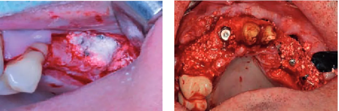
For this purpose, guided bone regeneration (gBr) methods were used. This technique was used in 12 patients (men) aged 20–56 years by implanting: 1) allogenic cortico-cancellous bone granules (50% of volume) with rapidly resorbable synthetic beta-tricalcium phosphate (BTCp) (50% of transplant volume) into bone defects in 6 pa-tients (Fig. 1 top); 2) allogenic cortico-cancellous bone granules (50% of volume) with slowly resorb-able xenogenic bovine mineral (50% of transplant volume) into bone defects in 6 patients (Fig. 1 bot-tom).
The main volume of transplants consisted of allogenic bone in the form of granules taken from one lot from the Central Tissue Bank. each im-plantable material that was used made up 50% of the 3 cm3 of transplant volume. patients were
giv-en pharmacotherapy: sumamed 1 × 0.5 mg daily for 6 days, metronidazole 3 × 0.25 g daily, aescin 3 × 1 daily (antioedema action), analgesics (Keton-al) and a probiotic (Trilac) for 6 days.
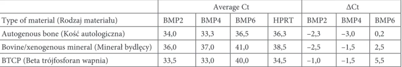
sutures were removed 10 days after surgery in all patients. 6 months following implantation of
guided bone regeneration material, cylinder bone samples were collected using a trephine bur dur-ing implant bed preparation after muco-periosteal flaps were elevated (Fig. 2). 18 samples in total (10 samples collected after implantation of allo-genic bone + bovine mineral, 8 samples collected after implantation of allogenic bone with BTCp) were placed in extraction reagents for BMp-2, –4 and –6 rna isoforms.
Real Time-PCR Test
Tissue Sampling
samples (5 in each group) collected after the explantation was immediately placed in tubes and immersed in 1 ml of Tri-reagent (sigma-aldrich), a liquid preventing the rna from being destroyed by rna-ses and enabling isolation of rna. Tubes were transferred to –22oC and after initial freezing
moved to –70oC until further processing. applied
methodology was described by Chomczynski et al. recently [18, 19].
Total RNA Extraction
Tissues with Tri-reagent in which they were stored were individually homogenized in cryomill in tubes washed with DepC-treated water to inac-tivate rna-ses. For total rna extraction a method was applied with the use of Trireagent (sigma-al-drich), Tri reagent is an improved version of the single-step total rna isolation reagent developed by Chomczynski [18, 19]. Homogenized tissue was placed in 1.5 ml centrifuge tube (eppendorf). Tri- -reagent was added up to 1 ml. Tubes were shak-en in an ambishak-ent temperature for 5 minutes. Thshak-en, the samples were allowed to stand for 5 minutes. 0.2 ml of chloroform was added (sigma-aldrich). Tubes were shaken vigorously and allowed to stand at room temperature for 10 minutes. Then, tubes were centrifuged at 12 000 g at 4oC for 15
min-utes. aqueous phase was transferred to a fresh tube (eppendorf) and 0.5 ml of isopropanol (sigma-al-drich) was added. samples were mixed and allowed to stand at room temperature for 10 minutes, then centrifuged at 12 000 g at 4oC for 15 minutes.
su-pernatant was removed and the pellet was washed by adding 1 ml of 75% ethanol (poCH). samples
Fig. 1. Jaw bone augmentation – surgical procedure graft consist of: BTCp + allogenic bone granules (up), xenogenic mineral + allogenic bone granules
Ryc. 1. augmentacja kości szczęk – zabieg chirurgiczny: przeszczep zawiera: BTCp + granulat kości alogenicznej (góra), minerał ksenogenny + granulat kości alogenicznej
Fig. 2. augmented jaw bone site: (core) bone microbiopsy taken by the trephine before implant installation/sample of augmented bone tissue inside the trephine
Ryc. 2. augmentowana tkanka kostna szczęk: biopsja tkanki kostnej pobrana trepanem przed instalacją implantu próbka–rdzeń tkanki kostnej wewnątrz trepanu
were vortexed and centrifuged at 7500 g at 4oC for
5 minutes. supernatant was removed and the sam-ples dried in ambient temperature for 20 minutes. The pellets were resuspende in 0.5 ml water for mo-lecular biology (sigma-aldrich) and the amount of rna was measured with the use of Biophotome-ter (eppendorf) in order to optimize the volume of sample used for reverse transcription.
Reverse Transcription
For obtaining cDna further used for real time pCr procedures, High Capacity cDna tran-scription Kit (applied Biosystems/life Technolo-gies) was applied according to the manufacturer’s protocol. Kit components included: buffer, dnTp mix, random primers, reverse transcriptase, rna-se inhibitor, nuclearna-se-free water. solution of rna isolated in the first step was added to the reverse transcription mix in concentrations allowing us to obtain up to 2 ug of rna per reaction tube which was controlled with the use of a spectrophotom-eter (Biophotomspectrophotom-eter, eppendorf). reverse tran-scription reaction was performed in thermal cy-cler (realplex2, eppendorf) according to the
pro-gram recommended by the producer of the reverse transcription kit. Tubes with the products of re-verse transcription were stored overnight in –22oC
for further processing.
Real Time PCR
real time pCr was carried out with the use of TaqMan gene expression assays and Taq-Man gene expression Master Mix (applied Bio-systems/life Technologies) according to the man-ufacturer’s protocols, on realplex 2 cycler (ep-pendorf). a single reaction tube contained 1 ul of TaqMan gene expression assays, 9ul of cDna template, 10 µl of TaqMan gene expression
Mas-ter Mix. Target genes were BMP-2, BMP-4, BMP-6. reference gene was HprT (hypoxanthine-gua-nine phosophoribosyltransferase). 40 cycles were performed, each including a 15 s phase at 95oC and
1 min phase at 60oC.
Results and Discussion
Clinical examination showed total wound healing in all patients 6 months after surgery. There are no differences in the gingival profile after the application of different grafting materi-als. a radiological and histological study will be publishing soon. During the next stage of implant therapy, all bone preparation was similar, the dif-ferences in hardest of bone core during bone
mi-crobiopsy by the trephine were not noticed. im-plants were installed in augmented sites with good primary stabilization in all patients.
PCR Results
according to the manufacturer’s instructions, CT values above 40 were considered as negative, i.e. no amplification of cDna could be confirmed.
analysis of detection pattern of BMps rna after implantation of guided bone regeneration materials revealed (Table 1, Figs. 3–5):
The expression of BMp-2 and BMp-4 genes was higher than the expression of the house-keep-ing gene HprT in case of both applied materials: bovine mineral, BTCp as well as the control, i.e. orthotopic autogenous bone.
The expression of BMp-6 gene was in all cas-es lower than the exprcas-ession of HprT and target genes for BMp-2,4, reaching values on the edge of detectability.
BMp-2 rna amount was comparable in case of orthotopic autogenous bone and bone formed after bovine mineral application; its expression was significantly lower in case of the use of BCTp as an osteoconductive scaffold.
The expression of BMp-4 rna was two times higher in autogenous bone in comparison to bone formed after application of BTCp and bovine mineral.
allogenic bone granules, characterised by min-imal osteogenic properties, were used with synthet-ic (BTCp) or natural (bovine mineral) implanted materials for promotion of bone formation.
The results achieved indicate that in humans: Bovine mineral seems to be more effective than BTCp in bone inducing properties through synthe-sis of BMp-2, the main osteogenic growth factor in human jaw bone augmentation methodology
BMp isoform tests revealed significant con-centration of BMp-2, lower expression of BMp-4 and trace amounts or no presence of BMp-6 mr-na in all induced bone samples
BTCp has shown low biological efficacy in the bone augmentation process, which occur through the inducing of expression of BMps isoforms in the envi-ronment of orthotopic autogenic jaw bone as well al-logenic bone granules. This observation could be ex-plained by BTCp properties i.e. fast resorbability.
The role of BMp-2 and BMp-4 in osteogenesis was confirmed, while BMp-6 isoform seems not to be involved in bone regeneration
Both implanted scaffolds: synthetic BTCp and natural bovine mineral are well described as os-teoconductive materials with no osteoinductive properties. However, when implanted with allo-genic bone granules into the autoallo-genic jaw bone
site – can improve not only bone shape/profile, but also cascade of new bone formation through os-teogenic growth factors.
The importance of BMps for the process of os-teogenesis was proved in several animal studies. Heterotopic bone formation in an animal model can be induced by the expression of BMp-2, 4 and acts on local stem cells present in the muscle tis-sue, out of periosteum [20].
BMps are a morphogen in the following pro-cesses: embryonic bone development, skeletal growth, bone remodeling, bone fracture healing as well as bone graft remodeling. The number of
local stem cells for osteogenesis that respond to BMps is not known, the application alone of 1 mil-lion human stem cells per graft for the treatment of bone defect – is not effective [3]. it seems that also local BMps concentration plays an important role. The golden standard, i.e. autologous bone im-plantation is the best type of bone graft but in that case an additional donor site is needed. allogenic bone, from a tissue bank, has very low osteoinduc-tive properties, and the concentration of BMps is estimated in picograms. The application of a com-bine graft: allogenic bone with bovine mineral or with BTCp still is advantageous as it filled the
de-Table. 1. Ct values observed in tissues obtained after application of bovine mineral and BTCp and control material – autogenous bone (minus values of the ∆Ct indicate an expression lower than the expression of the reference gene)
Tabela 1. Wartości Ct zaobserwowane w tkankach pozyskanych po zastosowaniu minerału bydlęcego, BTCp (beta trój-fosforanu wapnia) i materiału kontrolnego – kości autologicznej (wartości ujemne Ct świadczą o ekspresji większej od ekspresji genu referencyjnego)
average Ct ∆Ct
Type of material (rodzaj materiału) BMp2 BMp4 BMp6 HprT BMp2 BMp4 BMp6 autogenous bone (Kość autologiczna) 34,0 33,3 36,5 36,3 –2,3 –3,0 0,2 Bovine/xenogenous mineral (Minerał bydlęcy) 36,0 37,0 41,0 38,5 –2,5 –1,5 2,5 BTCp (Beta trójfosforan wapnia) 33,5 33,0 40,0 34,5 –1,0 –1,5 5,5
Fig. 3. average ∆Ct values as determined by real-time pCr by subtracting the average HprT Ct value from the average Ct for each target gene. series: 1 – BMp2, 2 – BMp4, 3 – BMp6; aB – autog-enous bone, BM – bovine mineral, BT – BTCp (minus values of the ∆Ctindicate an expression higher than the expression of the reference gene)
Ryc. 3. Średnie wartości ∆Ct wyliczone z wartości obserwowanych w real-time pCr, uzyskane przez odjęcie średniego Ct dla genu HprT od średniej wartości Ct dla każdego genu badanego. serie: 1 – BMp2, 2 – BMp4, 3 – BMp6; aB – kość autologiczna, BM – minerał bydlęcy, BT – BTCp, (beta-trójfosforan wapnia, wartości ujemne Ct świadczą o ekspresji wyższej od ekspresji genu refe-rencyjnego)
6 5 4 3 2 1 0 –1 –2 –3 –4
Av
er
ag
e
D
C
t v
al
ue
fect better within bone spaces than soft, gingival tissue. That type of a graft can induce wound heal-ing in a faster and biologically more effective man-ner. it seems that BTCp undergoes a fast degrada-tion, a process that does not take place normally
in a newly formed bone. it is possible that the sur-face of BTCp granules does not attract cells/stem cells as opposed to natural bovine bone mineral which shows better cell adhesion and spreading properties. The degradation of BTCp may not
in-Fig. 4. Collective diagram. cDna amplification for BMp-2, BMp-4, BMp-6 (target genes) and HprT (reference gene) sequence. The study samples underwent 45 pCr cycles. Detection (Ct) above 40 indicates a number of present tran-scripts on the verge of detectability
Ryc. 4. Diagram zbiorczy. amplifikacja cDna dla genów badanych BMp-2, BMp-4, BMp-6 oraz genu referencyjnego HprT. Badane próbki podlegały 45 cyklom pCr. Detekcja (Ct) powyżej 40. cyklu jest traktowana jako graniczna, nie potwierdza obecności badanej sekwencji
Fig. 5. a typical result from amplification observed in the study BMp-2 and BMp-4 transcripts detectable, BMp-6 transcripts undetectable
Ryc. 5. Typowy wynik amplifikacji obserwowany w badaniu. Transkrypty BMp-2, BMp-4 oznaczalne, BMp-6 nieoznaczalne
volve the activity macrophages/monocytes osteo-clastic cell line and hence BMp-6 pathways that are hypothetically co-dependent on the presence of macrophages/monocytes osteoclastic cell line in the site of tissue remodeling. it seems that the deg-radation of BTCp can proceed via simple dissolu-tion and without or with a poor activadissolu-tion of mac-rophages. author hypothesis is that BMp-6 can interfere in bone resorption. The clinical and ra-diological (not publish yet) results of authors study confirm the observation that the bovine mineral is refractory to resorption. The role of BMp-6 in bone metabolism is still not clear; BMp-6 was not expressed significantly in the human skeleton [21]. The biological and genomic bases for creeping sub-stitution of the allogenic, xenogenic and alloplastic
grafts are different. it seems that their combina-tion allows for better perspectives for oral, regen-erative medicine [22–23].
The authors concluded that though the appli-cation of BMps is successful in orthopaedic and maxillofacial surgery, human recombinant BMps are still not available for the dentists. an alterna-tive source of BMps is autologous bone, consid-ered as a golden standard – this, however, applied seldom, rather in maxillofacial cases. The combi-nation of allogenic bone from a tissue bank with alloplastic or xenogenic materials, especially long-resorbed materials is an effective method for bone augmentation in small/dental bone defects.
The project was approved by Bioethical Com-mittee WUM.
References
Davies s.D., ochs
[1] M.W.: Bone Morphogenetic proteins in Craniomaxillofacial surgery. oral Maxillofac. surg. Clin. n. am. 2010, 22 (1), 17–31.
Dmitriev a.e., Farhang s., lehman Jr. r.a., ling g.s.F., symes a.J
[2] .: Bone morphogenetic protein-2 used in spinal fusion with spinal cord injury penetrates intrathecally and elicits a functional signaling cascade. spine J. 2010, 10 (1), 16–25.
Wojtowicz a., Chaberek s., Urbanowska e., ostrowski, K
[3] .: Comparison of efficiency of platelet rich plasma, hematopoieic stem cells and bone marrow in augmentation of mandibular bone defects. n. Y. state Dent. J. 2007, 73 (2), 41–45.
Jeong B.-C., Kim H.-J., Bae i.-H., lee K.-n., lee K.-Y., oh W.-M., Kim s.-H., Koh
J.-[4] T.: CoMp-ang1, a chimeric
form of angiopoietin 1, enhances BMp2-induced osteoblast differentiation and bone formation. Bone 2010, 46 (2), 479–486.
Wikesjö U.M.e., Qahash M., polimeni g., susin C., shanaman r.H., rohrer M.D., Wozney J.M., Hall J
[5] .:
alveolar ridge augmentation using implants coated with recombinant human bone morphogenetic protein-2: Histologic observations.. J. Clin. periodontol. 2008, 35 (11), 1001–1010.
leknes K.n., Yang J., Qahash M., polimeni g., susin C., Wikesjö
[6] U.M.e.: alveolar ridge augmentation using implants coated with recombinant human bone morphogenetic protein-2: radiographic observations. Clin. oral implants res. 2008, 19 (10), 1027–1033.
Kubota K., iseki s., Kuroda s., oida s., iimura T., Duarte W.r., ohya K., Kasugai s.
[7] : synergistic effect of
fibroblast growth factor-4 in ectopic bone formation induced by bone morphogenetic protein-2. Bone 2002, 31 (4), 465–471.
emans p.J., spaapen F., surtel D.a.M., reilly K.M., Cremers a., van rhijn l.W., Bulstra s.K., Kuijer r.
[8] :
a novel in vivo model to study endochondral bone formation; HiF-1α activation and BMp expression. Bone 2007, 40(2), 409–418.
lim T.Y., Wang W., shi Z., poh C.K., neoh K.g.:
[9] Human bone marrow-derived mesenchymal stem cells and osteoblast differentiation on titanium with surface-grafted chitosan and immobilized bone morphogenetic pro-tein-2. J. Mat. sci.: Mater. Med. 2009, 20 (1), 1–10.
Kinoshita a
[10] .: pre-clinical and clinical studies on bone morphogenetic proteins for regenerative therapies in den-tistry field. Clin. Calcium 2006 16(2), 109–115.
Kim C.-s., Choi s.-H., Choi B.-K., Chai J.-K., park J.-B., Kim C.-K., Cho K.-s
[11] .: The effect of recombinant human bone morphogenetic protein-4 on the osteoblastic differentiation of mouse calvarial cells affected by porphyromo-nas gingivalis. J. periodontol. 2002, 73 (10), 1126–1132.
Deifenderfer D.l., osyczka a.M., reilly g.C., leboy p.s
[12] .: BMp responsiveness in human mesenchymal stem cells. Connective Tis. res. 2003, 44 (suppl. 1), 305–311.
luppen C.a., smith e., spevak l., Boskey a.l., Frenkel B.:
[13] Bone Morphogenetic protein-2 restores Mineralization in glucocorticoid-inhibited MC3T3-e1 osteoblast Cultures. J. Bone Min. res. 2003, 18 (7), 1186–1197.
osyczka a.M., Diefenderfer D.l., Bhargave g., leboy p.s
[14] .: Different effects of BMp-2 on Marrow stromal Cells from Human and rat Bone. Cells Tissues organs 2004, 176 (1–3), 109–119.
Hong J.H., lee g.T., lee J.H., Kwon s.J., park s.H., Kim s.J., Kim i.Y
[15] .: effect of bone morphogenetic protein-6 on macrophages. immunology 2009, 128 (1 part 2), e442–e450.
gutwald r., Haberstroh J., stricker a., rüther e., otto F., Xavier s.p., oshima T., sauerbier s.:
[16] influence
of rhBMp-2 on bone formation and osseointegration in different implant systems after sinus-floor elevation. an
el-amin s.F., Hogan M.V., allen a.a., Hinds J., laurencin C.T
[17] .: The indications and Use of Bone Morphogenetic proteins in Foot, ankle, and Tibia surgery. Foot ankle Clin. 2010, 15 (4), 543–551.
Chomczynski p., sacchi n
[18] .: single-step method of rna isolation by acid guanidinium thiocyanate-phenol-chloroform extraction. anal. Biochem. 1987, 162, 156.
Chomczynski p
[19] .K a reagent for the single-step simultaneous isolation of rna, Dna and proteins from cell and tissue samples. BioTechniques 1993, 15, 532–537.
Kochanowska, i.e., Wlodarski, K., Wojtowicz, a., niemira, K., ostrowski, K
[20] .: osteogenic properties of
various Hela cell lines and the BMp family genes expression. ann. Transplant. 2002, 7, 4, 58–62
Kochanowska i., Chaberek s., Wojtowicz a., Marczyński B., Włodarski K., Dytko M., ostrowski K
[21] .:
expression of genes for bone morphogenetic proteins BMp-2, BMp-4 and BMp-6 in various parts of the human skeleton. BMC Musculoskeletal Disord. 2007, 8, 12.
Krasny K., Kaminski a., Krasny M., Zadurska M., piekarczyk p., Fiedor
[22] p.: Clinical use of allogeneic bone granulates to reconstruct maxillary and mandibular alveolar processes. Transplant. proc. 2011, 43(8), 3142–3144 olender e., Uhrynowska-Tyszkiewicz i., Kaminski a
[23] .: revitalization of biostatic tissue allografts: new per-spectives in tissue transplantology. Transplant. proc. 2011, 43(8), 3137–3141.
Address for correspondence:
andrzej Wojtowicz Department of oral surgery Warsaw Medical University nowogrodzka 59
02-006 Warszawa poland
Tel.: +48 22 502 1242
e-mail: [email protected] received: 3.08.2012
revised: 11.09.2012 accepted: 14.09.2012
praca wpłynęła do redakcji: 3.08.2012 r. po recenzji: 11.09.2012 r.