AN EXAMINATION OF NP-P AND NP-V
INTERACTIONS WITHIN THE SIMIAN VIRUS 5 (SV5)
REPLICATION COMPLEX
Alison Bermingham
A Thesis Submitted for the Degree of PhD
at the
University of St Andrews
1998
Full metadata for this item is available in
St Andrews Research Repository
at:
http://research-repository.st-andrews.ac.uk/
Please use this identifier to cite or link to this item:
http://hdl.handle.net/10023/13882
An Examination of NP-P and NP-V
Interactions within the Simian Virus 5 (SV5)
Replication Complex
By Alison Bermingham
Division of Cell and Molecular Biology
School of Biological and Medical Sciences
University of St. Andrews
A thesis presented for the degree of Doctor of Philosophy in the
Faculty of Science at the University of St. Andrews
ProQuest Number: 10166286
All rights reserved
INFORMATION TO ALL USERS
The qua lity of this reproduction is d e p e n d e n t upon the qua lity of the copy subm itted.
In the unlikely e v e n t that the author did not send a c o m p le te m anuscript and there are missing pages, these will be noted. Also, if m aterial had to be rem oved,
a n o te will in d ica te the deletion.
uest
ProQuest 10166286
Published by ProQuest LLO (2017). C o pyright of the Dissertation is held by the Author.
All rights reserved.
This work is protected against unauthorized copying under Title 17, United States C o d e M icroform Edition © ProQuest LLO.
ProQuest LLO.
789 East Eisenhower Parkway P.Q. Box 1346
DECLARATION
i)
ii)
I, Alison Bermingham, hereby certify that this thesis, which is
approximately 50,000 words in length, has been written by me, that
it is the record of work carried out by me and that it has not been
submitted in any previous application for a higher degree.
Signed.
I was admitted as a part-time research student in October 1992 and as
a candidate for the degree of PhD in October 1993; the higher study
for which this is a record was carried out in the University of
S t Andrews between 1992 and 1997.
Signed.
iii) I hereby certify that the candidate has fulfilled the conditions of the
Resolution and Regulations appropriate for the degree of PhD in the
University of St. Andrews and that the candidate is qualified to submit
this thesi.s4n^pplication for that degree.
Signed.
Date. ...{.4
/ Â .L
iv) In submitting this thesis to the University of St. Andrews I understand
that I am giving permission for it to be made available for use in
accordance with the regulations of the University Library for the time
being in force, subject to any copyright vested in tlie work not being
affected thereby. I also understand that the title and abstract will be
published, and that a copy may be made and supplied to any
hona fide
library or research worker.
Acknowledgements
First, I would like to thank my supeiwisor, Dr. Rick Randall, for his patience and guidance, especially during the writing of this thesis. 1 would also like to thank Bernie Precious for aU his help with the reverse genetics studies and subsequent interesting discussions on the million reasons for failure. 1 would like to thank Dan Young for isolating and supplying the inducible cell lines used in many of the experiments presented here, and especially for regularly working out the missing clues to the prize 'Scotland on Sunday' crossword, which 1 haven't won yet.
1 would like to take this opportunity to show my appreciation to Prof. Bob Lamb and the members of his lab, for their generosity and kindness, both professional and personal, during my 4 month collaborative trip to Northwestern University, Evanston, Illinois, USA. 1 would especially like to thank Dr. Carol Ward for her words of encouragement in the face of disaster. Thanks also to Peg, Karen and Diana for showing me what life in Chicago is all about.
1 am indebted to all technicians in the Iiwine building who have helped me throughout my time in St. Andrews and to Dave Roche for his photographic skills. Thanks must also go to the members of Lab 28, past and present, for their help and advice on issues as diverse as 'What's best to line your stomach before a lab night out' to 'Possible substitutions for a macaroni pie after a lab night out'. Special thanks go to Jane P for proof-reading and filling me with caffeine on a regular basis. Et mon ami français, merci a toi. Courage pour ton these, le fin aiiivera!
My thanks must especially go to my mother for proof-reading and not being too disappointed to find that my university education didn't teach me to spell. Without the total support of my family, this thesis would never have been completed.
ABSTRACT
The aim of this study was to examine the mechanisms of transcription and replication of the paramyxovirus, simian virus type 5 (SV5). This was initially attempted using reverse genetics techniques and subsequently examining specific viral protein : protein interactions within the replication complex.
A cDNA clone encoding a synthetic negative-sense RNA genome analogue was constructed. Reverse genetics techniques were used to attempt to characterise
conditions which supported the transcription and replication of this genome analogue, with or without the use of wild-type helper virus but were unsuccessful.
During the course of these studies, a number of mammalian cell lines inducibly expressing SV5 proteins were isolated. These cell lines were subsequently used to examine viral protein : protein interactions within the replication complex.
When expressed alone, both P and V proteins exhibited diffuse cytoplasmic
lluorescence and V was also found in the nucleus. However, when NP was expressed alone, it was seen as punctate and granular cytoplasmic fluorescence. The distribution patterns of the proteins changed when expressed in combination. Large cytoplasmic aggregates similar to those at late times in an SV5 infection were seen in cells which co-expressed NP and P. When NP was co-expressed with V, however, NP was partially redistributed to give diffuse cytoplasmic and nuclear fluorescence. This showed that both P and V proteins could interact with NP and suggested that V may play a role in keeping NP soluble prior to an ordered encapsidation process.
Extracts from these cell lines were then used in a novel protein : protein capture assay and demonstrated that NP could interact with both P and V proteins. NP expressed by the cell line was shown to contained both soluble and polymeric forms of NP. P was shown to bind both forms of NP, while V could only bind soluble NP. Since P and V proteins are amino co-terminal, the site of interaction between P and polymeric NP was predicted to be in the P unique C-terminus. This was strengthened when a P-specific C- terminal mAh was found to block the binding of P with polymeric NP. Deletion mutant analysis in the C-terminus of the P protein showed that the mAh binding site was at the extreme C-terminus of the protein suggesting this is the point of interaction between P and polymeric NP. Possible roles for these protein : protein interactions and
ABBREVIATIONS
NUCLEIC ACIDS
DNA
RNA
A
C
G
T
U
2' deoxyribonucleic acid
ribonucleic acid
adenine (base in DNA or RNA)
cytosine (base in DNA or RNA)
guanine (base in DNA or RNA)
thymine (base in DNA)
uracil
(base in RNA)
NTP(s)
ATP
CTP
OTP
UTP
ribonucleoside triphosphate(s)
adenosine 5' triphosphate
cytidine 5' triphosphate
guanosine 5' triphosphate
uridine 5’ triphosphate
dNTP(s)
dATP
dCTP
dOTP
TTP
2' deoxyribonucleoside triphosphate(s)
2' deoxy-adenosine 5' triphosphate
2’ deoxy-cytidine 5' triphosphate
2' deoxy-guanosine 5' triphosphate
thymidine 5' triphosphate
ddNTP(s)
ddGTP
2',3' dideoxyribonucleoside triphosphate(s)
2',3' dideoxy-guanosine 5' triphosphate
cDNA
mRNA
vRNA
complementary DNA
messenger RNA
PHYSICAL UNITS
o c8
g
mg
F gng
1
ml
[i\
Ci
mCi
\iCi
M
mM
|LlM
kDa
kb
pH
VmA
U
temperature in degrees Celcius
centrifugal force
gram mass
milk gram (10 g)
micro gram (10 -6 g)
nano gram (10 “9 g)
litre volume
milli litre
micro litre
Curie (measure of radioactivity=3.7 x 10 19
disintegrations per second)
milli Curie
micro Curie
molar concentration
milli molar
micro molar
kilodalton
kilobase (pairs)
-log io[H+]
volts
milk amperes
CHEMICALS AND REAGENTS
14c
35s
DAPI
EDTA
EGTA
FITC
GMEM
KAc
NaAz
NBCS
NP40
PBS
SDS
TEMED
Tris-HCl
TE
radioisotope cai'bon-14
radioisotope sulphur-35
4, 6, diamino-2-phenylindole
ethylenediaminetetra-acetic acid
ethylene glycol-bis((3-aminoethyl ether)
N,N,N',N’-tetra-acetic acid
tluorescein-isothiocyanate
Glasgow modified Eagle's medium
potassium acetate
sodium azide
Newborn calf serum
nonidet p40
phosphate buffered saline
sodium dodecyl-sulphate
N,N,N',N’-tetramethylethylenediamine
tetra-acetic acid
tris-hydroxymetliyl-aminomethane, pH adjusted
with HCl
tris-EDTA
CDV
EMCV
FMDV
hPIVl
hPIV2
hPIV3
bPIV3
MeV
MuV
NDV
RSV
RV
SeV
SV5
V VVacT7
vsv
VIRUSES
canine distemper virus
encephalomyocarditis virus
foot and mouth disease virus
human parainfluenza virus type 1
human pai'aintluenza virus type 2
human parainfluenza virus type 3
bovine parainfluenza virus type 3
measles virus
mumps virus
Newcastle disease virus
respiratory syncytial vims
rabies virus
Sendai virus
simian vims type 5
vaccinia virus
MISCELLANEOUS
%
percent
%
v/v
%
volume of total volume
% w/v
%
weight of total weight
% w/w
% weight of total weight
BHK
baby hamster kidney (cells)
C-terminus
carboxy terminus
CAT
chloramphenicol acetyl-transferase
DI
defective interfering
E.coli
Escherichia coli
ECL
electrochemical Hght
IF
immunofluorescence
Ig
immunoglobulin
IP
immunoprécipitation
X
bacteriophage lambda
m.o.i.
multiplicity of infection
mAb
monoclonal antibody
N-terminus
amino terminus
p.f.u.
plaque forming units
p.i.
post infection
PAGE
polyacrylamide gel electrophoresis
PCR
polymerase chain reaction
RNasin
placental ribonuclease inhibitor
RT
reverse transcription reaction
RT/PCR
coupled RT and PCR reaction
SSPE
subacute sclerosing panencephalitis
TK
thymidine kinase
GENETIC CODE
TTT phe F TCT ser S TAT tyr Y TGT cys C TTC phe F TCC ser S TAC tyr Y TGC ays C TTA leu L TCA ser s TAA OCH z TGA OPA Z TTG leu L TCG ser s TAG AMB z TGG trp wCTT leu L CCT pro p CAT his H CGT arg R
GTC leu L CCC pro p CAC his H CGC arg R
CTA leu L CCA pro p CAA gin Q CGA arg R
CTG leu L CCG pro p CAG gin Q CGG arg R
ATT lie I ACT thr T AAT asn N AGT ser S
ATC lie I ACC thr T AAC asn K AGC ser S
ATA lie I ACA thr T AAA lys K AGA arg R
ATG met M ACG thr T AAG lys K AGG arg R
6TT val V GCT ala A GAT asp D GGT gly G GTC val V GCC ala A GAC asp D GGC g iy G
GTA val V GCA ala A GAA g in E GGA gly G
GTG val V GCG ala A GAG gin E GGG gly G
AMINO ACIDS
A Ala alanine M Met methionine
c
Cys cysteineN
Asn asparagineD Asp aspartate P Pro proline
E
Glu glutamate Q Gin glutamineF
Phe phenylalanineR
Arg arganineG Gly glycine S Ser serine
H His histidine
T
Thr threonineI Ile isoleucine V Val valine
K
Lys lysineW
Trp trj^ptophanL Leu leucine
Y
Tyr tyrosineMONOCLONAL ANTIBODIES
The following monoclonal antibodies were used during the course of this study :
SV5 NP-a - detects NP in immunofluorescence and immunoprécipitation experim ents.
SV5 NP-d -detects NP in western blots.
SV5 P-k - 9 amino acid epitope in N-terminal domain common to both P and V proteins.
- reacts in western blots, immunoprécipitation and immunofluorescence.
SV5 P-a - epitope in C-terminal domain of P
- reacts in wesem blots and immunofluorescence. SV5 P-d - epitope in C-terminal domain of P
- reacts in wesem blots and immunofluorescence. SV5 P-e - epitope in C-terminal domain of P
Table of Contents
C h ap ter 1 : In tro d u c tio n ... 1
1 Classification, Molecular Structure and Replication of the Parainyxoviridae...2
1.1 Classification of the Paramyxoviridae... 2
Family Paramyxoviridae...3
Subfamily Paiamyxovirinae...3
Genus M orbillivirus... 3
Subfam ily Pneum ovirinae...3
1.2 V irion S tructure... 4
1.3 Genomes and Encoded Proteins...7
1.3.1 The Nucleocapsid Protein (NP)... 7
1.3.2 Proteins encoded by the P G ene... 11
1.3.3 The Lar ge (L) Protein... 18
1.3.4 Matrix (M) Protein...19
1.3.5 The Envelope Glycoproteins...20
1.4 Genom e R eplication...27
1.4.1 Virus Adsorption and E ntry... 29
1.4.2 Viral Transcription...29
1.4.3 Genome Replication...31
1.4.4 Virus Maturation and Release... 34
2 Genetic Manipulation of Non-Segmented Negative-Strand RNA viruses 34 2.1 Reverse Genetics Approaches...35
2.2 RNPs derived from cDNA components... 37
2.2.1 The Use of vTF7-3 for the Generation of Genome Analogues...39
2.2.2 Transcriptionally Active Genome Analogues...40
2.3 Recovery of Infectious Virus from cDNA clones...41
3 Aims of this project... 42
3.1 Development of a Reverse Genetics System for SV5...42
3.2 Development of Inducible Cell Lines Expressing Viral P ro te in s ... 43
3.3 Viral Protein : Protein Interactions... 43
Chapter 2 : M aterials and M ethods... 45
1 Cells and viruses...45
1.1 Maintenance of mammalian cell lines...45
1.2 Preparation of SV5...45
1.2.1 Preparation of SV5 W3 working stock...45
1.2.2 Plaque assay titration of SV5 stock... 46
1.2.3 Infection of mammalian cells with SV5 W 3... 47
1.3 Preparation of vaccinia virus vTF7-3 (VacT7)... 47
1.3.1 Small scale preparation of VacT7... 47
1.3.2 Large scale production of VacT7...48
1.3.3 Plaque assay titration of VacT7 stock...48
1.3.4 Infection of mammalian cells with VacT7... 49
1.4 DAPI staining of cells and virus...49
2. Transfection of mammalian cells...50
2.1 Calcium phosphate mediated transfection... 50
2.1.1 Preparation of BBS for transfection...50
2.1.2 Preparation of HBS for transfection...50
2.1.3 Transfection of mammalian cells with BBS or H B S ...50
2.2 DEAE-dextran mediated transfection...51
2.3 Liposome mediated transfection...51
2.3.2 Liposome mediated transfection...52
3 Recombinant DNA Technology...52
3.1 Large scale production and purification of plasmid D N A ... 52
3.2 Small scale production of plasmid DNA...54
3.3 Restriction digestion of plasmid D N A ... 54
3.4 Agai'ose gel electrophoresis of digested D N A ...55
3.5 Purification of DNA fragments from agarose g els... 55
3.6 Ligation of DNA fragments... 56
3.7 Bacterial strains and culture... 57
3.8 Preparation of competent E.coli cells...57
3.9 Transformation of competent bacterial cells...57
3.10 Preparation of oligonucleotides... 58
3.11 In vitro transcription reactions... ...58
3.12 Prepai ation of total RNA from SV5 infected cells...59
3.13 Reverse transcription of cDNA from viral R N A ... 60
3.14 Polymerase chain reaction (PCR) amplification of DNA... 60
3.15 Preparation of plasmid DNA for dideoxyiiucleotide sequencing... 61
3.16 Dideoxyiiucleotide sequencing reactions... 61
3.17 Electrophoresis of sequencing reactions... 62
4, Generation of inducible cell lines expressing SV5 proteins...63
4.1 Transfection of BalbC cell for the generation of inducible cell lines...63
4.2 Isolation of Geneticin resistant colonies...64
4.3 Screening of new cell line clones... 64
4.4 Subcloning of positive clones ... 64
4.5 Ring cloning of single cell colonies...65
4.6 Freezing down of tissue culture cells...65
5 Protein A nalysis...65
5.1 Antibodies... 65
5.2 35 S labelling of proteins...6 6 5.3 In vitro Transcription / Translation...6 6 5.4 Immunoprécipitation... 67
5.5 SDS-Polyacrylamide Gel Electrophoresis... 67
5.6 W estern B lotting...6 8 5.7 Caesium chloride (CsCl) gradients... 69
5.8 SPOTS analysis...69
5.9 Immunofluorescence microscopy...70
5.10 Chloramphenicol Acety 1-Transferase (CAT) A ssays... 71
5.11 b-Galactosidase Assays...72
5.12 Preparation of an Infected Cell Extract (ICE)... 72
5.13 C apture A ssays...73
5.13.1 Initial Capture Assay Protocol... 73
5.13.2 Amended Capture Assay Protocol... 74
5.14 Preparation of cell extracts for capture assay...74
5.14.1 Preparation of extracts from cell lines inducibly expressing P or V proteins...74
5.14.2 Preparation of extract from cell line inducibly expressing NP protein... 75
5.14.3 Preparation of P-deletion mutant extracts from VacT7 infected cells... 75
5.14.4 Prepaiation of P extract from bacterial cells...76
5.14.5 Preparation of V extract from bacterial cells...76
Chapter 3 : R esults...78
1 Reverse Genetics of S V 5 ...79
1.1 Overview of mini-genome rescue system... 81
1.1.1 Transfection strategy for CAT rescue system ... 81
1.1.3 Comparison of transfection methods by CAT assay 82 1.1.4 Monitoring transfection efficiencies by (3-
Galactosidase assay...84
1.2 Rescue of mini-genomes by SV5 helper virus...8 6 1.2.1 VacT7 derived CAT activity from input pU C SV ST K C A T ...8 6 1.2.2 Construction of pUCSV5CAT... 89
1.2.3 Input DNA versus RNA from pUCSVSCAT...91
1.3 Cloning of SV5 NP, P, V and L ...93
1.3.1 Construction and expression of pGEM -BLNP... 93
1.3.2 Sequencing of pGEM -BLNP... 97
1.3.3 Carboxy-terminal repair of pGEM -BLNP ... . 97
1.3.4 Increase in NP expression after the addition of the |3- Globin leader sequence... 101
1.3.5 Cloning of other SV5 proteins... 104
1.4 Addition of SV5 proteins in trans... 106
2 Inducible expression of SV5 proteins in mammalian cell lines...117
2.1 Overview of expression from tTA system... 117
2.1.1 How does tTA w ork?... 117
2.1.2 Construction of responder plasmids for tTA system 121 2.2 Generation of cell lines expressing SV5 proteins... 121
2.2.1 BalbC cell lines expressing NP, P or V ... 123
2.2.2 Isolation of cell lines expressing P+NP and V +N P 123 2.2.3 Further isolation of NP+V cell lines... 126
2.2.4 BalbC cell lines co-expressing V+P and V +NP+P 128 2.3 Uses for the inducible cell lines... 132
2.3.1 CAT rescue in V+NP+P cells...132
2.3.2 Analysis of NP-P and NP-V complexes by CsCl g ra d ie n ts...133
2.4 Conclusions from the Generation of Inducible Cell Lines...133
3 SV5 Protein : Protein Interactions... 134
3.1 NP-P and NP-V interactions examined by capture assay... 134
3.1.1 Overview of capture assay...134
3.1.2 Direct interaction of NP with both P and V ... 136
3.1.3 V binds a subset of the available N P ...138
3.1.4 P binds soluble and polymeric NP while V binds only soluble N P ...140
3.1.5 mAb SV5 P-e blocks P binding to polymeric NP...142
3.2 Mapping of the mAb SV5 P-e epitope ...142
3.2.1 mAb epitope mapping using SPOTS membrane... 144
3.2.2 Cloning of C-terminally deleted mutants...146
3.2.3 Modification to the original capture assay... 146
3.2.4 Expression of P deletion mutants ... 148
3.2.4 Mapping of SV5 P-e binding site with deletion mutants...148
3.3 NP interactions with bacterially expressed P and V ...150
3.3.1 Construction and Expression of pET-P and pE T -V ...152
3.3.2 Bacterially expressed P and V bind N P ... 152
3.3.3 SV5 P-e blocks bacterially expressed P binding to polymeric N P... 154
3.4 Summary and Conclusions from Capture Assays... 155
Chapter 4 : D iscussion...158
1 Reverse Genetics of Negative Strand...159
RNA viruses...159
1.1 Problems associated with our SV5 rescue system... 159
2 ProteiniProtein Interactions within th e ...162
R eplication C om plex...162
2.1 Protein : Protein interactions seen by IF ...162
2.2 Protein : Protein interactions by capture assay... 163
2.3 P interaction with polymeric N P... 165
2.4 The nuclear localisation signal...166
3 A model for RNA Replication...168
3.1 Initiation of RNA synthesis...170
3.2 Initiation of RNA encapsidation... 170
3.3 Elongation of RNA encapsidation... 171
3.4 Possible roles for V in replication... 173
4 The future for negative-sense RNA ...175
viruses... 175
Appendix 1... 78a Appendix 2 ...177
LIST OF FIGURES
F ig .l : A schematic diagram of the Rubulavirus SV5...5
Fig,2 : Overview of the transcription and replication steps for the Paramyxoviridae...6
Fig.3 : Genome organisation in the Paramyxoviridae...8
Fig.4 : Schematic diagram of measles virus N protein... 10
Fig.5 : Schematic diagram of SeV P protein... 13
Fig. 6 : Consensus sequence for P mRNA editing... ... 16
Fig.7 : Schematic diagram of the paramyxovirs glycoproteins...21
Fig. 8 : Schematic diagram of integral membrane proteins...22
Fig.9 : Schematic diagram of the paramyxovirus life-cycle... 28
Fig.lO : Model for paramyxovirus replication... 33
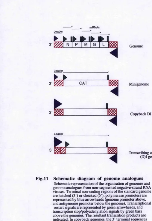
Fig. 11 : Schematic diagram of genome analogues...38
F ig.l2 : Transfection strategy for CAT rescue...80
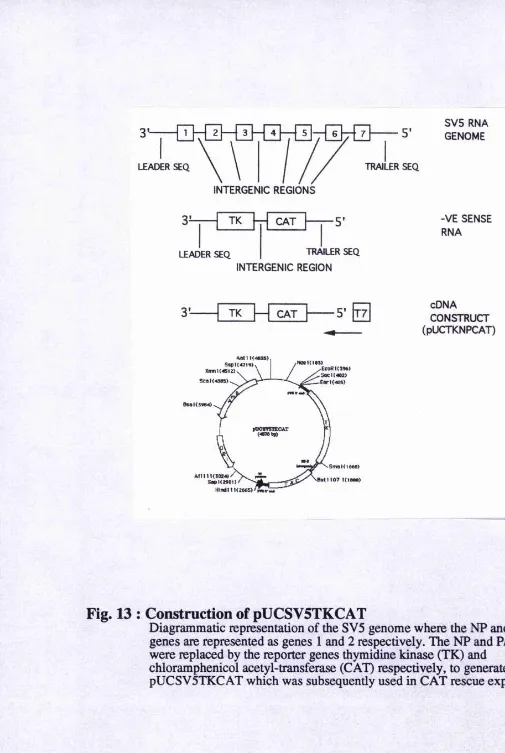
Fig.13 : Construction of pUCSV5TKCAT...83
Fig.l4: Comparison of transfection methods by CAT assay...85
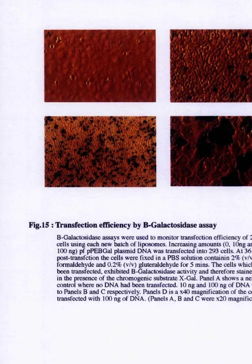
Fig.15 : Transfection efficiency by P-Galactosidase assay...87
F ig .l6 : Transfection of BHK cells with pUCSV5TKCAT after infection with VacT7 and SV5...8 8 Fig. 17 : Construction of pUCSV5CAT... 90
Fig.18 : Transfection of pUCSV5CAT DNA or RNA into BHK TK(-) C ells...92
F ig.l9 : Amended transfection strategy for CAT rescue... 94
Fig.20 : Construction of pG E M B L N P ...95
Fig.21 : Expression of NP protein from pGEM vectors... 98
Fig.22 : Sequencing of pGEM BLNP...99
Fig.23 : C-terminal repair of pGEMBLNP... 100
Fig.24 : Sequencing of pGEMBLNP-end repaired... 102
Fig.25 : Construction of pGEMpGlobinNP... 103
Fig.26 : Expression of NP protein after C-terminal repair and addition of the pGlobin leader sequence... 105
Fig.27 : Construction of pTM-P and pTM-V... 108
Fig.28 : Transient SV5 protein expression in VacT7 system... 110
Fig.29 : Western Blot of SV5 protein co-expression...111
Fig.30 : Construction of pPanHan... 113
Fig.31 : Digestion of pPANHAN constructs...115
Fig.32 : Overview of tTA transcriptional control...118
Fig.33 : Construction of pTET-pclobinNP (pTET-NP)... 120
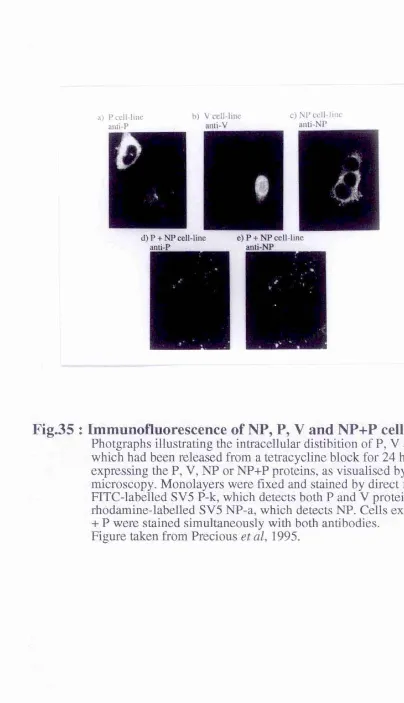
Fig.35 : Immunofluorescence of NP, P, V and NP+P cell lines ... 124
Fig.36 : Immunofluorescence of NP+V and NP cell lines...125
Fig.37 : Immunofluorescence of NP+V and NP+P+V cell lines...127
Fig.38 : Time course of induction for BalbC NP+V, NP and NP+P cell lines... 129
Fig.39 : Protein expression from BalbC V+P sub-clones... 130
Fig.40 : Protein expression from BalbC NP+P+V cell line sub-clones...131
Fig.41 : Protein : protein capture assay...135
Fig.42 : NP can bind to both P and V proteins... 137
Fig.43 : V binds a subpopulation of the available NP... 139
Fig.44 : P binds both soluble and polymeric NP while V binds only soluble NP...141
Fig.45 : mAb SV5 P-e blocks P binding to polymeric NP... 143
Fig.46 : Mapping of mAb binding sites in C-terminus of P...145
Fig.47 : Construction of P deletion mutants... 147
Fig.48 : VacT7 expression of P deletion mutants in capture assay...149
Fig.49 : Mapping of SV5 P-e binding site by deletion mutant analysis...151
Fig.50 : Bacterially expressed P and V can bind NP... 153
Fig.51 : Binding of bacterially expressed P to polymeric NP can be blocked by mAb SV5 P-e...156
Fig.52 : NP:P and NP:V interactions...164
Fig.53 : Initiation of RNA encapsidation...169
Chapter 1 : INTRODUCTION
This thesis aims to explore the mechanisms involved in the transcription and replication
of simian virus 5 (SV5), a member of the paramyxovirus family. These mechanisms were investigated utilising both reverse genetics techniques and by examining specific
viral protein : protein interactions within the replication complex. This chapter is
therefore divided into three main sections: the first, gives an overview of what is
currently known about the Paramyxoviridae transcription / replication mechanisms; the second, documents the development of genetic manipulation techniques for the
examination of the biology of negative stranded RNA viruses; the third outlines the
aims of the project presented in this thesis. Chapter 2 documents the materials and
methods employed during the course of this work. The results are presented in Chapter
3 where they have been divided into 3 sections. The first deals with the attempted development of a reverse genetics system for SV5; the second documents the development of cell lines expressing SV5 proteins; the third examines SV5 protein :
protein interactions. In Chapter 4, the results, which give some insight into the possible
1 Classification, Molecular Structure and
Replication of the
Paramyxoviridae
The Paramyxoviridae is a family of enveloped viruses, with a single-stranded, non segmented, RNA genome of negative polarity. They resemble two other families of negative-stranded RNA viruses, namely the Rhabdoviridae, for a similarity of genome organisation and expression, and the Orthomyxovindae, for a similarity in the
biological properties of the envelope glycoproteins.
The negative sense RNA genome has a dual purpose: firstly, it is the template for mRNA synthesis, and secondly, is a template for antigenome (full length positive strand copy of the genome) synthesis, which in turn, acts as a template for the synthesis of genomic (negative) sense RNA. These viruses encode their own RNA-dependent RNA polymerase, which acts as both a transcriptase, during the generation of mRNA, and a leplicase, when synthesising genomes (or antigenomes). mRNA is generated once the virus has been uncoated in the infected cell and is followed by viral replication, which requires continuous synthesis of viral proteins.
1.1 Classification of the
Paramyxoviridae
In 1995, the International Committee on Taxonomy of Viruses, reclassified the
Paramyxoviridae family into 2 subfamilies, namely, the Paramyxovirinae and the
Pneumovirinae. The Paramyxovirinae contains three genera, Parainfluenzavirus, Rubulavirus and Morbillivirus, while the Pneumovirinae contains only the
Pneumovirus genus. This classification was based on morphological similarities, antigenic cross-reactivity between members of a genus, and the coding organisation of the P genes. Examples of viruses found in each genus aie given in Table 1. The
Parainfluenzaviruses and Rubulaviruses are differentiated from the Morbilliviruses on the basis of biological activity of the attachment protein. In addition, the Rubulaviruses
Family
Paramyxoviridae
Subfamily
Paramyxovirinae
Genus
Parainfluenzavirus
Sendai virus (mouse parainfluenza virus type I)
Human parainfluenza virus type 1 and type 3
Bovine parainfluenza virus type 3
Genus
Rubulavirus
Simian virus 5 (Canine parainfluenza virus type 2)
Mumps virus
Newcastle disease virus
Human parainfluenza virus type 2, type 4a and 4b
Genus
Morbillivirus
Measles vims
Dolphin morbillivirus Canine distemper virus
Peste-des-petits-ruminants virus
Phocine distemper vims Rinderpest virus
Subfamily
Pneumovirinae
Genus
Pneunovirus
Human respiratory syncytial virus
Bovine respiratory syncytial virus
Pneunonia virus of mice Turkey rhinotracheitis virus
Table 1 Members of the
Paramyxoviridae
The Pneumaviriuae are very dilTerenl from the Paramyxovirinae in that, although their gene order is similar, they encode a number of additional genes, (N Sl, NS2 and M2),
and their attachment protein is different in terms of structure and biological activity.
1.2 Virion Structure
The Paramyxoviridae. although pleomorphic in shape, are generally spherical. They consist of a single stranded RNA genome, encapsidated in a helical core stmcture
known as the nucleocapsid. This is surrounded by matrix protein (M) and a host derived
lipid bilayer, through which two glycoprotein (HN and F) protrude. A schematic
diagram of the Rubulavirus. SV5, is given in Fig.l.
The infectious cycle begins with virus atttachment to the target cell surface and fusion with the cell membrane, and is mediated by the glycoproteins. The attachment protein (H, HN or G), interacts with a specific receptor on the host cell surface whereupon the
fusion protein (F) fuses with the cell membrane facilitating the release of the
nucleocapsid into the cell cytoplasm (Reviewed in Choppin and Compans, 1975).
The nucleocapsid is comprised of the negative sense RNA genome in tight association
with the nucleocapsid protein (NP) in a helical core structure which is resistant to nuclease attack. To this, the phosphoprotein (?) and the large protein (L) are attached. It has been reported that V protein is also in association with the nucleocapsid of SV5
(Paterson et aL 1995). This nucleocapsid core serves as the template for both the
generation of mRNA (transcription) and genomic/anti-genomic RNA (replication) as
shown in Fig.2. Transcription and replication are functions of the viral polymerase,
which requires both P and L proteins to constitute an active complex (Hamaguchi et al,
1983).
Paramyxovirus nucleocapsids are not rigid structures, as they coil and uncoil in
response to changes in salt concentration (Heggeness et aL 1980). Furthennore, Sendai
virus has been shown to exist in several distinct morphological states at normal salt
>
c/3
I
a "§
i
*o
I
Primary transcription
5'—
Secondary
Transcription
5* Input nucleocapsid (-RNP)
mRNAs
Viral proteins
I
Progeny Virions
Replication
+ RNP
-R N P
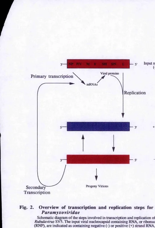
Fig. 2. Overview of transcription and replication steps for the
Paramyxo viridae
Schematic diagram of the steps involved in transcription and replication of the
Rubulavirus SV5. The input viral nucleocapsid containing RNA, or ribonucleoprotein (RNP), are indicated as containing negative (-) or positive (+) strand RNA, as discussed
[image:24.614.12.527.11.770.2]nucleocapsid core may be necessary for the viral polymerase to gain access to the RNA
bases. Therefore, the most extended form of the nucleocapsid structure, where the helix
is almost completely unwound, may represent the structure adopted during the copying
of the RNA template.
1.3 Genomes and Encoded Proteins
The genomes of the Paramyxoviridae are single stranded, non-segmented RNAs of
negative polarity, and are approximately 15 kb in length. The genome encodes 6-10
genes (Fig.3) depending on the virus being examined, where a 'gene' refers to a region
of genomic RNA which encodes one (or more) specific mRNA(s). Flanking the genes,
is a 3' extracistronic sequence known as the leader region and a 5' extracistronic
sequence known as the trailer region. Both are around 50 nucleotides in length, are well
conserved within the Paramyxoviridae, and contain conserved sequences at each end of
the genome (Galinski and Weschler, 1991). In human parainfluenza virus type 3
(hPIV3), 33 of the first 39 nucleotides are conserved in the leader and trailer sequences
(Galinski, 1988 ) suggesting these sequences contain signals enabling polymerase attachment to the template and encapsidation of the nascent strand (Blumberg et al.
1991). There are sequences found between the genes (i.e. after one gene end sequence
and before the next gene start sequence) known as intergenic regions. These are
variable in length for the Rubulaviruses (1-47 nucleotides) 'ànd Pneumoviruses (1-56
nucleotides) but are exactly three nucleotides long in both the Parainfluenzaviruses and
the Morbilliviruses. In measles virus (MeV), Sendai virus (SeV) and hPIV3, the intergenic sequences are GAA, GGG and GCT respectively (Galinski and Wechsler,
1991). The influences on viral transcription of these intergenic sequences, and the leader and trailer sequences, are discussed further in section 1.4.2.
1.3.1 The Nucleocapsid Protein (NP)
Pamitÿluenzavirus - Sendai virus
1682 1894 1173 1846 1891
NP P/aW M F HN
6799
L
Rululavirus - SV5
I I I
I I I
1787 1298 1371 1709 292 1869
NP P/V M F SH HN
Morbillivirus - Measles virus
6823
L
1688 1657 1473 2377 1949
N P/C/V M F H 6639L
Pneumovirus - Respiratory Syncytial Virus
528
499 907 405 1899
NSl NS2N P M SH G F M2
6 8
Kilobases
10 12 14 16
Fig.3 Genome organisation in the
Paramyxoviridae,
Schematic diagram showing the genome organisation of a representative member from each genus of the Paramyxoviridae. The L gene of the
Pneumoviruses ovCTlaps with that of the M2 gene and is therefore shown as staggared. Adapted from Lamb and Kolakofs^ (1995).
firstly encapsidates genomic (and anti-genomic) RNA, giving protection from nuclease
attack (Banerjee, 1987), and also gives lise to the characteristic helical structure of the
nucleocapsid (Das and Banerjee, 1993; Books et ciL 1993; Biichholtz etal, 1993). NP interacts with both the polymerase complex (Kingsbury, 1974) and M protein
(Markwell and Fox, 1980), suggesting a role for NP in transcription, replication and vims assembly. Furthermore, it is thought that the intracellular concentration of NP is a major factor in the switch from transcription to replication (Vidal and Kolakofsky,
1989).
The sequences of many NP proteins have been predicted (e.g. Neubert etal, 1991;
Parks etal, 1992; Rozenblatt é'f r//,1985; Collins etal, 1985) and range from 489-533 amino acids (a a ) in length. Protein sequence alignments have suggested that NP
contains 2 domains, a large globulai* N-terminal domain of around 500 amino acids which is relatively conseiwed between related viruses, and a small, hyper-variable, C-
terminal tail region extending from the globular body of the protein (Heggeness, 1981).
Trypsin digests of the NDV nucleocapsid (Kingsbury and Darlington, 1968)
demonstrated that the C-terminal domain could be cleaved off without affecting the
ability of NP to fomi RNase resistant nucleocapsids. These nucleocapsids were then examined by EM and were found to retain their characteristic helical confomiation. This suggested that both the RNA-binding domain and the residues involved in the
interactions resulting in helical nucleocapsid formation, were to be found in the globular N-terminal domain. A conserved hydrophobic motif of FX4YX4SYAMG
(where X is any residue), is found in the middle of the protein, suggesting this may be
the site of NP:NP interaction (Morgan et al, 1984). However, since this motif contains a number of aromatic amino acid residues (Tyr, Phe or Trp), it has been suggested that
this could be the RNA binding site (Morgan etal, 1984).
The divergent C-ierminus of NP contains many negatively charged residues and the
major sites of phosphorylation for the protein. In Sendai virus, a stretch of 7 acidic
Deletion of a a 4-188 and a a 374-492 caused formation of unstructures
aggregates
Requned for formation of soluble N:P complexes
304-373
a a 4-188
a a 374-492
Deletion here has no effect on N:P interaction but abolishes N:N interaction
Necessary for the stability of
the protein
a a 240-303
a a 189-239
‘525
Amino acids 1-398 are necessary for the formation of
nucleocapsid-like structures
Fig. 4 Schematic diagram of measles virus N protein
Sites of proteiniprotein interaction on MeV N protein were identified by deletion mutant analysis. Two non contiguous regions (amino acids 4-188
and 304-373) are required for the formation of soluble N:P complexes, while deletion of amino acids 189-239 did not affect N:P binding. Amino acids 240-303 appear necessary for the stability of the protein. The N-terminal 398 amino acids are all required for the formation of organised nucleocapsid-like particles, since deletion of the central region from amino acids 189-373 completely abolish N:N interaction, and deletion of amino acids 4-188 and 374-492 caused the formation of unstructured aggregates.
[image:28.612.11.525.22.645.2]Structure / function analysis of Sendai virus NP protein found that the C-terminal tail
was not essential for encapsidation of a synthetic defective interfering (DI) particle but
was required for this template to be functional in subsequent replication assays (Curran
et aL 1993). The role of the NP C-terminus remains unclear, but since NP protein with C-terminal deletions can form nucleocapsid-like structures containing cellular RNA, it may play a role in the specificity of RNA assembly (Buchholz et al, 1993).
During viral replication, soluble (unassembled) NP must be transported to the site of
nucleocapsid assembly since genome replication and encapsidation are concurrent
(Vidal and Kolakofsky, 1989). There is evidence to suggest that P protein chaperones
NP to the polymerase complex (Horikami etal, 1992; Buchholz etal, 1993; Cuiran et
al, 1993) and in doing so, prevents NP from self-aggregating. Evidence presented in this thesis, and elsewhere, suggests that V protein may play a similar role in preventing NP from self-aggregating (Randall and Bermingham, 1996; Horikami etal, 1996). A
further interaction, probably with the viral polymerase complex, must take place, which
allows NP to specifically assemble the nascent RNA and which, presumably also
releases P.
Deletion mutant analysis on both Sendai virus and measles virus, have identified areas of NP:NP and NP:P interaction (Buchholz et al, 1993, Bankamp et al, 1996) as shown in Fig.4. The N-terminal 398 amino acids were required for the formation of
nucleocapsid-like structures and deletion of amino acids 189-373, abolished NP:NP
interaction. Two regions (amino acids 4-188 and 304-373) were also identified as being
required for soluble N:P complex formation. Measles N also undergoes a conformational maturation upon phosphorylation, whereupon it is available for
assembly into nucleocapsid structures (Gombart et al, 1993).
1.3.2 Proteins encoded by the F Gene
When comparing the P genes, a major difference is found between the Pneumovirinae
multiple reading frames. These reading frames are accessed during viral transcription
either by the use of a variable number of initiation codons or by a process known as
RNA editing. For the latter, a frameshift is generated by the addition of non-templated
residues due to a polymerase stuttering mechanism. Thus, proteins with common N- terminal sequences but unique C-termini are generated. These proteins, and their
generation, are discussed in more detail below.
1.3.2.1 P Protein
The sequences of many paramyxovirus P proteins have now been predicted and are
found to be variable in length within the family (e.g. Bellini et al, 1985, Thomas et al,
1988, Shioda et al, 1986, Southern et al, 1990, Spriggs and Collins, 1986). The P
proteins, named because they are highly phosphorylated, play a pivotal role in RNA
synthesis. P functions as a subunit of the viral polymerase as both P and L proteins are necessary to constitute an active polymerase complex (Hamaguchi et al, 1983). It also
binds soluble (unassembled) NP, forming an NP-P complex which prevents NP from self aggregating and possibly delivers NP to the nascent RNA chain for encapsidation during viral replication (Curran et al, 1995a). As previously mentioned, there is some evidence to suggest that NP-P complexes prevent NP from non-specifically
encapsidating cellular RNA (CuiTan et aL 1993, Buchholz et al, 1993, Masters and
Banerjee, 1988).
The P protein can be divided^into an N-terminal domain, a C-terminal domain and a
hypervariable region in between as shown in Fig.5, The N-terminal region of P proteins contain the phosphorylation sites (Das etal, 1995; Byrappa etal, 1995;Vidal etal,
1988) and areas involved in RNA synthesis (CuiTan etal, 1994 and 1995a).
The immediate N-terminal residues (amino acids 1-77) in Sendai virus (SeV) were shown to be essential for RNA encapsidation (Curran et al, 1994). This study also
demonstrated that 9 amino acids within this region (amino acids 33-41) were required
Soluble NP binding site 33,--- ,41
Nucleocapsid binding Trimérisation domain site
344 411 479 568
+ G 412 445 L binding site
100 200 300
No. of amino acids
400 500 568
Fig.5 Schematic diagram of SeV P protein
Illustration of the areas of contact be ween P and the other viral proteins indicated. Residues 33-41 are required for the chaperoning of unassemled NP during the nascent chain assembly step of genome replication. Two blocks within the C-terminal 40% of the protein (ao344-411 and 479-568) are involved in binding to nucleocapsids. Residues 344-411 are also required for
trimérisation and only trimers bind to the nucleocapsid template. Residues 412-478 represent the stable binding site for the L protein. The site for the addition of a non-templated G residue at codon 308 during mRNA synthesis is also indicated.
Adapted from Curran (1996)
The C-terminal portion of SeV P protein contains 2 nucleocapsid (assembled NP)
binding sites at amino acids 345-411 and 479-568 (Ryan and Kingsbury, 1988; Ryan eî
aL 1991), one of which (amino acids 344-411) is also the region essential for
oligomerisation of the P protein (amino acids 344-411) into homotrimers (Curran etaL
1995) and only trimers bind to the nucleocapsid template. Between the NP binding
sites, lies the L binding site (amino acids 412-445) which loops out to contact L and does not overlap with either of the NP binding sites (Curran et aL 1994; Smallwood et
a/, 1994).
1.3.2.2 C Proteins
Only members of the Parainfluenzavirus and Morbillivirus genera express small basic
C proteins from their P genes (Barrett etaL 1985, Bellini et aL 1985, Spriggs and Collins, 1986). SeV encodes 4 'C-ltke' proteins, namely C , C. Y1 and Y2 (CuiTan and
Kolakofsky, 1989) while hPIVl expresses C , C and Y1 (Power etatl 1992). These
proteins are expressed both in vivo and in vitro and are generated from a variable number of start codons, including non-AUG initiation codons (reviewed in Kolakofsky
et al, 1991). SeV C and C (But not Y 1 or Y2) proteins have been shown to inhibit
mRNA synthesis and genome replication by possibly forming inactive complexes with
L protein. It has been suggested that the C proteins exhibit a negative regulatory role
during virus infection to limit both genome amplification and mRNA synthesis (Cadd et aL 1996; Curran etaL 1992). Given the very different levels of C expression among the
different Paramyxoviridae, the viruses may have differing requirements for down- regulation of genome amplification and expression in the later stages of infection. A C- deleted measles virus has recently been found to be viable and essentially without
mutant phenotype in cell culture (Radecke and Billeter, 1996) as has the analogous
rhabdovirus, vesicular stomatitis virus (VSV; Kretzschmar et aL 1996). However,
1.3.2.3 V, W and I Proteins
The P genes of most of the Paramyxovirinae are able to express two mRNA species by a mechanism of RNA editing which inserts non-templated G residues. It is a virus specific event which takes place co-transcriptionally by reiterative copying of a short C-
stretch at a specific region on the genome template (Vidal et al, 1990a). The
septanucleotide consensus motif (3 - UUU/CUCCC -5'), (Fig. 6) including the C-
stretch, has been proposed as a requirement for editing (Park and Krystal, 1992). A
'stuttering' model proposes that the polymerase pauses at this site, and when the pause is sufficiently long, slippage of the nascent mRNA occurs by one or two nucleotides, thereby reiteratively inserting one or two Gs (Vidal et al, 1990b).
For the parainfluenzaviuses (SeV, hPIVS) and Morbillivirus es (measles virus), the unedited mRNA that is a faithful copy of the genome, encodes llie P protein while the
edited mRNA, with IG addition, encodes the V protein (Vidal et al, 1990a; Galinski et
al, 1992). However, for the Rubulaviruses (SV5, MuV), the unedited mRNA encodes the V protein while the edited mRNA, with a 2G insertion, encodes the P protein (Thomas etal, 1988; Southern etal, 1990; Paterson and Lamb, 1990; Ohgimoto etal,
1990). In either case, the P and V proteins are amino-coterminal, while the -1 or -2
frame is used to generate unique C-termini from the edited mRNA.
For the Morbilliviruses and some of the SeV group which require a IG insertion to
access the V open reading frame (ORF), a 2G iiisertional event (at a much reduced frequency) leads to a protein called W. This is essentially the N-terminal part of the P protein up to the editing site as the downstream ORF is quickly closed by a stop codon
(Vidal etal, 1990a,b; Kolakofsky et al, 1991). The remaining P gene product of the
Rubulaviruses, represents tlie N-terminal part of the V or P protein up to the editing
site, referred to as the I protein, is accessed by the insertion of 4Gs rather than 2Gs
(Paterson and Lamb, 1990).
Of the distinct P gene products, the V protein is of particular interest because of its almost universal conservation in all three genera of the Paramyxovirinae. To date, human parainfluenza virus type 1 (hPIVl) seems to be the only exception, as it has
MeV
A A U U U U U C C C G U g U C
5‘
SeV
g u U U U U U C C C G U a U C
5'
MuV
A A A U U C U C C C C c c g g
5'
SV5
A A A U Ü C U C C C C g u c C
5’
consensus
U U Y U C C C
Fig. 6 Consensus Sequence for P mRNA Editing
The four paramyxovirus minus-strand genome sequences where G
insertion are known to occur, written 3' to 5' are shown. Exact homologies are highlighted in underlined bold script and other homologies are given in capital letters. The consensus sequence is given underneath.
1992; Rochat et al, 1992). The V protein is structurally characterised by a highly
coiisei*ved cysteine-rich domain in its carboxy-terminal half, which is fused to the amino-terminal half of the P protein. The V proteins of MeV, SV5 and NDV have all been shown to bind zinc (Liston and Briedis, 1994; Paterson etal, 1995; Steward etal,
1995), although the functional significance of this remains unclear.
The V proteins of SeV and measles virus (MeV) are not present in virions and are not
associated with the RNP, probably because these V proteins are missing the
trimérisation site, L binding site and assembled NP (nucleocapsid) binding sites, which are located in the C-terminal half of the protein (see 1.3.2.1 for further details).
However, the V protein of the Rubulaviruses does appear to be present in virions
(Paterson et al, 1995; Takeuchi etal, 1990, Kawano et al, 1993) and unlike the
Parainfluenza- and Morbilliviruses, their N-terminal halves are predicted to be basic
rather than acidic in character. The strong conseivation of the V ORF throughout
almost all the Paramyxovirinae suggests that it may interact with a structurally
invariant surface e.g. a cellular protein which is highly conserved from chickens (NDV) to man. Interactions between V and cellular proteins have been reported for MeV
(Liston et al, 1995) and SV5 (Precious et al, 1995) although the cellular proteins
involved were of markedly differing size.
The function of V protein has been studied using SeV defective interfeiing (DI)
particles as a genome analogue. The V (and W) protein was found to inhibit DI genome replication (Curran et al, 1991a) possibly by interfeiiing with the RNA encapsidation step (Curran et al, 1994) but did not appear to affect viral mRNA synthesis (CuiTan et
al, 1994). However, when a V-minus (but W-augmented) SeV was engineered from
cDNA, it was found to replicate normally in cell culture and embryonated chicken eggs (Deienda et al, 1997). Moreover, when a similar V-minus SeV was generated which
also did not produce W protein, it displayed markedly increased gene expression and
cytopathogenicity at the cellular level in vitro, but was strongly attenuated in
pathogenicity for mice. Therefore, V may act in animal infections to avoid the anti-viral
The recovery of V-minus viruses from cDNA indicaies that V protein is not an essential factor for SeV replication. This suggests that V may be an accessory protein, not
absolutely required by all viruses, and therefore some viruses have adapted to do without them e.g. hPlV l. Similar conclusions have been drawn for the role of C protein
in MeV and VSV (Radecke and Billeter, 1996; Kretzschmar er al, 1996; and section
1.3.2.2).
1.3.3 The Large (L) Protein
The large (L) protein is, as its name suggests, the largest and least abundant protein in
the virion. Both P and L proteins are necessary to constitute an active polymerase
complex (Hamaguchi et al, 1983), although L is thought to be responsible for the
majority of enzymatic activities involved in viral transcription and replication. Many of
these activities have been demonstrated in genetic or biochemical studies on the L protein of the rhabdovirus VSV. Viral mRNA capping, methyl-transferase activities and polyadenylation have all been attributed to VSV L protein (Abraham etal, 1975;
Hercyk et al, 1988; Hunt et al, 1984). The L proteins of VSV and SeV also have kinase
activity since they phosphorylate both NP and P proteins (Sanchez etal, 1985;
Einberger et al, 1990).
Sequence alignments with several L proteins from rhabdoviruses and paramyxoviruses have shown six conserved regions, separated by variable regions, suggesting a structure
of concatenated functional domains (Poch etal. 1990). FurtheiTnore, four short
conserved sequence motifs were found in domain III of the L proteins which were also
conserved in the RNA-dependent RNA polymerases of other virus families (Poch et al,
1990). These motifs surrounded an invariant pentapeptide (QGDNQ) and were suggested to represent important elements of the active site for template recognition and/or phosphodiester bond formation by the L proteins.
The N-terminal of both SV5 and MeV L proteins have been shown to contain
sequences important for stable L-P complex formation (Parks, 1994; Horikami etal,
L protein, showed that this region was important for both transcription and replication functions of the protein (Chandiika et al, 1995).
1.3.4 Matrix (M) Protein
The matrix (M) protein is the most abundant protein in the virion and lies between the
nucleocapsid and the glycoprotein-containing envelope. The sequences of many of the
Paramyxoviridae M proteins have been predicted (Bellini et al, 1986; Blumberg etal,
1984; Satake and Venkatesan, 1984; Sheshberaderan and Lamb, 1990), indicating they
are basic proteins of between 341-375 amino acid residues. M protein is hydrophobic in
nature but does not have any hydrophobic stretches long enough to span a lipid bilayer
(Reviewed in Peeples, 1991). Very little is known about the structure of the M protein, but a recent report on the M protein of the rhabdovirus VSV suggested a rod-like shape (Barge et al, 1996).
It is generally accepted that M protein is the central organiser of virion assembly, as its hydrophobic character allows it to interact with the glycoprotein-containing plasma
membrane and its positively charged residues allows interaction with the newly
synthesized (and negatively charged) nucleocapsids (Yoshida etal, 1976;
Sheshberaderan and Lamb, 1990; Morrison, 1988). M protein can also bind to itself and forms a paracrystalline anay with defined periodicity on the inner side of the infected cell plasma membrane (Bachi, 1980). This self-association and its contact with the
nucleocapsid may be the driving force in forming a budding virus particle (Peeples,
1991). It has also been reported that M protein binds actin (Giuffre etal, 1992; Bohn et al, 1986) which may also prove important for virus budding.
There is clear evidence that M proteins of VSV and influenza virus down-regulate transcription from RNP cores (Ye et al, 1989; Zvinarjev and Ghendon, 1980; Ye and
Wagner, 1992; Black etal, 1993) and there is some evidence to suggest measles virus M protein is an endogenous inhibitor of RNP transcription (Suryanarayana et al, 1994).
This study, and elsewhere (Cattaneo etal, 1988a; Cattaneo etal, 1988b) suggested that
an inability of the M protein to bind RNPs or associate with budding structures. Therefore, the striking abundance of RNP cores in brain cells of patients with the fatal persistent measles vims infection, subacute sclerosing panencephalitis (SSPE), could
possibly result from unrestricted transcription and replication of RNP cores as a result
of certain mutated M proteins. These mutated M proteins could either be defective in
transcription inhibition, or may just have lost their ability to bind RNP cores
(Suryanarayana et al, 1994).
1.3.5 The Envelope Glycoproteins
The membranes of Paramyxoviridae virions are studded with spike structures which
can readily be visualised by electron microscopy. Biochemical characterisation of these
structures has revealed there to be two different glycoprotein spikes on the surface of the virion. One acts as an attachment protein which serves to bind the vims to the cell suface and is composed of a protein termed haemagglutinin-neuraminidase (HN) protein in the Parainfluenza- and Rubulavirus genera, Haemagglutinin (H) protein in
the Morbillivirus genus and G protein in the Pneumovirus genus. As their names
suggest, the attachment protein of the Parainfluenza- and Rubulavirus genera also
contains a neuraminidase activity not demonstrated for the attachment proteins of the other genera (Scheid et al, 1972). The other spike is composed of the fusion protein and mediates the fusion of the viral membrane with that of the host cell, facilitating the
release of the nucleocapsid into the cell cytoplasm. This protein also mediates cell-to-
cell spread of the genetic information, and a prominant feature of the cytopathic effect of paramyxoviruses is syncytium formation (reviewed in Choppin and Compans, 1975).
The virus utilises the host cell pathways for synthesis, transport and post-translational modifications of the surface glycoproteins for insertion into the plasma membrane. Once the two glycoproteins are inserted into the plasma membrane of infected cells,
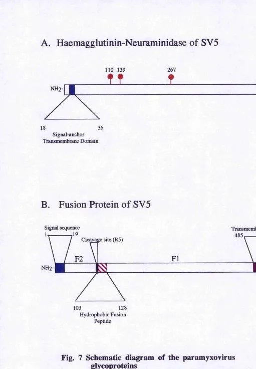
A. Haemagglutinin-Neuraminidase of SV5
n o 139 267 504
565
36 18
Signal-anchor Transmembrane Domain
B. Fusion Protein of SV5
Signal sequence Transmembrame Domain 485 509 Cleavage site (R5)
529
128 103
Hydrophobic Fusion Peptide
Fig. 7 Schematic diagram of the paramyxovirus
glycoproteins
(A) Haemagglutinin-neuraminidase attachment protein of SV5
indicating the signal anchor transmembrane domain and the sites used for the addition of N-linked carbohydrates.
(B) Fusion protein of S V5 indicating the position of the signal sequence, the transmembrane domain, the cleavage site and the
hydrophobic fusion peptide.
[image:39.612.15.526.16.752.2]ER lumen, cell surface or virion surface
Fusion Pqjtide
Cytoplasm
Fig.8 Schematic diagram of integral membrane proteins
The type I and type II integral membrane proteins F and HN of the Paramyxoviridae. Of the Paramyxovirinae, only SV5 has been shown to encode an SH protein, although there is an
SH ORF in mumps virus.
1.3.5.1 A ttachm ent Protein
The HN protein of the Parainfluenzavinises and the Rubulaviruses is a multifunctional protein. It is responsible for the binding of the virus to sialic acid-containing cellular
receptors such as glycoproteins or glycolipids, and mediates enzymatic cleavage of
sialic acid from the surface of virions and the surface of infected cells (Morrison and
Portner, 1991). By analogy to the influenza virus neuraminidase, it seems likely that the
neuraminidase activity of HN prevents self-aggregation of the virus particles when budding from the plasma membrane.
The sequences of many attachment proteins have been predicted (Blumberg et al,
1985a; Hiebert etal, 1985a; Alkhatib and Briedis, 1986; Wertz e ta l 1985) indicating
that they are 565-582 amino acids in length. HN proteins are type II integral membrane proteins, which span the membrane once, and contain a single hydrophobic-domain at
the N-terminus. This acts as both a signal and anchorage domain, leaving a long C- terminal ectodomain (Reviewed in Moii’ison, 1988). Conserved cysteine, proline and glycine residues between paramyxovirus HN proteins, has suggested a similarity in
protein structure across the family (Figs. 7A and 8).
HN exists as a disulphide linked dimer which forms a non-covalently linked tetramer
(Ng gf a I 1989), The stable formation of the tetramer is dependent on the
transmembrane domain and the cytoplasmic tail of the protein (Ng et a l 1989; Parks and Lamb, 1990; McGinnes e ta l 1993). The intramolecular disulphide bonds are formed during folding of the protein (McGinnes and Morrison, 1994a) forming the
covalently linked dimer (McGinnes and Morrison, 1994b). This disulphide bond formation has recently been shown to be cotranslational and important to the
subsequent folding of a conformationally normal and active HN protein (McGinnes and
Morrison, 1996). Furthermore, during its synthesis, HN binds the cellular protein GRP78-B1P (Ng et a I 1989) which prevents transport of defective proteins from the endoplasmic reticulum to the Golgi network (Pelham, 1986; Hurtley et aL 1989).
HN has 4-6 sites for the addition of N-linked carbohydrate chains (Ng et a l 1990)
which, in the case of MuV and SeV, is necessary for incorporation into virions (Herrler
where glycOvSylation is required for virus infectiviiy but not incorporation into virions (Morrison et al, 1981). These glycosylation sites are found towards the C-terminal end of HN (Monison, 1988), suggesting the protein consists of a globular head, containing
the enzymatic activités, supported by a stalk-like structure which is inserted into the
plasma membrane (Thompson etal, 1988). In addition, HN contains a conserved
sequence, NRKSKS, which is similar to the known sialic acid binding site of influenza
vims neuraminidase (Monison and Portner, 1991). On comparing the primary protein sequences of the paramyxovirus HN proteins with influenza virus NA, four regions
containing conserved motifs were found. These motifs in NA, and by analogy in HN, were postulated to be brought together to form the neuraminidase active site in the
conformationally mature protein (Colman et al, 1993). Supporting this hypothesis was
the finding that measles virus H, which does not have neuraminidase activity, contains only one of these conserved motifs.
There is still some disagreement whether the sites of haemagglutinin and neuraminidase
activities are combined (Scheid and Choppin, 1974) or are separate. If the analogy with influenza virus NA can be continued to include an NA with haemagglutinating activity,
then the HN activities would be expected to be located at separate sites (Laver et al,
1984).
As mentioned previously, the H protein of the Morbilliviruses had no neuraminidase activity (Morrison and Portner, 1991 and references therein). It specifically interacts with the CD46 cellular receptor molecule (Dorig etal, 1993; Naniche, 1993). Since
CD46 is expressed at a low level on the cell surface, aggregation during virus budding
is unlikely, thus negating the need for Morbilliviruses to exibit neuraminidase activity
to free themselves from the cell surface.
Like HN, measles virus H is a type II integral membrane protein and also forms disulphide linked dimers which are also possibly homotetramers. They also contain N-
linked carbohydrate chains which are located in the N-terminus of the protein (Alkhatib and Briedis, 1986). unlike HN where the glycosylation sites are distributed towards the
C-terminus. These H glycosylation sites are collectively required for folding of the
The G protein of the Pneumovirinae is, like HN and H, a type II integral membrane protein but has neither haemagglutinin nor neuraminidase activities. At 298 amino acids, it is much smaller than either HN or H (Wertz et al, 1985) and is found in
infected cells in both a proteolytically cleaved, soluble form, and membrane bound. The protein is extensively modified by the addition of both 0-linked and N-linked
oligosaccharides (Wertz etal, 1985; Grober and Levine, 1985). These differences have led to speculation that G protein has a different evolutionary ancestry to HN and H, and is more structurally similar to a group of cellular mucinous proteins (Wertz et al, 1985; Sullender and Wertz, 1991).
1.3.5.2 Fusion (F) Protein
The fusion (F) protein of the Paramyxoviridae mediates fusion of vims to the host cell membrane and between the infected ceU to the adjacent cell, promoting viral spread (Choppin and Compans, 1975). F is synthesised as an inactive precursor Fq, and subsequently cleaved to Fi and F^ subunits, which are disulphide linked in the
biologically active form (Scheid and Choppin, 1974, and 1977). The protein sequences of the F proteins have been predicted (Blumberg et al, 1985b; Paterson et al, 1984a; Richarson et al, 1986; Elango et al, 1985) indicating the encoded proteins contained 540-580 amino acid residues (Figs. 7B and 8).
F is a type I integral membrane protein that spans the membrane once and contains a cleavable signal sequence at the N-terminus of the protein. It has a typical three-domain structure, consisting of a large, relatively hydrophilic domain external to the virion, a second domain of 20 or more uncharged residues that anchors the protein to the lipid bilayer, and immediately adjacent, a hydrophilic C-terminal domain, which exists on the inner side of the virion bilayer or host cell plasma membrane (Monison and Portner, 1991). Although no overall homology between the different paramyxovirus F protein sequences was found, similar placement of cysteine, proline and glycine