PARAMYXOVIRUS PERSISTENCE
RACHEL FEARNS
A Thesis Submitted for the Degree of PhD at the
University of St Andrews
1995
Full metadata for this item is available in St Andrews Research Repository
at:
http://research-repository.st-andrews.ac.uk/
Please use this identifier to cite or link to this item: http://hdl.handle.net/10023/10973
PARAMYXOVIRUS PERSISTENCE
By Rachel Feams
Division of Cell and Molecular Biology
University of St. Andrews
DECLARATION
(a) I, Rachel Fearns hereby certify that this thesis has been composed by myself, that it is a record of my work and that it has not been accepted in partial or complete fulfillment of any other degree or professional qualification.
Signed .... Date .... f..t?: •• t?. (. : ..
9.£
..
:
....
..
... .
(b) I was admitted to the Faculty of Science of the University of St. Andrews under Ordinance General No. 12 on 1st October 1991 and as a candidate for the degree of Ph.D. on 1st October 1992.
Signed ... Date ....
!.!?
:
..
c:.
.
~
.
:
.
1.t:
...
.
... .
(c) I hereby certify that the candidate has fulfilled the conditions of the Resolutions appropriate to the Degree of Ph.D.
Signature of Su
C : : :
Date ...~9.
(!/r.
.f..
...
.
... .
ACKNOWLEDGMENTS
First and foremost I would like to thank my supervisor, Dr. Rick Randall, for his
patience and encouragement during my time at St Andrews. I wish to thank Dr. Ian Kerr
for providing the materials used to assay interferon activity and to Alison Bermingham for
providing me with the components for the 17 expression system. I am indebted to all the
technicians who have helped me during my studies here, particularly to Bill Blyth for the
photographic work, and to the members of lab. 28 for their help and advice. In particular, I
wish to thank Dan Young for many hours of patient explanation and for carrying out the
animal work involved in the elL assays and Bernie Precious for many hours of interesting
and thought provoking discussion. Thanks are due to Matt for his proofreading assistance
and patience, and to my family for their support during my studies. Finally, I am grateful to
the Medical Research Council for providing financial support during the course of this
ABSTRACT
In this study, SV5 infection of Balblc mouse fibroblast cells has been used as a
model system to investigate the possible mechanisms underlying the establishment and
maintenance of Paramyxovirus persistence.
It was found that following entry to these cells, the virus initiated a wave of
transcription and replication, similar to that of a permissive infection, in which normal
levels of each of the virus proteins were synthesized. However, by 48-72 hours post
infection (pj.) there was an almost complete cessation of virus mRNA and protein synthesis. Despite the decrease in virus activity, full length viral genome RNA and P and
NP, the proteins involved in transcription and replication, could be detected at consistently high levels up to 5 days pj., although the levels ofHN, M, F and V declined.
Immunofluorescence analysis supported these data showing that at later times pj. although there were some cells positive for all the viral proteins, a high proportion of cells
were strongly positive for NP, L and P, but negative for M, F and HN. In these cells, NP, L
and P were often located in discrete cytoplasmic foci. These results suggested that the
persistently infected cell population consisted of some cells in which the virus was active
and other in which it was quiescent within cytoplasmic inclusions.
A series of cell lines was established from a monolayer of Balb/c cells that had been
infected at a high multiplicity. Immunofluorescence studies showed only a minority of
cells in these clones to be infected with virus, indicating that during division, not all
daughter cells became infected. Of the infected cells, some were positive for all the viral proteins, while others were positive for only NP and P. Co-cultivation of the cloned cells
with Vero cells, which are permissive for SV5 replication, rapidly yielded non-defective
virus, suggesting that the virus was active in some cells. These results suggested that the
persisting virus was in a state of flux, able to reside as inclusions of inactive nucleocapids
from which it could reactivate to initiate a new round of infection.
Experiments aiming to determine if the persistently infected cells were resistant to immune attack demonstrated that cells at 5 days pj., in which the majority of cells were
quiescently infected, were less susceptible to immune lysis than cells at I day p.i. in which
there was ongoing protein synthesis.
Further experiments were carried out both to try to determine what had induced the
persistent state in mouse cells and also to examine factors which might induce a similar
NUCLEIC ACIDS DNA RNA A G C T U cAMP NTP(s) ATP GTP CfP UTP dNTP(s) dATP dGTP dCfP TTP ddNTP(s) ddGTP cDNA mRNA vRNA cRNA
ABBREVIA TIONS
2' deoxyribonucleic acid
ribonucleic acid
adenine (base in DNA/RNA)
guanine (base in DNA/RNA)
cytosine (base in DNA/RNA)
thymine (base in DNA) uracil (base in RNA)
cyclic 3', 5' adenosine monophosphate
ribonucleoside triphosphate(s)
adenosine 5' triphosphate guanosine 5' triphosphate
cytidine 5' triphosphate
uridine 5' triphosphate
2' deoxyribonucleoside triphosphate(s)
2' deoxy adenosine 5' triphosphate 2' deoxy guanosine 5' triphosphate
2' deoxy cytidine 5' triphosphate
thymidine 5' triphosphate
2',3' dideoxyribonucleoside triphosphate(s)
2', 3' dideoxy guanosine 5' triphosphate
complementary DNA
messenger RNA
viral genomic RNA
AMINO ACIDS
A Ala alanine M
C Cys cysteine N
D Asp aspartate P
E Glu glutamate Q
F Phe phenylalanine R
G Gly glycine S
H His histidine T
I De isoleucine V
K Lys lysine W
L Leu leucine y
PHYSICAL UNITS
·C temperature in degrees Celsius
g mg ~g ng I ml
gram mass or centrifugal force milli gram (10-3 g)
micro gram (10-6 g) nano gram (10-9 g)
litre volume
milli litre
micro litre
Met methionine Asn asparagine
Pro proline
GIn glutamine
Arg arginine
Ser serine Thr threonine
Val valine
Trp tryptophan
Tyr tyrosine
~ Ci rnCi ~Ci M mM ~M leD kb pH V rnA
Curie (measure of radioactivity = 3.7x 10¹⁰ disintegrations/second)
milli Curie ~ U micro Curie molar concentration milli molar micro molar kilodalton kilobase (pairs) -loglO [H+] volts milli amperes micro farads
CHEMICALS AND REAGENTS NBCS GMEM EDTA EGTA NP-40 SDS KAc TEMED DATD DAPI FITC PBS SSC Tris-HCI TE MOPS 32p 35S 51Cr VIRUSES hPIVl hPIV3 bPIV3 SV5 SV40 SV41 NDV MY SV
CDV
VSVRS virus
LCMV
HIV
Newborn calf serum
Glasgow modified Eagle's medium
ethylenediaminetetra-acetic acid
ethylene glycol-bis (~aminoethyl ether) N, N, N', N'-tetra-acetic acid
nonidet p40
sodium dodecyl sulphate
Potassium acetate
N, N, N', N'-tetramethylethylenediamine N, N-diallyltartardiamide
4, 6 diamidino-2-phenylindole
fluorescein isothiocyanate phosphate buffered saline
standard citrate saline
tris-hydroxymethyl-aminomethane, pH adjusted with HCI tris EDTA
3-(N-morpholino) propane sulphonic acid radioisotope phosphorous -32
radioisotope sulphur -35
radioisotope chromium -51
human parainfluenza virus type 1 human parainfluenza virus type 3
bovine parainfluenza virus type 3 simian virus type 5
simian virus type 40 simian virus type 41
Newcastle disease virus
measles virus
Sendaivirus
canine distemper virus
vesicular stomatitis virus
respiratory syncytial virus
lymphocytic choriomeningitis virus
MISCELLANEOUS
N-tenninus
C-tenninus GRP78BiP
E. coli
CNS CSF
scm
IFN DI MIBE SSPE CILMIle
ll..-2 Ig>
<A
% %v/v %w/v PAGE PCR RNA sin BHK BF moi pfu p.i. c.p.m. uv MAb ECL amino terminus carboxyl terminusglucose responsive protein (mw 78) binding protein
Escherichia coli
central nervous system cerebrospinal fluid
severe combined immunodeficiency interferon
defective interfering
measles inclusion body encephalitis subacute sclerosing panencephalitis cytotoxic T cell
major histocompatibility complex interleukin 2 immunoglobulin greater than less than approximately bacteriophage lambda percent
% volume of total volume
% weight of total volume
polyacrylamide gel electrophoresis
polymerase chain reaction placental ribonuclease inhibitor baby hamster kidney (cells) Balb/c mouse fibroblast (cells) multiplicity of infection plaque forming units post infection
counts per minute ultraviolet
monoclonal antibody
CONTENTS
INTRODUCI10N ...
1PART I: PARAMYXOVIRIDAE CLASSIFICATION, MOLECULAR STRUCTURE AND REPUCATION ... 2
1.1 nASSIFICA TIONS ... 2
1.2 PARAMYXOVIRUS STRUCIURE AND REPLICATIVE CYnE ... 6
1.3 THE PARAMYXOVIRUS GENOME ... 8
1.4 THE PARAMYXOVIRUS ENCODED PROTEINS ... 9
1.4.1 The attachment protein ... 9
1.4.2 The fusion protein ... 13
1.4.3 Small hydrophobic protein ... 15
1.4.4 Matrix protein ... 15
1.4.5 Nucleoprotein ... 16
1.4.6 The L protein ... 19
1.4.7 The phosphoprotein ... 20
1.4.8 V protein ... 22
1.4.9 C protein ... 23
1.5 THE PARAMYXOVIRUS REPLICATION CYnE ... 24
1.5.1 Virus entry ... 24
1.5.2 Adaptation of the nucleocapsid for transcriptional activity ... 25
1.5.3 Virus transcription ... 26
1.5.4 Genome replication ... 28
1.5.5 Virus maturation and release ... 30
PART
2:
VIRAL PERSISTENCE ... 322.1 DEFINITION OF VIRUS PERSISTENCE ... .32
2.2 DAMAGE LIMITATION ... 33
2.3 THE IMMUNE RESPONSE TO VIRAL INFECTION ... 34
2.3.1 Launching an immune response against viral infection ... 34
2.3.2 Recognition of viral antigens by effectors of the immune system ... 35
2.3.3 Effects of components of immune response ... 36
2.4 MEANS BY WHICH VIRUSES CAN OVERCOME THE HOST IMMUNE RESPONSE ... 37
~.4.3 MIlC haplotype of the host animal ... .39
2.4.4 Immune suppression: infection of immune cells ... .41
2.4.5 Immune suppression: antagonism of the CIL response ... .42
2.4.6 Summary ... 42
2.5 FACTORS mAT INFLUENCE PERSISTENCE: HOST FACTORS ... .43
2.5.1 Tissue tropism ... 43
2.5.2 Cell cycle and differentiation ... .45
2.5.3 Antibody mediated antigenic modulation ... .46
2.5.4 Interferon ... 48
2.5.5 Host cell evolution ... 50
2.6 FACTORS WHICH INFLUENCE PERSISTENCE:VIRAL FACTORS ... 50
2.6.1 Defective interfering (01) particles ... 51
2.6.2 Mutations leading to abortive infections ... 53
2.6.3 Mutations affecting virus virulence ... 55
2.6.4 Generation of mutant viruses and their role in persistence ... 56
2.7 CHRONIC DISEASES ASSOCIATED Willi PERSISTING P ARAMYXOVIRUSES ... 58
2.7.1 Subacute sclerosing panencephalitis (SSPE) ... 58
2.7.2 Paget's bone disease ... 60
2.7.3 Crohn's disease ... 61
2.8 PERSISTENCE: IMPLICATIONS FOR THE VIRUS AND HOST ... 61
2.9 OBJECTIVES OF WORK ... 62
MATERIALS AND METIIODS.
1. CElLS AND VIRUS. 1.1 Maintenance of mammalian ceUlines ... 641.2 Preparation of a working stock of SV5 ... 64
1.3 DAPI staining of cells and virus ... 65
1.4 Plaque assay titration of virus stocks ... 65
1.5 Virus infection of cells ... 66
1.6 Single cell cloning of infected BF cells ... 67
2 RNA ANALYSIS. 2.1 Extraction of total cellular RNA ... 67
2.2 Northern blotting of RNA ... 68
2.3 Dot blotting of RNA ... 69
2.4 Probe preparation. 2.4.1 End labelled oligonucleotide probe ... 70
2.5 Hyb}idisation ... 71
3. PROTEIN ANALYSIS 3.1 Antibodies ... 73
3.2 [35S] labelling of cells ... 73
3.3 Immune precipitation of viral proteins ... 74
3.4 SDS polyacrylamide gel electrophoresis ... 75
3.5 Western blotting of viral proteins ... 75
3.6 Dot blotting of viral proteins ... 76
3.7 Immunofluorescence microscopy ... 77
4. CITOTOXICITY ASSAYS. 4.1 Priming of cytotoxic T lymphocytes (CfLs) ... 78
4.2
en..
assays. 4.2.1 [51Cr] Labelling of target cells ... 794.2.2 Preparation of effector cells ... 79
4.2.3 Estimation of CfL activity ... 80
5. RECOMBINANT DNA TECHNIQUES. 5.1 Overview of the cloning stategy for SV5 ... 80
5.2 Preparation of oligonucleotides ... 82
5.3 Reverse transcription of cDNA from viral RNA ... 83
5.4 Polymerase chain reaction (PCR) amplification of DNA ... 83
5.5 Restriction enzyme digestion ofDNA. ... 84
5.6 Agarose gel electrophoresis of DNA ... 85
5.7
Purification of DNA fragments from agarose gels ... 855.8 Ligation of DNA fragments ... 86
5.9 Bacterial strains and culture ... 87
5.10 Preparation and transformation of competent E.
coli
cells ... 875.11 Colony peR amplifications ... 88
5.12 Small scale preparation of plasmid DNA ... 89
5.13 Large scale preparation of plasmid DNA. ... 89
6. SEQUENCING OF RECOMBINAN!' DNA CWNES. 6.1 Preparation of single stranded DNA. ... 90
6.2 Dideoxy sequencing of the V insert ... 91
6.3 Electrophoresis of sequencing reactions ... 92
7. EXPRESSION OF FOREIGN DNA IN MAMMAliAN CELLS. 7.1 Transient transfection of 293 and Balb/c fibroblast cells ... 93
7.2 Electroporation of BF cells ... 94
7.3 Isolation of histidinol resistant clones ... 94
8.1 Overview of method ... , ... 95
8.2 Collection of medium from infected BF cells ... 95
8.3 Antiviral assay from mouse interferon ... ~ ... 96
RESUL
1'S ...
97PART 1: CHARACTERISATION OF SV5 PERSISTENCE ... 97
1.1 CHARACfERISATION OF SV5 INFECTION OF CULTURED BALB/C CEIJS ... 99
1.1.1 RNA levels in infected cells ... 99
1.1.2 Comparison of viral protein synthesis in infected BF and BHK cells ... 101
1.1.3 SV5 protein synthesis in BF cells over time ... 104
1.1.4 Levels of virus released from infected BF cells ... 106
1.2 CHARACfERISA nON OF THE PERSISTENT ST A TE ... 107
1.2.1 Immunofluorescence studies of infected BF cells ... 108
1.2.2 Relative protein levels in infected BF cells ... 114
1.2.3 Pulse chase analysis of viral proteins ... 118
1.2.4 Passage of persistently infected cells ... 122
1.2.5 Single cell cloning of infected cells ... 123
1.2.6 Susceptibility of persistently infected cells to CIL attack ... 126
1.2.7 Summary ... 1 28 PART 2: FAcrORS INFLUENCING SV5 PERSISTENCE ... 129
2.1 P AND V IN INFECfED CEL~ ... 129
2.1.1 Potential roles for P and V in the establishment of persistence ... 129
2.1.2 Approach for studying the roles of P and V ... 130
2.1.3 Cloning of V ... 131
2.1.4 Sequencing of the V fragment. ... 131
2.1.5 Transient expression of V ... 133
2.1.6 Stable expression of V in BF and 293 cells ... 133
2.1.7 PCR of DNA within the transfected cells ... 136
2.1.8 Conclusion ... 136
2.2 THE ROLE OF INTERFERON DURING SV5 INFECTION OF BF CELLS ... 136
2.2.1 Release of interferon by SV5 infected BF cells ... 137
2.2.2 Effects of exogenous interferon of SV5 synthesis in BF cell cultures ... 139
2.2.4 Can interferon induce the fonnation of inclusions in HeLa
cells? ... 143
2.2.5 Effects of interferon of SV5 protein synthesis in HeLa cells ... ~~ ... 146
DISCUSSION ...
1471. A MODEL FOR PARAMYXOVIRUS PERSISTENCE ... 147
1.1 Can the virus exist in a transcriptionally inactive state? ... 147
1.2 The inclusions may consist of stable aggregates of nucleocapsids ... 149
1.3 Was the virus active in
some
cells? ... 1501.4 Reactivation from transcriptional quiescence ... 151
1.5 Could the model be established under different conditions? ... 153
2. POTENTIAL FACfORS AFFECflNG THE ESTABLISHMENT OF PERSISTENCE IN THE SV5/BF SySTEM ... 153
2.1 Could V induce persistence? ... 154
2.2 Interferon ... 157
3. IMPLICATIONS OF THE MODEL DURING IN VIVO INFECflON ... 159
3.1 Could the model be established by other factors known to influence persistence? ... 159
3.2 Avoidance of the host immune response ... l61 3.3 Can the model be applied to chronic disease? ... 162
4. FU1URE STUDIES ... 163
Figure 1 Figure 2 Figure 3 Figure 4 Figure 5 Figure 6 Figure 7 Figure 8 Figure 9
Figure 10 Figure 11
Figure 12
Figure 13
Figure 14
Figure 15
Figure 16 Figure 17 Figure 18 Figure 19 Figure 20 Figure 21 Figure 22 Figure 23 Figure 24 Figure 25 Figure 26 Figure 27 Figure 28 Figure 29
LIST OF FIGURES
Replication strategy of the Paramyxoviridae ... 3
Genome organisation of the different genera of the Paramyxoviridae ... 5
Structure of a Paramyxovirus particle ... 7
Notable features of the attachment proteins... ... ... .... ... ... 11
The structure of the fusion protein of SV5 ... 14
The structure of the nucleoprotein of Sendai virus... .... ... 18
Characterised domains of the P and V proteins of Sendai virus ... 21
Possible interactions of the P, L and NP proteins ... 21
Synthesis of a probe specifically against negative stranded viral RNA ... 72
Cloning strategy for the V gene of SV5 ... 81
Immunofluorescent staining of BF cells 1 day following infection with SV5 ... 98
The levels of viral RNA in SV5 infected BF cells ... 100
Comparison of viral protein synthesis in BF and BHK cells ... 103
Analysis of SV5 protein synthesis in BF cells from 1 to 5 days p.i.. ... 105
Distribution of viral proteins in BF cells at 1 and 5 days p.i... ... 109
Immunofluorescence analysis of BF cells at 1 day p.L ... 111
Immunofluorescence analysis of BF cells at 5 days p.L ... 112
Distribution of P, NP and L in infected BF cells ... 113
Analysis of the relative amounts of viral proteins in infected cells ... 115
Western blot analysis of P, V and M proteins in infected BF cells ... 117
Pulse chase analysis of NP protein ... 119
Pulse chase analysis of P, V, M, F and HN proteins ... 120
Distribution ofHN and P proteins in cloned BF cells ... 124
Distribution of P and HN in Vero cells co-cultivated with cloned BF cells ... 125
Cell mediated lysis of BF cells infected with SV5 for 1 and 5 days ... 127
Nucleotide and anticipated peptide sequences of the cloned V ... 132
Western blot analysis showing expression of V from SV5 infected cells and recombinant V from the transient expression vector ... 134
Analysis by PeR of transfected BF cell clones ... 135
Figure 31
Figure 32
Figure 33
Table 1
Table 2 Table 3 .
Table 4
~V 5 in the presence of interferon ... 140
Assay showing effect of IFN pretreatment on EMC virus infection ... 142
Immunofluorescence analysis of HeLa cells treated with IFN prior to infection with SV5 ... 144
Effects of IFN pretreatment on SV5 protein synthesis in infected HeLa cells. .... .... ... ... ... ... ... ... ... 145
LIST OF TABLES
Organisation of the family Paramyxoviridae ... .4V specific primers used for sequencing ... 91
Levels of virus release from SV5 infected BF and Vero cells ... 107
CORRIGENDUM
Since this thesis was written, the International Committee on Taxonomy of Viruses has
made recommendations for virus identification (the International Committee On Taxonomy of
Viruses: Virus Taxonomy. The Sixth Report of the International Committee on Taxonomy of
Viruses, Springer-Verlag, Wein/ New York, 1995). The following nomenclature has been agreed;
in formal taxonomic usage, the first letters of virus family, subfamily and genus are capitalized and
the terms are italicized. Species terms are not capitalized (unless they are derived from a place name
or a host family or genus name) nor italicized; for example: family Paramyxoviridae, genus
Morbillivirus, measles virus. Having been defined in formal tenns, a virus can be referred to by
infonnal vernacular terms. In this case, the virus family. subfamily, genus and species names are
written in lowercase Roman script; they are not capitalized nor italicized. However, ambiguity
should be avoided by following the taxonomic unit with the name of the taxon; for example: the
paramyxovirus family. This thesis was written before the above was published and therefore the
INTRODUCTION
This thesis aims to explore how Paramyxoviruses may be able to persist in vivo.
Persistence by Paramyxoviruses is a widely acknowledged phenomenon: Paramyxoviruses
are implicated in a number of chronic diseases and one, measles virus, is known to be the
aetiologic agent of the disease subacute sclerosing panencephalitis (SSPE). Although it is
generally accepted that Paramyxoviruses are able to give rise to persistent infections, it is
unclear how an RNA virus is able to be maintained in a host without being cleared by the
host's immune system. In the results section of this thesis, evidence is presented which
suggests a possible model to explain how a virus could persist in the face of the immune
response, whilst still being able to cause disease.
As an introduction to the thesis, two subjects are considered. The ftrst section is a
review of the current literature concerning the molecular biology and replicative cycle of
Paramyxoviruses. The second section deals with viral persistence. This section covers a
number of topics, including ways in which persisting viruses may be able to avoid immune
clearance, factors which are known to influence viral persistence and a review of some
PART
1: PARAMYXOVIRIDAE CLASSIFICATION, MOLECULAR
STRUCTURE AND REPLICATION
1.1 CLASSIFICATION
The Paramyxoviridae are a family of enveloped, non-segmented, negative stranded
RNA viruses. They bear resemblances to two other RNA virus families, namely the
Orthomyxoviridae (eg. influenza A), with whom their envelope glycoproteins share
common features (Lamb, 1993) and the Rhabdoviridae (eg vesicular stomatitis virus,
VSV), which have a similar genome organisation and mode of replication (Pringle, 1987).
Like the Rhabdoviruses, the Paramyxovirus genome acts as a template both for mRNA
transcripts and for the synthesis of a full length positive stranded RNA species, which acts
as an intermediate during RNA replication (see figure 1).
The Paramyxoviridae family is subdivided into two subfamilies, the
Paramyxovirinae and the Pneumovirinae. The Pneumovirinae contains the genus
Pneumovirus, and the Paramyxoviruses are divided into three genera, the Parainfluenza
viruses, the Rubulaviruses and the Morbilliviruses. Examples of viruses found within each
genus are shown in table 1 (Lamb and Kolakofsky, in press). The Parainfluenza and
Rubulaviruses are very similar to one another and were previously classified in a single
genus, Paramyxovirus (Kingsbury
et al.,
1978). These viruses are differentiated on thebasis of their cross reactivity with antibodies and on the coding organisation of their P
genes . The Parainfluenza and Rubula viruses are distinguished from the Morbilliviruses
on the basis of the biological activity of the attachment protein (Matthews, 1982). The
Pneumoviruses are quite different from the Paramyxovirinae. Although their gene order is
similar, these viruses encode a number of additional genes, NS 1, NS2 and M2, and their
attachment protein is very different both in terms of biological activity and structure. The
w
3' • • • • • • • • • • • • • • • • • • • • • • • • • • • • 5' genomic
-ve sense RNA
5' AAAAA 3' 5' • • • • • • • • • • • • • • • • • • • • • • • • • • • • 3' antigenomic
+ve sense RNA mRNA
j
3' • • • • • • • • • • • • • • • • • • • • • • • • • • • • 5' -ve sense RNA genomIc
Figure 1: A schematic diagram showing the replication strategy of the Paramyxoviridae.
FAMILY: PARAMYXOVIRIDAE
Subfamily: Paramyxovirinae
Genus: Parainjluenzavirus
Sendai virus (SV)
Human parainfluenza virus type 1 (hPIV1) Human parainfluenza virus type 3 (hPN3) Bovine parainfluenza virus type 3 (bPN3)
Genus: Rubulavirus
Simian virus type 5 (SV5) Simian virus type 41 (SV41)
Human parainfluenza virus type 2 (hPN2)
Human parainfluenza virus type 4a and 4b (hPN 4a and hPN 4b) Newcastle disease virus (NDV)
Mumps virus (Mu V)
Genus: M orbillivirus
Measles virus (MV)
Canine distemper virus (COV)
Phocine distemper virus (PDV)
Rinderpest virus (RV)
Peste-des-petits-ruminants virus (PPRV)
Subfamily: Pneumovirinae
Genus: Pneumovirus
Human respiratory syncytial virus (RS virus)
Bovine respiratory syncytial virus (bRS virus)
Pneumonia virus of mice (PVM)
Turkey rhinotracheitis virus (TRTV)
Table 1: Table showing the organisation of the family Paramyxoviridae. Examples of
the family members are shown. with the abbreviations by which they are referred to
Parainfluenza virus- SV
3'
I
NP P/CN M F HN LI
5'Rubulavirus- SV5
3'1
NPI
VIPI
M FIsHi
HN LI
5'MOIbillivirus- MY
3'
I
NPI
P/cN M F H LPneumovirus-RS virus
3'!NS4NS~
NP!
P!
MISH!
GI
F 22KI
L
Figure 2: A schematic diagram showing the genome organisations of the different genera of the Paramyxoviridae. It should be noted that these are examples from each genus and that there can be differences between different members of the same genus.
5'
1.2 PARAMYXOVIRUS STRUCTURE AND REPLICATIVE CYCLE
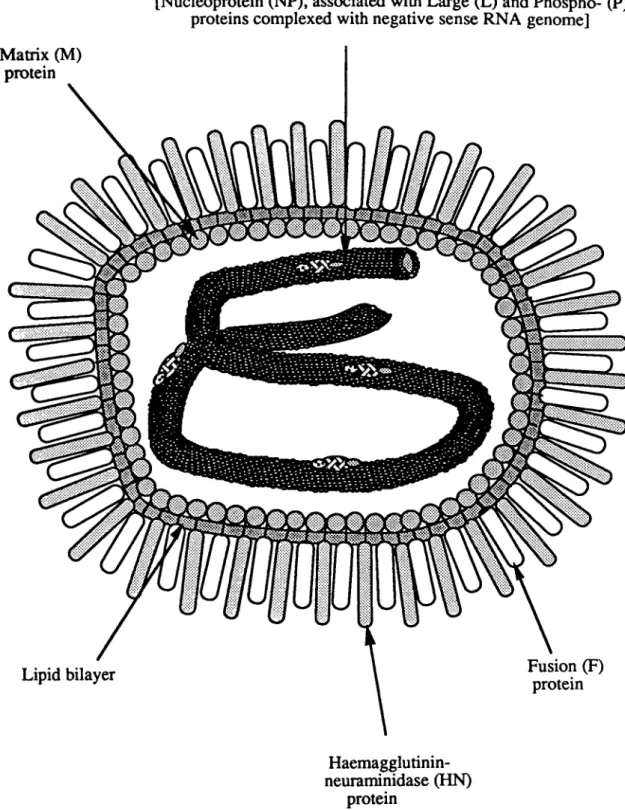
Paramyxovirus particles are pleiomorphic structures comprised of a helical
nucleocapsid core surrounded by a matrix protein (M) and a lipid envelope through which
two viral glycoproteins project as spikes (figure 3). The infectious cycle of
Paramyxoviruses begins with the attachment and fusion of the virion with the host cell
plasma membrane. This step is mediated by two glycoproteins, the attachment protein
(HIHN or G) and the fusion protein (F). One interacts specifically with receptors on the
surface of the host cell and the other fuses with the membrane, thus allowing release of the
viral nucleocapsid into the cell cytoplasm (reviewed by Choppin and Compans, 1975). It is
in the cytoplasm of the cell that transcription and replication of the viral genome take
place. The nucleocapsid is the fundamental unit required for viral transcription and
replication (Hamaguchi et al., 1983). It is comprised of the viral genome !ightly encased in
a nucleoprotein (NP). More loosely associated with the nucleocapsid are the polymerase
proteins, the large protein (L) and the phosphoprotein (P). The NP protein protects the
viral RNA from nuclease attack and facilitates recognition of the template by the viral
polymerase. The P and L proteins function together as a polymerase, using the viral
genome RNN NP complex as a template.
Following entry to the cell, a round of primary transcription is initiated. When there
are sufficient levels of nucleoprotein in the cytoplasm in order to encapsidate RNA,
replication and secondary transcription, from the newly generated genomes, can begin.
Following their synthesis, the nucleocapsids are transported to the cell surface. The M
protein is responsible for the alignment of the nucleocapsids with the viral glycoproteins
which localise in patches in the cell membrane (Yoshida et al., 1976). Once the virus
particle has been assembled, the structure buds from the surface of the cell and is released.
Alternatively, the virus can spread by fusion of the infected cell with its neighbour. This
Matrix (M)
protein
Nucleocapsid:
[Nucleoprotein (NP), associated with Large (L) and Phospho- (P) proteins complexed with negative sense RNA genome]
Haemagglutinin-neuraminidase (HN)
protein
Figure 3: A
schematic diagram showing the structure of a Paramyxovirus particle
1.3 THE PARAMYXOVIRUS GENOME
The Paramyxovirus genome is approximately 15 kb in length and encodes 6 to 10
genes, depending on the virus (figure 2; Pringle, 1991). The viral genome is in the negative
sense, meaning that it cannot be directly utilised as mRNA. Instead, the nucleocapsid
template is transcribed sequentially to yield capped and polyadenylated mRNA copies of
each of the viral genes. These are subsequently translated into proteins on host cell
ribosomes and the glycoproteins are modified by cellular enzymes. Viral replication occurs
using the same template RNA as for transcription. However, whereas during transcription,
each gene is copied in isolation (there are exceptions, described below), during replication,
a copy of the entire genome is made. The fact that the RNA genome can direct the
synthesis of two different sets of RNA products would suggest that there are a number of
cis acting signals inherent to the genome which are selectively acknowledged or ignored,
and that the differential recognition of cis acting sequences by the pol~rase
distinguishes between transcription and replication. The genomes of a number of
Paramyxoviruses have now been sequenced (reviewed by Galinski, 1991). Using this
information, several predictions have been made as to what sequences comprise the
coding and non-coding regions of the genome. Furthennore, by comparing regions of
sequence homology and by analogy with other negative stranded viruses, putative
functions have been assigned to some of the non coding sequence.
At the 3' and 5' ends of the genome are extracistonic sequences which act as
templates for positive and negative strand leaders. These sequences are well conserved
between the Paramyxoviruses (Galinski, 1991) and also between the very 3' and 5' ends of
the genome; for example, 33 of the first 39 nucleotides are conserved in the positive and
negative strand leaders of hPIV3 (Galinski et al., 1988). Because of their conservation and
position on the genome, these regions are suspected to contain signals pertaining to
polymerase attachment to the template and encapsidation of the nascent strand (Blumberg
et aI., 1991).
With the exception of the L gene of RS virus (Collins et al., 1987), each of the
composed of a gene end signal, a non transcribed intergenic region and a gene start signal.
The gene end and gene start signals comprise the ends and beginnings of the mRNA
transcripts. These sequences are semi-conserved within the different viruses, suggesting
that they may have an important regulatory role during transcription (Galinski, 1991). The
gene end signal contains a run of U residues which are presumably part of the
polyadenylation signal. Apparently, the polymerase occasionally does not efficiently
recognise the gene junction sequences, and rather than synthesising a monocistronic RNA
species, it produces a polycistronic transcript (Collins et al., 1982, Wilde and Morrison,
1984; Gupta and Kingsbury, 1985; Spriggs and Collins, 1986; Elango et ai, 1988;
Castaneda and Wong, 1989).
The level of conservation of the intergenic region is dependent on the virus, in the
cases of measles virus, Sendai virus and PIV3, the intergenic regions are consistently
trinucleotides of either GAA, GGG or GCf (Galinski, 1991). In contrast, the other
Paramyxovirus intergenic regions can vary in sequence and in length, with RS virus
intergenic sequences ranging from 1 to 52 nucleotides (Collins, 1991). Thus the influences
of the intergenic regions on the activity of the polymerase complex may vary from virus to
virus.
1.4 THE PARAMYXOVIRIDAE ENCODED PROTEINS
1.4.1 The Attachment Protein
The attachment protein of the virus can have several functions. It is responsible for
binding the virus particle to receptors expressed on the surface of host cells and therefore
is responsible to some extent for the host range specificity of the virus. The attachment
proteins of some Paramyxoviruses also have a neuraminidase activity which enables newly
released virus particles to dissociate from the cell membrane and from each other upon exit
from the cell. In addition, some Paramyxoviruses require the attachment protein in order to
negotiate cell fusion.
The different genera of the Paramyxoviridae family possess attachment proteins
HN, has both haemagglutinating and neuraminidase activities, the Morbilliviruses have a
haemagglutination protein, H, with no neuraminidase activity and the Pneumovirus
attachment protein, G, does not possess haemagglutinating nor neuraminidase activities
(Choppin and Compans, 1975; Matthews, 1982; Gruber and Levine, 1983). These
functional differences are reflected in the primary sequences of the proteins. There is poor
amino acid sequence conservation across the Paramyxoviridae family, although structural
features are conserved within the different genera. The different attachment proteins of the
Paramyxoviridae do have one shared feature in that, like the Influenza virus neuraminidase
protein, they are all type II integral membrane proteins. Thus these proteins are anchored
to the membrane of the virus and cell via a sequence at their N-termini, and have long
C-terminal ectodomains (reviewed by Morrison, 1988; figure 4).
The HN protein exists as disulphide linked dimers that fonn either non-covalently
linked or disulphide linked tetramers (Ng et ai., 1989). The stable formati.on of a tetrameric
structure is dependent on the transmembrane domain and the cytoplasmic tail of the
protein (Ng et ai., 1990; Parks and Lamb, 1990; McGinnes et al., 1993). During its
synthesis, HN interacts with the cellular protein GRP78-BiP. It is thought that this
interaction allows the protein to attain its correct oligomeric structure prior to export to the
Golgi network (Ng et ai., 1989). The HN protein is glycosylated by N-linked
oligosaccharides. In the cases of Sendai virus and mumps virus, glycosylation is necessary
for virion incorporation (Nakamura et ai., 1982; Herrler and Compans, 1983). In contrast,
the NOV HN protein does not require glycosylation to be incorporated into virions, but is
required for virus infectivity (Morrison et ai., 1981). The glycosylation sites are found
towards the C-terminus of the protein (Morrison, 1988) which suggests that HN consists of
a globular head containing the regions of enzymatic activity, supported by a stalk -like
structure which inserts into the plasma membrane (Thompson et al., 1988).
The three dimensional structure of HN remains to be elucidated and as yet, the
different functions of the protein have not been assigned to regions of the primary
sequence. However, a number of studies have compared the structures of the
~ ~
SV5HN
o 000 000
o
00 000 00,
,
,
,
"
NH21
11 ________________
---'
Measles virus H
00
o
00 000o
000 0•
nY •
NH21
III~__________________________ __'
RS virusG
0000
o
cysteine residues• y y y
• • •
NH2c=J1~
____________
~,
N-linked glycosylation sitesIII
hydrophobic stretchesFigure 4: A diagram showing the notable features of the attachment proteins of the genera of the Paramyxoviridae. The attachment protein of the Rubula and Parainfluenza viruses is similar and is represented here by SV5.
dimensional structure of the Influenza virus NA protein has been resolved, and the amino
acids involved in the active site determined (Colman, 1989; Colman et al., 1983).
Comparison of the sequences of Influenza virus NA and Paramyxovirus HN proteins
shows that the primary sequences of the proteins can be aligned. This study suggests that a
number of amino acids known to reside in the active site of the neuraminidase, and which
contact the substrate sialic acid residue, are conserved in the lIN protein (Colman et al.,
1993). Most of these amino acids are found in four conserved motifs, three of which are
absent in the H protein of MY, supporting the hypothesis that they are involved in the
neuraminidase activity of the protein. The motifs are located in different regions along the
length of the primary sequence and presumably are brought together to form the
neuraminidase active site in the conformationally mature protein.
Despite the low levels of sequence similarity, there are elements of the
Parainfluenza! Rubulavirus and Morbillivirus attachment proteins which are common.
Like the lIN protein, the measles virus H protein forms disulphide linked dimers and also
possibly homotetramers. Alignment of sequences of the two genera shows that a number
of residues potentially important for the three dimensional structure of the protein, ego
glycine and cysteine residues, are conserved (Morrison and Portner, 1991). Mutation
analysis of the cysteine residues of MY have shown that a number of these are essential for
the correct folding and transport of the protein (Hu and Norrby, 1994). Each of these
structurally important cysteine residues corresponds with an equivalent residue in the HN
protein, suggesting that the structural frameworks of the proteins are similar. Like HN, the
H protein is decorated with N-linked glycans. However, whereas in Parainfluenza virus
and Rubulavirus lIN they are distributed towards the C-terminus of the protein, in the MY
H protein they are located closer to the N-terminus (Alkhatib and Briedis, 1986). Four of
the five potential N-linked glycosylation sites in H of measles virus are utilised. It was
found that removal of individual sites had little effect on the folding, oligomerization or
biological activity of the molecule, but it is thought that they are collectively required to
Although the G protein of the Pneumoviruses is a type II integral membrane
protein, in all other respects it is quite different to the attachment proteins of the
Paramyxovirinae. Its primary sequence is significantly shorter than that of the HN and H
proteins (Wertz et ai., 1985) and no alignments can be made (Morrison and Portner, 1991).
Furthennore, the glycosylation patterns of the G proteins are different, with evidence that
this protein is modified by addition of O-linked, as well as N-linked, oligosaccharides
(Gruber and Levine, 1985; Wertz et ai., 1985). Because of its differences from the other
Paramyxoviridae attachment proteins, it has been hypothesised that the Pneumovirus G
protein has a different ancestry to HN and H. Instead, it has been speculated that G is more
similar in structure to a group of cellular mucinous proteins (Wertz et ai., 1985; Sullender
and Wertz, 1991).
1.4.2 The Fusion Protein
The fusion protein of Paramyxoviruses mediates both the entry of the virus into the
host cell, and viral spread by cell to cell fusion (Choppin and Compans, 1975). The
primary sequences of the Paramyxoviridae fusion proteins are similar and there are several
structural features which are conserved throughout the family (Morrison, 1988; Morrison
and Portner, 1991). An example of a Paramyxovirus fusion protein is shown in figure 5.
The fusion proteins are synthesised as a precursor polypeptide, FO (Scheid and Choppin,
1974). This polypeptide has a membrane anchor domain at its C-terminus and an
N-terminal signal sequence domain, which is cleaved following synthesis. In addition, there
is a third hydrophobic domain located in the N-terminal portion of the polypeptide and
lying immediately following a basic peptide cleavage site (paterson et ai., 1985; Spriggs et
ai., 1986; Kawano et ai., 1990; Bando et ai., 1991). An essential step in the maturation of
the fusion protein is cleavage at this site, usually by cellular proteases located in the Golgi
apparatus, to produce a heterodimer composed of the peptides F 1 and F2 linked by a
disulphide bond (Schied and Choppin, 1977; Hsu et ai., 1981). The cleavage process
exposes the hydrophobic fusion sequence and is prerequisite for the fusion protein to be
.F2
Flheptad
repeal cleavage heptad
site
oJ
J
0--oTo
I '
,
"
,
~2~1~~
__
~
__
~jj~~M~I
__________________
~j~::i~4~.~1
t
1
hydrophobic fusion sequence
transmembrane anchor domain
, hydrophobic domains
o cysteine residues
Figure S: A diagram showing the structure of the fusion protein of SV5
location until a late stage in the protein's synthesis, it is prevented from acting as a stop
transfer signal during translocation of the FO peptide across the membrane of the
endoplasmic reticulum. This mode of maturation thus allows the fusion domain to
ultimately be expressed, externally to the cell (Paterson and Lamb, 1987).
Immediately adjacent to the fusion sequence, is an amphipathic alpha helix motif
(Chambers et al., 1990). This domain is thought to be important in either folding or
transport of the protein to the trans golgi (Wang et aI., 1992). The arrangement of a
hydrophobic domain followed by a heptad repeat sequence is a feature common to other
viruses and may represent a structure vital to the mechanism of viral fusion. In addition,
there is a second amphipathic alpha helix located at the C-terminal end of Fl. This domain
has also been shown to be critical for measles virus fusion (Buckland et al., 1992).
In addition to the domains that are described above, the fusion proteins contain a
number of conserved cysteine residues. As previously mentioned, the mature fusion
protein comprises of a heterodimer linked by disulphide bonds. Therefore, at least one
cysteine in each subunit of F must contribute to disulphide bond formation. There are a
bonds. Like th~attachment proteins, F is glycosylated by N-linked oligosaccharides
(Morrison and Simpson, 1980; Herrler and Compans, 1983). The finding that
unglycosylated F is only cleaved inefficiently, suggests that the sugar additions may
maintain the precursor polypeptide in the correct confonnation to expose the cleavage
peptide (Collins and Mottet, 1991).
1.4.3 Small Hydrophobic Protein
The Pneumoviruses, mumps virus and SV5 each encode a third integral membrane
protein, called the small hydrophobic (SH) protein, from a gene which lies between F and
HN on the viral genome of SV5 and mumps virus (Hiebert et 01., 1985; Elango et 01.,
1989) and M and G of RS virus (Collins et 01., 1986; Olmstead and Collins, 1989). The
predicted amino acid sequence of the SV5 SH protein suggests that it is comprised of 44
amino acids and consists of two domains; an N-terminal hydrophilic domain and a slightly
longer C-tenninal hydrophobic domain. The hydrophobic domain of the protein spans the
plasma membrane such that the hydrophilic domain lies in the cytosol and a short region of
5 amino acids protrudes extracellularly. The protein has been found to co-localise with HN
in infected cells but is not detected in virions, suggesting that it is selectively excluded
from the membrane of the budding virus particle (Hiebert et 01., 1988).
The function of the SH protein is not yet known. It has been found that the RS
virus SH protein can enhance the fusion properties exhibited by F and G, or F alone.
However, the same study established that no such role exists for the SV5 SH protein
(Hemin way et 01., 1994).
1.4.4 Matrix Protein
In the virus particle, the matrix protein lies between the glycoprotein containing
envelope and the nucleocapsid. In the infected cell, it is thought to act as the orchestrator
of virus maturation, by acting as a bridge between the viral glycoprotein containing plasma
membrane and the newly synthesised viral nucleocapsids (Yoshida et 01., 1976). It is
leads to an accumulation of nucleocapsids within the cell (Wechsler and Fields, 1978; Hall
et al., 1979). The primary sequences of the Morbilli, Parainfluenza and Rubulavirus matrix
proteins are similar, but there are no obvious structural features of note. In each case it is a
basic protein, with a number of paired basic residues. It is hydrophobic in nature, but does
not have any hydrophobic stretches long enough to traverse a membrane (reviewed by
Peeples, 1991). It is thought that its hydrophobic character allows it to associate with
membranes and that its positively charged residues facilitate binding to the negatively
charged nucleocapsid (Morrison, 1988; Sheshberadaran and Lamb, 1990). M protein is
also able to bind to itself, to give rise to a paracrystalline sheet structure and is reported to
bind to actin (Giuffre et al., 1982; Morrison and McGinnes, 1985; Bohn et al., 1986). This
interaction may be important for virus budding.
1.4.5 Nucleoprotein
The nucleoprotein is the most abundant protein in the viral nucleocapsid (Lamb et
01., 1976). This protein is responsible for the characteristic helical structure of
nucleocapsids (Spehner et al., 1991; Das and Banerjee, 1993; Fooks et al., 1993) and
protects the viral genome from nuclease attack (Banerjee, 1987). In addition to protecting
the viral RNA, the NP enables the genome to act as a template (Kingsbury, 1974),
probably by acting as a foundation on which the polymerase complex can attach and
possibly by ironing out the secondary structure of the RNA which might otherwise hinder
the progress of the polymerase along the RNA template. The nucleoprotein also has a role
in viral assembly as it is this component of the nucleocapsid which contacts the matrix
protein (Markwell and Fox, 1980).
In the infected cell, NP exists in two fundamental forms; either in the context of the
viral nucleocapsid or as a soluble unit During replication, the soluble units of NP are
presumably recruited to the polymerase complex and, together with the newly synthesised
RNA, incorporated into a growing nucleocapsid. Several studies in which NP has been
expressed in cells, independently of the virus, have found that is able to self assemble into
with one another which gives rise to the nucleocapsid morphology (Blumberg et ai., 1983;
Spehner et aI., 1991; Buchholz et ai., 1993; Fooks et ai., 1993). In some cases, the density
of these nucleocapsids was found to be the same as of nucleocapsids derived from virally
infected cells (Blumberg et ai., 1983; Spehner et aI., 1991; Buchholz et ai., 1993). From
this finding, the investigators suggested that. in the absence of other viral components, the
NP non-specifically uses cellular RNA as a basis upon which to build the nucleocapsid
structure. Deletion analysis has shown that the N-terminal three quarters of NP is sufficient
and necessary for nucleocapsid assembly, suggesting that this region holds domains for
NP-NP and possibly NP-RNA interactions (Buchholz et aI., 1993). In the case of Sendai
virus, this region is composed of two domains. The N-terminal domain is enriched in
positively charged residues which may interact with the negative charges of the
phosphodiester backbone of the viral RNA (Morgan et al., 1984). The second domain is in
the central region of the protein, and involves a sequence that is highly conserved between
the Paramyxoviruses (figure 6; Parks et al., 1992). This domain is hydrophobic and it has
been suggested that it may promote NP-NP interactions during nucleocapsid assembly
(Morgan et ai., 1984). During viral replication, soluble NP has to be sequestered to the site
of nucleocapsid assembly. There is evidence to suggest that it is the P protein which guides
the NP to the polymerase complex (Horikami et aI., 1992; Buchholz et ai, 1993; Curran et
ai, 1993). Deletions in the N-terminal three-quarters of the Sendai virus NP protein
abolishes P binding (Homann et al., 1991). Possibly there is a binding site for P in this
region which overlaps with the regions responsible for NP-NP and NP-RNA interactions.
It may be that P binding to this site prevents self assembly of NP until a further interaction
with the polymerase releases the P protein, allowing the NP to specifically assemble the
nascent RNA.
Although the N-terminus of NP carries sufficient structural information to mediate
nucleocapsid assembly, the reaction carried out by this portion of the protein is
non-specific. In vitro Sendai virus replication systems have shown that NP lacking the
C-terminal 124 amino acids forms nucleocapsid structures more readily in the absence of
suggests that the specificity of encapsidation is dependent, in part, on the C-terminal
quarter of NP.
a
E:m::!~:m] postively charged region, possibly contacts RNA
III
hydrophobic conserved region possibly responsible for NP-NP interactions• negatively charged region required for the nucleocapsid to serve as a template
a region necessary for nucleocapsid assembly
Figure 6: A schematic diagram of the nucleoprotein of Sendai virus, with regions that are conserved between the Paramyxoviruses shown.
The C-terminus of NP has also been attributed with other functions. It has been
found that when antibodies against NP are reacted with nucleocapsids, most antibodies
bind to the C-terminus of NP, suggesting that this region is exposed at the surface of the
nucleocapsid and is not directly involved within the nucleocapsid structure (Ryan et al.,
1993). In keeping with its proposed location, the C-terminal tail of NP seems to be
responsible for a number of processes involved in coordinating viral replication. The
encapsidation property of NP is distinct from its ability to enable the RNA to act as a
template. In addition to conferring the specificity of encapsidation, it has been found that
the C-terminus of NP is also required for the nucleocapsid to function as a template for the
viral polymerase. A block of negatively charged residues, proximal to the C-terminal end
of NP are well conserved throughout the Paramyxoviruses (Parks et ai., 1992). It has been
mUltiple rounds of genome synthesis (Curran et a/., 1993). Possibly this site provides a
transient anchorage for the viral polymerase as it moves along the template.
As well as having a role in genome replication, the NP protein is the nucleocapsid
constituent that is suspected to bind to the M protein, thus enabling the assembly of newly
fonned nucleocapsids into viral particles (Markwell and Fox, 1980). The sites on NP
which are responsible for such interactions have not been characterised, but given that M is
positively charged, it has been speculated that the block of negatively charged residues
described above, might be involved in this interaction (Parks et a/., 1992).
1.4.6 The L Protein
Due to its large size and low abundance within the viral particle and the infected
cell, the catalytic functions required for RNA synthesis have been attributed to L.
Biochemical and genetic studies of the VSV L protein confmns that poly~denylation,
capping and methyltransferase activities are all associated with this protein (Abraham et
a/., 1975; Hunt et a/., 1984; Hercyk et a/., 1988). Sequence alignment of the L proteins of
several Rhabdoviruses and Paramyxoviruses has identified six conserved regions. It has
been suggested that the protein is made up of several distinct domains, each responsible for
the different functions involved in RNA transcription and replication (Poch et a/., 1990;
Parks et al., 1992). The active site for phosphodiester bond formation and/or template
recognition has been suggested to lie within region
m.
The basis for this hypothesis is thesimilarity between the conserved motifs found in this region, QGDNQ, and the GDD motif
found in RNA dependent RNA polymerases of a variety of viruses (poch et ai., 1989; Poch
et a/., 1990; Sleat and Banerjee, 1993). In agreement with this suggestion, it has been
reported that in the case of Sendai virus L protein, any change to this consensus
pentapeptide motif ablates polymerase activity, whereas changes outside this region, did
not affect replicative activity (Graef et ai., 1994).
In addition to its polymerase activity, the presence ofL leads to a higher level of
phosphorylation of P (Einberger et ai., 1990). In the case of VSV, phosphorylation of P by
Paramyxovirus~s, a kinase site was proposed to reside at region VI, as this site resembles
an A TP binding motif (poch et al., 1990). However, in the case of Sendai virus, it has been
found that although the presence of L is required to phosphorylate P, region VI can be
deleted without abolishing kinase activity. This finding suggests that, either there is an
alternative, previously unacknowledged, kinase motif in the Paramyxovirus L proteins or
that L is an essential co-factor for a cellular kinase (Graef et al., 1994).
1.4.7 The Phosphoprotein
Although L has been assigned the catalytic properties necessary for transcription
and replication, the phosphoprotein, P, is absolutely required for the polymerase to be
functional (Curran et al., 1991). Surprisingly, given its pivotal role, the P proteins of the
Paramyxoviruses are each quite different, with no obvious regions of conservation. This
may be because the protein acts in a structural capacity; P seems to act as a mediator
between the different components of the polymerase complex as it has been shown to bind
to both NP and L (Ryan and Kingsbury, 1988; Ryan and Portner, 1990; Horikami et al.,
1992; Parks, 1994; Smallwood et al., 1994). The binding sites for L and NP have been
mapped by deletion mutation analysis and have been found to lie in the C-terminal region
of the Sendai virus P protein (see figure 7). Two noncontiguous regions within P enable it
to bind both to soluble NP, and to the viral nucleocapsid (Ryan and Kingsbury, 1988; Ryan
and Portner, 1990; Homann et al., 1991). A single smaller region lying between the NP
binding domains allows binding with L (Curran et al., 1994; Smallwood et al., 1994). P
seems to be able to confer encapsidation specificity on NP. Co-expression of NP with P
maintains the solubility of NP, presumably the P protein maintains NP in a soluble state
until it contacts the nascent RNA being synthesised by the RNA polymerase (Horikami et
al., 1992; Buchholz et aI., 1993). Thus the P protein seems to act as an escort for the NP,
bringing it into juxtaposition for encapsidation. Also, as P interacts with NP within the
nucleocapsid structure (Ryan and Portner, 1990), it may be the component of the
P
NH2v
NH2a
b
~ most heavily phosphorylated
~ regionofP
_ residue 1-77, required
Ell for encapsidation
IIITITIII cysteine rich
II1llIlII conserved domain
I11111111111111111
II
regions required for nocleocapsid and nucleoprotein bindingD
L binding domainFigure 7: A figure showing the characterised domains of the P and V proteins of Sendai virus. Region a is necessary for RNA synthesis activity of the polymerase. P and V are amino co-terminal proteins, the residues common to both
are
the bracketed regions, marked b.P/NP
complex
newly synthesized RNA encapsidated in NP
NP/RNA template
Figure 8::A schematic diagram showing the possible interactions of P with L and NP In this scheme, one P or group of P molecules binds to L and anchors the polymerase to the
Althou&h the C-tenninus of P is the region of the protein responsible for binding to
NP and L. the terminal region of the protein is also important. Deletion of the
N-terminal 77 amino acids of Sendai virus P yields a protein which is able to participate in
transcription. but is incompetent for replication. Although this part of the protein does not
bind in a stable manner to NP, it appears that these residues are essential for genome
encapsidation (Curran et al., 1994). The N-terminal region of P has another interesting
property. either residues 1-77 or residues 78-145 are required for RNA synthesis, however,
these regions appear to be able to substitute for each other to restore wild type like activity.
Strangely. the middle region of P serves no obvious function, although this is the most
highly phosphorylated part of the protein (Vidal et al., 1988). Possibly this region has a
more subtle role in coordinating virus transcription and replication.
1.4.8 V Protein
Due to a frameshifting event which sometimes occurs during transcription, the P
genes of most Paramyxoviruses are able to specify two different mRNA molecules. The
two mRNAs code for proteins, P and V, which are amino co-terminal but which differ in
their C-tenninal sequences (Thomas et al., 1988; Cattaneo et al., 1989a; Paterson and
Lamb, 1990; Southern et al., 1990; Vidal et ai., 1990a; Samson et ai., 1991; Kawano et ai.,
1993; figure 7). Interestingly, despite the very weak levels of conservation between the P
proteins of the Paramyxoviruses, the C-terminal regions of the V proteins, encoded by the
same nucleotides, are well conserved, with an abundance of cysteine residues being the
most striking feature. The necessity for V during viral infection is in dispute. The fact that
the protein shares sequences with P, and is highly conserved, suggests that V may have an
important role in transcription and/or replication. However, in disagreement with this
conjecture. is the finding that hPIV 1 does not code for a V protein, showing that for this
virus, V is not an essential element (Matsuoka et ai., 1991). It may be that V fulfills a
"luxury" role during a viral infection, possibly by enabling an additional level of control
over replication and transcription. Evidence in support of this notion comes from
dose dependenrfashion but does not affect transcription (Curran et aI., 1991; 1994).
During a viral infection, the effect of high concentrations of V could be to promote
transcription over replication. That V might fulfIll some role during viral infection is
possibly demonstrated by the characteristics of hPIVl infection. This virus only replicates
to low levels and infected cells show no obvious cytopathic effects (Matsuoka et aI.,
1991). It may be that the restricted nature of infection is due to a lack of coordination of
the relative levels of transcription and replication.
1.4.9 C Proteins
In addition to being able to code for two different mRNA molecules, the P gene of
some viruses can yield several other proteins by the use of alternative initiation sites in an
overlapping reading frame. These are known as the C proteins and are found during
infection with Sendai virus, HPIV3, MV and COV (Dethlefsen and Kolakofsky, 1983;
Barrett et al., 1985; Bellini et al., 1985; Spriggs and Collins, 1986). The Sendai virus C
proteins are the best characterised. In this virus, the C open reading frame lies at the
N-terminal region, in the + 1 frame, relative to P. From this reading frame, four different
proteins are expressed, each from a different start codon (reviewed by Kolakofsky et al.,
1991). An interesting property of these proteins, which suggests a possible role during
infection, is that they have a strong inhibitory effect on transcription, with little effect on
genome replication (Curran et al., 1992). The mechanism for repressing transcription has
not been elucidated, although it is known that inhibition depends on a region lying near the
N-terminus of the C protein chain and on co-expression with P and L. There are apparently
no C proteins synthesised during infection with the Rubulaviruses, implying that for these
viruses they are not essential. Similarly to V, this group of proteins may serve a "luxury"
1.5 THE PARAMYXOVIRUS REPLICATION CYCLE
1.5.1 Virus Entry
The initial stage in viral entry is the attachment of the virus particle to a specific
receptor molecule on the surface of the host cell. This interaction is mediated by the
attachment protein of the virus. Different receptors are used by different viruses. In the
case of the parainfluenza viruses, mumps virus, NOV and RS virus, the receptors are
molecules containing sialic acid residues. This has been demonstrated by experiments in
which neuraminidase treatment renders normally susceptible cells resistant to virus
infection (Markwell, 1991). The receptor for Sendai virus is the best characterised and has
been shown to be a ganglioside molecule (Markwell et al., 1981). Infections with the
Morbilliviruses are not affected by prior treatment with neuraminidase (Howe and Lee,
1972), demonstrating that these viruses interact with a different surface receptor. The
receptor for measles virus has recently been identified as CD46 (Dorig et aI., 1993;
Naniche et al., 1993). This protein is involved in regulation of complement activation and
is widely distributed throughout human tissue (Dorig et aI., 1993).
Following attachment to the receptor molecule, the viral membrane fuses with the
cellular membrane to release the viral nucleocapsid into the cell. An alternative means for
viral nucleocapsids to enter cells, is by fusion of an infected cell with its neighbour. As yet
the fusion process is poorly understood. It takes place at neutral pH and begins with the
formation of a 2nm wide pore, which dilates as fusion progresses (Spruce et al., 1991).
There has been some controversy as to the viral components that are necessary for fusion;
some viruses need the attachment protein, in addition to F, for cell fusion to occur. The
differing requirements for fusion presumably reflect slightly different mechanisms for the
process in different viruses, however, for each of the viruses examined, it has been found
that the presence of the attachment protein enhances the fusion process (reviewed by
Lamb, 1993).
To some extent, the role of the attachment protein can be attributed to the binding
of the protein to its receptor, thus bringing the fusion protein into close proximity to the
found that reconstituted virus envelopes fuse with erythrocytes in the presence of wheat
germ agglutinin (Hsu et al., 1979). However, this is not always the case. The F protein of
bPN3, hPIV3 and NDV can only induce fusion if co-expressed with HN (Sakai and
Shibuta. 1989; Ebata et al., 1991; Morrison et al., 1991) suggesting a more specific
association between the two proteins. Furthermore, Hu et aI. (1992) have found that the
homologous HN is required rather than HN from related viruses. One possible explanation
for the need for homologous F and HN/H proteins is that the F protein needs to be
precisely positioned in order to interact with the cellular membrane to induce fusion, and
that only the homotypic HN interacting with its cellular receptor will be able to achieve the
correct bridging distance.
The interaction of the attachment protein with its receptor may also be required to
activate the fusion protein. Lamb (1993) has hypothesised that F proteins undergo a
conformational change at the correct time and place in order to mediate fvsion. It is
possible that in the cases of viruses for which HN is absolutely required for fusion, binding
of lIN with the sialic acid receptor initiates a specific interaction of lIN with F causing a
conformational change in F and subsequently positioning the fusion domain in the correct
configuration for interaction with the plasma membrane. In the less stringent viruses,
possibly a less specific interaction of the fusion protein with the cell plasma membrane
triggers the prerequisite conformational change.
1.5.2 Adaptation of the nucleocapsid for transcriptional activity
Having fused with the cell membrane, the viral nucleocapsids are released into the
cytosol of the cell, the site of virus transcription and replication. Prior to transcription, the
nucleocapsid has to be depleted of the surrounding matrix protein. In the case of
Paramyxoviruses. this process takes place at neutral pH and may be linked to viral fusion
(Zhirnov, 1990). In addition, having entered the cell, the P protein on the nucleocapsid
changes from a random distribution to discrete clusters, often co-Iocalising with L.
suggesting a transition from a quiescent state to a transcriptionally active complex (portner
complexes are !lot characterised, but consist of at least the NP/RNA complex and the P and
L proteins. Host cell factors are thought to playa role during transcription, as demonstrated
by the necessity for the presence of uninfected cell extract during in vitro transcription
reactions (Moyer et ai, 1986; 1990; De et aI., 1991; Barik, 1992). In the case of measles
virus and Sendai virus, the critical cellular component was found to be tubulin (Moyer et
al., 1986; 1990), whereas PIV3 transcription is dependent on actin (De et al., 1991). De
and coworkers (1993) have suggested that actin polymerisation concomitantly induces a
confonnational change of the PIV3 viral nucleocapsid, and that the change of the
nucleocapsids from a loosely coiled to a condensed arrangement is necessary for
transcriptional activation.
1.5.3 Virus Transcription
As previously described, virus transcription involves synthesis of individual copies
of each of the viral genes. It is suspected that there are cis acting signals which direct the
viral polymerase to attach to the template genome and then to copy each viral gene, .
generating a capped and polyadenylated mRNA transcript. It has been found that the
relative levels of each of the mRNA transcripts reflects their order in the genome, with a
decrease in abundance from the 3' to the 5' ends of the template. Hence the NP proteins are
the most abundant and the L proteins the least so (Glazier et al., 1977). The level ofL
mRNA synthesised during RS virus infection, is even more heavily curtailed, as this gene
overlaps with the gene for the M2 protein and does not have its own independent start site
(Collins et al., 1987). Because of the attenuated gradient of mRNA species synthesised, the
currently favoured model for transcription is that the viral polymerase initiates
transcription at a single promoter site, at or near the 3' end of the genome (Galinski, 1991).
From here it sequentially synthesises the leader and each of the viral mRNAs. At each of
the gene junctions, the polymerase is thought to pause, possibly during polyadenylation
and/or capping (Iverson and Rose, 1981) at which points the polymerase complex may