AFFECTOR
ELECTRO IOTECHNOLOGY
T.
9aynor
BE
(Hons)
A thesis presented for the degree of Doctor of Philosophy
In
Electrical and Electronic Engineering at the
University of Canterbury, Christchurch, New Zealand.
ABSTRACT
In this thesis, aspects of mainly electric field effects on cellular systems are investigated, although electromagnetic effects on biological systems in general are also considered. The standard biotechnology processes of electroporation and electrofusion are shown to be electrically sub-optimal. Conventional d .c. pulses generate undesirable conditions such as asymmetrical membrane breakdown and cellular rotation. Normal electrical protocols also contribute to lower efficiencies by being sensitive to cell radii and other biological and physical variables.
Both physical and numerical models indicated that replacing d.c. pulses with a.c. pulses symmetrical about a zero potential axis, not only reduces the inherent problems of asymmetrical breakdown and cellular rotation, but also provides the means to reduce efficiency sensitivity to cell radii. This is important in fields such as human monoclonal antibody research and the generation of transgenic animals. In addition, improvements to existing d.c. multiple pulse systems can be made by using shorter time constants and reducing the magnitude of successive pulse electric fields.
The lysing effect of high magnitude electric fields are used in the study of disinfect-ing biologically contaminated liquids. It was earlier found that water could successfully be disinfected of the bacterial organism serratia marcescens. However, for higher liquid conductivities, _applied electric field frequencies had to be increased so that electrol-ysis effects could be controlled. There is an upper limit to the frequency that can be used due to the cell membrane capacitance, which correspondingly puts an upper limit on the conductivity of the liquids that can be treated. Dead band regions where the applied a.c. electric field does not induce membrane dielectric breakdown, must be minimised. This suggests that a square wave should be used. Lower conductivity liquids are shown to provide the best opportunities for practical applications.
Cancer cell growth modification unique to the combined effects of low magnitude d.c. electric fields and silver ions was found to exist. The morphological changes suggest that possible dedifferentiation of the cells has been achieved. Other metal ions tested did not produce the same results. Silver ion effects also provided evidence of disinfection properties that can be utilised in some water treatment applications.
IV ABSTRACT
ACKNOWLEDGEMENTS
In this section it is expected that thanks will be offered to many people. Be that as it may, I would still be acknowledging what the following people have done for me even if it was not traditionally part of a thesis.
I am extremely grateful for the good fortune of securing Patrick Bodger (Pat) as my supervisor. Apart from the unerring professional guidance, Pat always proved to be inspirational whenever I felt that maybe I was not cut out for a Ph.D. or that the project was faltering. Furthermore I am very happy to be able to consider Pat as a valued friend.
Heaven knows what my wife Donna has put up with during my postgraduate years. She has endured periods of grumpiness, depression, anger, misfortune and a fair amount of financial drought, all usually with a smile. I seriously believe that if Donna had not been by my side, the chances of me accomplishing what I have, would have been severely reduced.
My parents brought me up believing that the only limits in my life would be put there by me, and have they ever paid for that (literally). I will always be indebted to them. Thanks also go to my sister who kept the proverbial wool out of my eyes with a skilful hand and a lethally sharp pair of scissors.
Breaking iRto the field of biology put me in great need of coaching, guidance and resources. The two people central to meeting those needs were Dr Mike Bodger (Pats big brother) of the Haematology Research Unit., Christchurch Public Hospital, and Dr Peter Elder of the Steroid Unit, Christchurch Public HospitaL Even though I undoubtedly tried their patience with incessant questions and bungling proficiency, they taught me well and made me feel like I might have a reasonable aptitude for this kind of research.
VI ACKNOWLEDGEMENTS
(which he somehow did anyway). Many thanks to Dermot Sallis for his multiple text book-like memory and willingness to share that knowledge. Other highly appreciated technical assistance came in the form of excellent electron microscope operation by Mike Flaws, Mike Cusdins' great wealth of practical experience and weekly thrashings at squash administered by Nick Smith.
Without the direct help of Darin Hutcheson and Peter Crellen on the dedifferenti-ation experimentdedifferenti-ation, Marc Palmer building the water disinfection power supply and Brandon Lancastle in the silver disinfection experimentation, my thesis would not be the document it is. I wish them well in their engineering futures.
I have seen quite a number of postgrads come and go during my studies and I got to know most of them. A few in particular stand out as they offered friendship, encouragement and help without any expectations. Members of this group are Ken Johnson, Dave Hawkins, Robert van Nobelen, Gary Ballantyne, Stu MacDonald, Dave Gilbert and Mike Livingstone. I will consider myself very lucky if I continue to meet people like this in my future years.
GLOSSARY
Terminology
Antibody
Antigen
B cells
Bacterial cells Biocompatible
Carcinogenic Cell membrane
Cell nucleus
Cell tissue Cell viability
Culture Culture dish Cytoplasm
Globular protein produced by activated B cells with extremely specific binding sites used in spe-cific immunological processes.
Complementary molecule to antibody binding site and trigger for antibody production by ac-tivated B cells.
B lymphocytes. Prokaryotic cells.
Does not induce an immune response m the organism.
Cancer inducing.
Phospho-lipid bilayer that surrounds the con-stituents of all cells.
Membrane bound region where genome is re-stricted to.
Structurally connecting cells.
Physical condition of a cell in its ability to pro-create. Viable: it can. Non-viable: it can not. An isolated solution of growing cells.
A type of container where cells can be grown. All constituents of a cell excluding the cell mem-brane and the genetic material.
Dedifferentiation Reversal of differentiation characteristics. Differential regulation Specific control of the genes in the genome
dur-ing differentiation.
Differentiation Changing cellular characteristics to a more spe-cialised state.
viii GLOSSARY
Electropermeabilization Process of electrical induction of membrane di-electric breakdown leading to a higher mem-brane permeability. Eukaryote Gene Genome Hybridoma In-vitro In-vivo
Ion pumps
Single or multi-celled organisms that have a dis-tinct nucleus.
DNA sequence that codes for an entire protein. The entire set of genes in an organism.
A cell that is the product of the fusion of two dissimilar cells.
Experiments performed on biological cells in culture.
Experiments performed on whole biological systems.
Transmembrane proteins that drive ions either into or out of the cell interior.
Maturity Degree of cellular differentiation.
Membrane permeability Transparency of membranes to material transport.
Metabolite Mitosis
Monoclonal antibodies Multipotent cells
Oncogene
Organelles
Ovum Pathological Pluripotent cells
Prokaryote
Restriction enzyme Sperm
Molecule used in the metabolism function. A form of cellular division and multiplication. Antibodies all with the same binding site. These cells have undergone a degree of differen-tiation past the pluripotent stage but still may differentiate into one of a number of different cells.
A naturally occurring gene, that when activated, has the potential to cause the cell to become cancerous.
Functional multi-component units In the cytoplasm.
Female gamete (or sex cell). Disease forming or inducing.
These cells belong to the ectoderm, mesoderm, and endoderm families and may differentiate into anyone of their possible products.
Single celled organisms that do not have a nucleus.
GLOSSARY IX
T cells T lymphocytes.
Tissue regeneration Reconstruction of lost or damaged tissue to a morphologically correct leveL
Totipotent cells These cells are able to produce an entire or-ganism if placed in an appropriate environment
(have undergone little or no differentiation). Transgenic
Virus
Yeast cell Zygote
An organism that has had foreign DNA incor-porated into its genome that is passed on to its progeny.
Metabolically inactive self contained units of ge-netic material that invade cells.
Single celled type of fungi.
Totipotent cell formed by the fusion of an ovum and sperm cell.
Abbreviations AIDS ASA {3 DMEM DNA D.T. EM ESR EXAFS FCS 'Y HIV H.M.E.F. IR MRI mRNA NMR Ql Q2 RNA RPMI
UV
XANESAlpha dielectric dispersion. Auto-Immune Disease Syndrome. American Standards Association. Beta dielectric dispersion.
Dubecco's Modified Eagles Medium. Deoxyribonucleic Acid.
Decay Time. Electromagnetic.
Electron Spin Resonance.
Extended X-ray Absorption Fine Structure. Fetal Calf Serum.
Gamma dielectric dispersion. Human Immunodeficiency Virus. High Magnitude Electric Field. Infra Red.
Magnetic Resonance Imaging. Messenger Ribonucleic Acid. Nuclear Magnetic Resonance. Source current transistor. Mirror current transistor. Ribonucleic Acid.
Roswall Park Memorial Institute. Ultra-Violet.
x
Mathematical Notation
a (cm)
em
(F/cm2)d (cm)
L'lUm(t) (V)
L'l3(t)
E (eV) Eo (V /cm) Ep (V /cm)
E(t) (V /cm)
E(O)
(V
/cm)EO (C2/(N m2))
F(a)
h (J s)
M(t)
(V) w (radians/s)¢
(radians)R2 (0)
Rm (Ocm2)
Pe (Ocm) Pi (Ocm)
a (S/cm)
ae (S/cm) ai (S/cm)
am (S/cm)
Tr (s)
t
(s)
o
(s) UmO (V)Cell or balloon model radius. Specific membrane capacitance. Membrane thickness.
Induced change in membrane potential at time
t.
Induced change in membrane structure at time
t.
Energy.
A.C. applied electric field amplitude.
Constant electric field magnitude or capacitive discharge peak electric field magnitude.
Excitation electric field at time t. Excitation electric field at time 0.
Permittivity of free space. Relative permittivity. Cond uctivity factor. Plank's constant.
Membrane dipole moment. Angular frequency.
Angle from a point on the membrane surface to the axis which is parallel to the applied electric field and passes through the origin of the cell or balloon model.
Current setting resistor. Specific membrane resistance.
External suspension medium resistivity. Internal resistivity of cell or balloon model. Variable of conductivity.
External suspension medium conductivity. Internal conductivity of cell or balloon model. Membrane conductivity.
Membrane relaxation time constant. Variable, of time.
Time variable of intergration.
Induced a.c. transmembrane potential amplitude.
Membrane potential at time t.
PREFACE
For a number of years, electric and magnetic fields have been major components of many processes in the medical arena. To name all of the processes here would be impractical if not impossible. Be that as it may, it is worth mentioning some of the more widely used and important techniques. The electrocardiogram (ECG or EKG), and electroencephalogram (EEG), have provided essential information on the functioning of the heart and brain [Cromwell et al. 1980]. Nuclear magnetic resonance (NMR)
[Hollas 1982], magnetic resonance imaging (MRI) [Morris 1986] and X-rays [Lange
et at. 1989] have resulted in the ability to determine molecular structures and to
non-invasively obtain detailed visual information on internal bodily objects. There is a common factor in these named processes. They are all diagnostic tools. This is by far the prevalent application of electromagnetics in medicine. Use of electromagnetics to affect biological systems is considerably less common.
Prior to the 20th century, many electrotherapeutic techniques existed [Becker 1990]. However, modern science indicated that most of these techniques were without any observable scientific foundation. A dogma resulted and any use of electro magnetics in therapies or to affect biological systems, was frowned upon. It was not until after World War Two that the scientific community began to relax its position. Advancements in microwave generation resulted in the obs€)rvation that soft tissue could be uniformly heated by its application [Field and Franconi 1987]. Exposure of living tissue to ,-rays indicated that there was a favourable killing rate differential between normal cells and cancer cells [Swan 1981, Mansfield 1983]. Laser technology was found to be useful in a number of applications from eye treatments to cauterising scalpels [Fuller 1987]. In virtually all cases where electric, magnetic or electromagnetic (em) effects could be used in a medical application, significant improvements over the practiced processes was achieved. In spite of this trend, very little research has been carried out in biology and medicine on specifically trying to find practical effects of electromagnetics. This is opposed to the substantial amount of work that has been and continues to be carried out on the causal effects of electro magnetics in biological systems. This research does not actively look for applications but ha..<; resulted in the applications mentioned above and others. Some of these applications are components of this thesis.
xu PREFACE
Its sole purpose is to provide brief background information on biology for those readers not conversant in that field.
Discovered high magnitude electric field effects on cellular membranes has directly led to three different applications. One known as electroporation is investigated Chapter 2. Electroporation utilises the high cellular membrane permeability (or poros-ity) state that is induced through the application of high magnitude electric fields, to selectively transport materials of interest across the membrane. A physical 'bal-loon' model of a cell is introduced and yields results that were previously unobservable. Modifications to the electroporation technique are then suggested.
Cells permeabilised similarly to electroporation that are brought into close contact, tend to fuse and form hybridomas. This effect is used in a process called electrofusion and is studied in Chapter 3. It is shown through numerical and physical modelling that a.c. signals symmetrical about a zero potential axis should result in more efficient electrofusion and electroporation. It is also indicated that using such waveforms should increase controllability of the techniques.
In both the electroporation and electrofusion processes, application of electric fields with excessively high parameters results in massive loss of cell viability. Chapter 4 looks at using this effect to disinfect biologically contaminated liquids. The major liquids concentrated on are water and hydrocarbons such as kerosene and diesel. The applicability of high magnitude electric field (h.m.e.f.) killing of liquid borne biological contaminates is shown to be mainly determined by the conductivity of the liquid which produces upper and lower bounds.
Perceived changes to cellular maturity and genetic expression by the application of very low magnitude d.c. electric fields in the presence of particular ionic compounds has indicated at possible applications in regeneration and cancer therapy. Initial in-vestigations and experimentation in this vain are described in Chapter 5. Cancer cells of morphologically different backgrounds, exposed to low level d.c. electric fields and silver ions can be seen to convert to a common morphology. This is proposed as being an indicator of cellular dedifferentiation.
In contrast to probing already existing processes, Chapter 6 is effectively an ex-ploration in the possibility of using resonant em energy transfer for various medical applications. More specifically in applications that require the ability to precisely tar-get certain biological aspects. These are discussed with respect to different energy and power levels which directly determines the mechanisms of action.
The thesis as a whole is concluded in Chapter 7. The main results are briefly summarised and future work is proposed.
PREFACE Xln
of explorative freedom, other avenues were travelled along. This is the main reason for the relatively diverse range of chapter topics. It is my belief that the result has provided me with a broader understanding in the field that interests me. Engineering philosophy encourages such understanding as it is the ability to bring together diverse ideas to form a functional and novel creation, that forms the foundation of a good engineer.
All of the biological experiments performed as a direct part of this thesis were co-ordinated and supervised by either Dr Michael Bodger of the Haematology Research Unit, or Dr Peter Elder ofthe Steroid Unit, Christchurch Public Hospital. Their exper-tise also ensured that results were not misconstrued and that unrealistic presumptions and expectations were pointed out.
All images in this thesis that originate from photographic sources, have been digi-tally scanned and contrasted. This was essentially done to make generation of multiple copies simple. Some of the figures in Chapter 2 and Chapter 3 had their contrast enhanced to highlight details that might otherwise be lost in reproduction.
Papers prepared during the course of this thesis are listed below in the approximate order of preparation.
Gaynor, P.T. and Bodger, P.S. 'Ionisation of dielectric spheroid membranes: A
balloon model of electl'oporation of biological cells', lEE Proceedings-A, VoL 141, No, 3, 1994, pp. 190-196.
Gaynor, P.T. and Bodger, P.S. 'Electrofusion processes: Theoretical evaluation of high electric field effects on cellular transmembrane potentials" lEE Proceedings-A,
accepted for publication, May 1994.
Gaynor, P,T. and Bodger, P.S. 'A balloon model of biological cell electropermeabi-lization in relation to the radius dependence of membrane dielectric breakdown', lEE Proceedings-A, accepted for publication, November 1994.
Bodger,
p.s.,
Gaynor, P.T., Hutchinson, D.G. and Crellin, P.J. 'In-vitro cell growth modification by combined d.c, and silver ion action', Submitted to Biophysical Journal,CONTENTS
ABSTRACT iii
ACKNOWLEDGEMENTS v
GLOSSARY vii
PREFACE xi
CHAPTERl BACKGROUND
1.1 The Cell
1 1
3 4
5
1.1.1 The Cell Membrane
1.1.2 Multicellular Organisms 1.2 DNA And Genetics
1.2.1 DNA
1.2.2 Genetics 1.3 Immunology
1.3.1 Non-Specific Immunity
1.3.2 Specific Immunity
5 6
7
8 8
CHAPTER 2 ELECTROPORATION 11
2.1 Introduction 11
2.2 Dielectric Membrane Responses To High Magnitude
Elec-tric Fields 12
2.2.1 The Induced Transmembrane Potential 12
2.2.2 The Inherent Transmembrane Potential Effect On
Membrane Dielectric Breakdown 14
2.2.3 Effective Breakdown Area Of Non-Spherical Cells 14
2.2.4 Electropermeabilization Dynamics 15
2.3 Physical Dielectric Spheroid Model 16
2.3.1 Balloon Model Experimental Setup 16
2.3.2
2.3.3
2.3.4 2.3.5
Balloon Model Experimental Results Water Droplet :Formations
Photographic Photon Capture Microscope Evaluation
2.4 Discussion
XVI CONTENTS
2.5 Conclusions 26
CHAPTER 3 ELECTRO FUSION 29
3.1 Introduction 29
3.2 Dielectrophoresis 30
3.3 The Induced Transmembrane Potential 33 3.3.1 Numerical Model Of Induced Transmembrane
Po-tential 34
3.3.2 Model Simulation Results 35
3.3.3 Electrofusion Dynamics 42
3.4 Balloon Model Applied To Electrofusion 46
3.4.1 Experimental Setup 46
3.4.2 Experimental Method 48
3.4.3 Experimental Results 48
3.4.4 Electroporation Of Organelles Within Cells 49 3.5 Biological Electrofusion Experimentation 51
3.5.1 Electrofusion Apparatus 51
3.5.2 Experimental Procedure 52
3.5.3 Experimental Results 53
3.6 A.C. Pulse Electrofusion/Electroporation System 53 3.6.1 A Simple A.C. Pulse System 54 3.6.2 An Optimisation Research Tool 55
3.7 Discussion 56
3.8 Conclusions 58
CHAPTER 4 H.M.E.F. KILLING OF LIQUID BORNE
BIOLOGICAL CONTAMINATES 61
4.1 Introduction 61
4.2 H.M.E.F. Water Disinfection 62
4.2.1 Primary Disinfection Design Specifications 62 4.2.2 Basic Electrode/Chamber Design 63 4.2.3 Basic Electrical Properties Required Of Water 63 4.2.4 Basic Electric Field Composition 64
4.3 Experimental Investigations 64
4.3.1 Previous Investigations 64
4.3.2 Effects Of Increasing Water Conductivity And De-creasing A.C. Waveform Dead Bands 66 4.4 Disinfection Of Hydrocarbon Liquids 66 4.4.1 Modified Electrode/Chamber Design 67 4.4.2 Pre-Treatment Of Hydrocarbon Liquids With Charge
Carriers 68
4.5 Discussion 68
4.5.1 Water Disinfection 69
CONTENTS
4.6 Conclusions
CHAPTER 5 COMBINED D.C. AND IONIC INDUCTION OF DEDIFFERENTIATION
5.1 Introduction 5.2 Cell Microbiology
5.2.1 Differentiation
5.2.2 D.C. Electric Fields And Currents During Differ-entiation And Growth Of Cells And Tissue 5.2.3 Dedifferentiation
5.2.4 Cancer
5.3 Experimental Materials And Methods 5.3.1 Very Low D.C. Current Source 5.3.2 Electrode/Culture Dish Arrangement 5.3.3 Cell Cultures
5.3.4 Experimental Procedure 5.4 Results And Discussion
5.4.1 Cell Viability 5.4.2 Silver Disinfection
5.4.3 Silver Disinfection Experiments 5.4.4 Dielectrophoresis
5.4.5 The Effect Of Silver Ions On Cellular Growth 5.5 Conclusions
CHAPTER 6 RESONANT EM ENERGY TRANSFER INTO BIO-SYSTEMS
6.1 Introduction
6.2 Resonant EM Energy Transfer
6.3 Resonant EM Energy Transfer Into Bio-Systems 6.3.1 High Energy Effects
6.3.2 Low Energy, High Power Effects 6.3.3 Low Energy, Low Power Effects 6.4 Discussion
6.5 Conclusions CHAPTER 7 CONCLUSIONS
7.1
Future WorkAPPENDIX A ROD SHAPED CELL MODEL OF
ELECTROPERMEABILIZATION AREA
Chapter 1
BACKGROUND
This chapter essentially acts as a basic terminology coach for those who have had little exposure to biological theory. Owing to the vast amount of even basic theory, only those topics relevant to the thesis will be backgronnded. The interested reader can find further comprehensive basic biology in the book by H. Curtis and N. S. Barnes, called 'Biology'. Each other chapter contains the relevant theory required independently of this one. Readers conversant in basic biology may, if they wish, pass over this chapter.
1.1
CELL
Most of the content of this thesis deals with electromagnetic characteristics of and effects on biological cells. More specifically, experimentation is conducted on some bacterial and mammalian cells. It is then pertinent to give a brief overview of cell physiology and dielectric properties.
There exists what is known as the 'cellular theory', which states that all organisms are composed oj cells. This is one of the few fundamental statements in biology [Hoppe
et al. 1983]. The cell is thermodynamically an open system which is in constant
ex-change with its environment. It is like a microscopic factory that takes in materials and uses its internal machinery to convert those materials into something else which may go into making new machinery, factory expansion and multiplication, power, exportation or defence from rival factories.
Cells are made up from a set of common components. Any particular cell type is determined by the combination and amount of those components. All cells have two components in common. These are the cell membrane and some form of genetic material [Curtis and Barnes 1989a].
2 CHAPTER 1 BACKGROUND
Prokaryotes (meaning before a nucleus) concentrate most of their genetic material to a loosely defined region called the nucleoid near the cell centre. Eukaryotes (meaning true nucleus) separate most of their genetic material into a well defined region, called the nucleus, with a double membrane sack known as the nuclear envelope. All bacteria are prokaryotic and all animal and plant cells are eukaryotic.
In addition to a cell membrane, prokaryotic cells also have a cell wall which is produced by the cell and resides on the outer surface of the cell membrane. Some eukaryotic cells also produce cell walls but are quite different to those of the prokaryotes. Animal cells do not have cell walls.
The rest of the components of a cell are grouped as constituents of the cytoplasm. The cytoplasm contains all the molecules required for existence and other well defined regions of function known as organelles (meaning small organs).
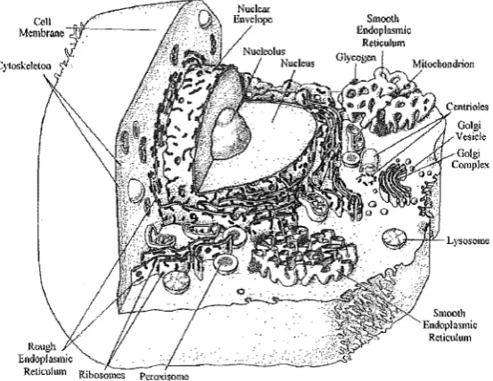
All cells contain very small organelles called ribosomes. Ribosomes are the con-struction sites of protein synthesis. Prokaryotes can contain a few other organelles such as storage granules (of substances like glycogen), flagella, dlla, microfilaments, micro-tubules and centrioles. Flagella and cilla are evaginations of cell membrane bound microfilaments and are involved with motility. Microfilaments, microtubules and cen-trioles are all protein based physical construction materials involved with cell shapes, movement, division and internal structure. Eukaryotic cells contain most of these and many other more complex organelles that usually involve membrane material. The major organelles of this type are, endoplasmic reticuli, golgi complexes, lyso-somes, peroxilyso-somes, mitochondria, and in plant cells plastids and vacuoles. A sectional representation of an animal cell is shown in Figure 1.1 [Curtis and Barnes 1989a]. The endoplasmic reticulum is a labyrinth of membrane material and is divided into two forms, rough and smooth. Rough endoplasmic reticulum has attached ribosomes whereas smooth endoplasmic reticulum does not. Molecules synthesised here are put into membrane bound vesicles and transported to the golgi complexes.
Golgi complexes are constructed as loosely stacked sacs. They act as packaging and distribution centres for molecules produced in the celL Large vesicles produced by the golgi complexes are lysosomes and peroxisomes.
Lysosomes contain hydrolytic enzymes that are used by the cell to break down imported proteins, polysaccharides and lipids. Peroxisomes contain lytic enzymes that break down purines and other similar compounds.
Mitochondria are large and very important membrane bound organelles that supply energy giving molecules for most cellular processes. The number of mitochondria in a cell is determined by the relative energy requirements of that cell. Mitochondria also contain their own genetic materiaL
1.1 THE CELL 3
Figure 1.1 Sectional view of a typical animal cell and its components
yellow pigments) and chloroplasts (chlorophyl-containing plastids where photosynthesis takes place).
The way a cell uses its molecular make up and organelles to function is beyond the scope of this chapter. SufIice to say that cell functioning as a whole is exceptionally complicated and affecting any of the organelles significantly alters those functions.
1.1.1
The Cell Membrane
Much of the thesis is concerned with the high magnitude electric field effects on cell membranes. Therefore, the basic physical structure and dielectric properties of most cell membranes are introduced here.
The fundamental building block of cell membranes is the phospho-lipid. These consist of a hydrophillic phosphoric acid and a pair of hydrophobic fatty acid chains. In
[image:21.595.165.442.124.338.2]4 CHAPTER 1 BACKG·ROUND
Figure 1.2 Fluid mosaic model representation of cell membrane material. The small spheres repre-sent the lipid phospho-heads and the thin tails reprerepre-sent the fatty acid sections. Also shown are typical placements of membrane globular proteins.
The lipids and proteins that make up a cell membrane are not fixed in their struc-tural positions. They can readily move transversely to the plane of the membrane and also have the ability to rotate or flip across the membrane plane (although this is much less common). These motional characteristics have resulted in the formation of the 'fluid-mosaic' model of the cell membrane.
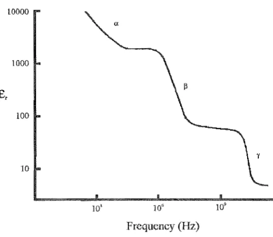
Comprehensive data has been produced on the passive dielectric properties of cel-lular membranes from about 1Hz to 100GHz [Pethig 1984, Schwan 1985, Pethig 1988]. As a simple approximation, the cell membrane can be dielectrically modelled by a capacitor in parallel with a high magnitude resistor. Thus, as frequency increases, the impedance of the membrane decrea.'3es. If impedances on either side of mem-brane remain relatively unchanged over a particular frequency range, then potentials developed across the membrane by an applied electric field will decrease. This effect is characterised by what is known as a Beta (,f3) dispersion in the relative permittivity (tl') for a bulk suspension of a particular cell type. The change in tl' of a standard colloidal cell suspension is s~own in Figure 1.3 [Schwan 1985]. There is also an alpha (0:) disper-sion due to the inherent transmembrane potential polarisation of associated molecules and a gamma (-r) dispersion due to the relaxation of water in the suspension. Most of the high magnitude electric field investigations deal with relative permittivities be-tween the 0: and (3 dispersions. More detailed information on the passive and dynamic dielectric behaviour of cell membranes is given in Chapter 2 and Chapter 3.
1.1.2 Multicellular Organisms
1.2 DNA AND GENETICS 5
LOOOO
1000
100
10
10' 10" 10~
[image:23.595.158.427.108.341.2]Frequency (Hz)
Figure 1.3 Relative permittivity dispersion curve for a colloidal suspension of E-coli cells. The main dispersion sites are labelled.
The behaviour of a single celled organism in an environment is quite different to the behaviour of a multicellular organism in the same environment. Interactions between cells can invoke responses to stimuli that have no effect on unicellular systems. In this manner, electromagnetic characteristics of effects on multicellular systems can be very different to those of unicellular systems.
1.2 DNA AND GENETICS
In Section 1.1 it was stated that all cells have some sort of genetic material. Genetic material is responsible for all the physical aspects of an entire organism. In the most basic terms, genetic material is a particular type of molecule that contains biological information in code. The most common form of this molecule is deoxyribonucleic acid (DNA) [Hoppe et al, 1983].
1.2.1
DNA
6 CHAPTERl BACKGROUND
Figure 1.4 Simple structural representation of the DNA molecule.
1.2.2 Genetics
When the code in DNA is to be expressed, the double helix is parted between the purine and pyrimidine bonds and a strand of ribonucleic acid (RNA) is produced. RKA is much like DNA except it is single stranded. Deoxyribose is replaced by ribose and one of the pyrimidine types is replaced by another. RKA is then 'read' by a ribosome which attaches amino acids together in the order stated to produce proteins.
The DKA sequence that codes for a complete protein is known as a gene. It is therefore genes or combinations of that characterise the entire physical makeup of an organism in a particular environment. determine the colour of eyes, the size of limbs and susceptibility to many ailments.
When cells divide, all of the DNA in the parent cell is copied (or replicated) so that both daughter cells have a complete copy of that DNA. As in all information stor-age systems, errors are made during replication. Inherent 'proof reading' and 'repair' molecules correct most of these errors. However, if there are too many breaks or errors in the DNA, some-will not be intercepted. This can lead to changes in genes. The result may be a mutation in some physical characteristic [Curtis and Barnes 1989a]. Mutations are most likely to be detrimental to an organism and may result in the loss of viability. In rare instances, mutations can be beneficial if the altered physical aspect makes the organi'3m more competitive against other organisms in its environment.
Many external conditions can cause genetic errors. Some chemicals and radiation produce specific mutations that can kill cells or disrupt their growth mechanisms which may lead to some forms of cancer. Changes in genes can also be induced by some viral bodies.
1.3 IMMUNOLOGY 7
cell usually dies due to excessive membrane damage. Some viruses can incorporate their genetic material into the host cells' genetic material. This can lead to genetic mutations with the associated problems described above. Normally though, the ge-netic incorporation has no adverse effects and is a state of latency for the virus. At a later time, the viral DNA usually dissociates from the host cell DNA and once again becomes active.
It is possible to incorporate foreign, yet specific, genetic material into cells and literally fool them into expressing for that DNA. This is known as transformation or transfection. In this manner, bacterial cells have been induced to produce human insulin in large quantities. If the genetic transformation is maintained and carried on to the organism's progeny. The organism can then be labelled as transgenic [Larrick and Burck 1991]. Transgenic fish are being produced that have had foreign genes added to their genetic material [Inoue et ai. 1990]. These genes express for attributes such as increased resistance to disease and size and are passed on to the fish offspringo
Every cell (bar some extremely specialised cells) in a multicellular organism carries a complete copy of the organism's genetic material (or genome). The genome contains all of the genes for the entire organism. Therefore, in order to have a muscle cell, all the genes that express for other cells must be 'turned off'. This is the case for all cell types and is known as differential regulation [Larrick and Burck 1991]. One of the common dogmas in biology essentially states that differential regulation can only switch genes off. That is, from sexual conception, cells can only become more specialised and specific in function. Recent studies have shown that under some conditions, genes can be turned back on, thus making the cell less specialised. The effect is called dedifferentiation.
Another way cells change their genetic characteristics is by fusing with other cells to form hybrids or hybridomas. Some different cell types with different characteristics can come into close contact and have their membranes fuse together to become one. The nuclei of the original cells fuse in a similar manner and lump their genomes together. The hybrid cell and its progeny then have the functional characteristics of the two original cells.
1.3 IMMUNOLOGY
In Chapter 3, a technique that has a substantial bearing on immunology research is presented and discussed. Basic immunological facts are introduced in the following sections. The mechanisms of immunological action are also addressed in Chapter 6.
Multicellular organisms have immune systems that guard against invasion by other organisms or foreign matter. In animals and especially mammals, the immune system is very complicated and sophisticated.
8 CHAPTER 1 BACKGROUND
1.3.1 Non-Specific Immunity
As a first line of defence against invasion by microorganisms or particles, a physical barrier exists in the skin and mucous membranes. If these initial barriers are breached, then an inflammatory response is engaged. Inflammatory responses are performed by general organism and particle engulfing white blood cells (phagocytic cells) known as granulocytes and monocytes. It is generally thought that these cells recognise the foreign bodies by their unusual chemical excrements. However, other stimuli may also be involved.
1.3.2 Specific Immunity
Organisms that evade the non-specific immunity system then encounter the specific immunity system. This system is based on the actions and interactions of two white blood cell types known as B lymphocytes and T lymphocytes (or commonly B cells and T cells).
B cells instigate what is known as an antibody response. Antibodies are globular proteins that have extremely specific binding sites. Specific antibodies bind to specific antigens (short for antibody-generating substance). Virtually all foreign proteins can act as antigens. For example, a bacterial cell is likely to have a number of different antigens on its surface.
B cells have on their surface the same type of binding site as antibodies. When a B cell comes into contact with an appropriate antigen, it turns into a plasma cell and starts producing large numbers of complementary antibodies for export. A purified population of one type of antibody is known as a monoclonal antibody population. When the antibodies bind to an antigen they act in one of three ways. They may coat the foreign bodies and make them clump together so that the inflammatory response can take over. Th€ antibodies may combine with the antigen in such a way as to disrupt some vital activity in the foreign organism. Finally, the antibodies could attract naturally occurring lysogenic substances in the body.
Antibody response normally acts against viruses, bacteria and the toxins they may produce. Immune response against other eukaryotic cells is mediated through T cells. Three types of T cells are evident, cytotoxic, helper and suppressor T cells.
Cytotoxic T cells seek out foreign or infected self eukaryotic cells recognised through similar mechanisms as those involved with antigens. When found, the cytotoxic T cells release either phagocytic cell attractor chemicals, or cytotoxic chemicals that lyse the target cells directly.
1.3 IMMUNOLOGY 9
Chapter
2
ELECTROPORATION
2.1
INTRODUCTION
During the formative years of modern micro and molecular biology, chemical and biolog-ical techniques were developed to transfer selected material through cellular membranes [Ausubel et at. 1990]. The ability to perform transmembrane transport of material is
critical to many areas of biological research. Much of this research requires trans-port of macromolecules such as DNA, RNA or antibodies, chemical drugs, metabolites, molecular probes and various vesicles.
For any particular application, choosing a given transfer process is based on its efficacy, ease of use and side effects (if any). A characteristic most of the chemical and biological techniques share is that they are usually cell-type dependent and have relatively poor efficiencies. As such, methods which are versatile yet efficient are always being searched for and investigated. Electroporation is such a method.
The possibilities of using the dielectric breakdown of cellular membranes as a transport mechanism had been postulated in the early 1970's [Coster and Zimmer-mann 1975a, ZimmerZimmer-mann et al. 1973]. Research pertaining to membrane dielectric
breakdown was carried out in the following years. The term electroporation wac'> coined as a result of the observation that dielectric breakdown of cell membranes appeared to generate holes or pores that material could pass through [Hofmann and Evans 1986]. It has subsequently been shown that the idea of a hole is overly simplistic and that the dielectric breakdown induces a state of membrane permeability [Chang 1992]. Thus, the process is commonly called electroporation whereas the act of membrane dielectric breakdown is usually referred to as electropermeabilization.
Over the past ten or fifteen years the electroporation process has been used to affect material transport in an extremely wide range of prokaryotic and eukaryotic cells [Chang et al. 1992b, Neumann et at. 1989]. These uses include DNA and RNA
trans-fection of bacterial, yeast, mammalian and plant cells. Uses also include introduction of molecules such as restriction enzymes [Morgan et al. 1990, Tsongalis et at. 1990],
12 CHAPTER 2 ELECTROPORATION
et al. 1989].
More recently, electroporation has been used in attempts to produce transgenic animals [Inoue et al. 1990] as an alternative to time consuming and extremely
spe-cialised microinjection techniques. Electroporation has been applied to in-vitro and in-vivo transient gene assays in animal and plant tissue [Dekeyser et al. 1990, Pu and
Young 1990]. Receptor proteins important to AIDS research have been 'electroinjected into cell membranes [Zeira et al. 1991].
Electroporation has been investigated and refined to an extent that, for most ap-plications, it is simpler and more efficient than rival chemical and biological processes [Chang et al. 1992b, Neumann et at. 1989]. However, the mechanisms of
electroperme-abilization are still not fully understood and there are aspects of the process which are, as yet, sub-optimal in their possible performance.
The scope of this chapter is to address some of these problems in an attempt to increase the understanding and efficiency of the electroporation process.
2.2
DIELECTRIC MEMBRANE RESPONSES TO
MAGNITUDE ELECTRIC FIELDS
2.2.1
The Induced Transmembrane Potential
In order to achieve permeabilization of the membrane, an external electric field of rela-tively high magnitude (kV
jcm
range) is applied to colloidal cells via parallel electrode pairs. For most applications, biologically compatible suspension media is used which is assumed to be conductive relative to the membrane. Due to the nature of the system, the applied electric Jield induces a dipole across the membrane as shown in Figure 2.1. The dipole moment M(t),
at timet
is calculated as [Holzapfelct
al. 1982],M(t)
=
(-2;:oa
3
)
J
E(O),('T:)dO
- 0 0
(2.1)
where fO is the permittivity of free space, a is the cell radius, B is the time variable of integration, E( B) is the excitation electric field at time Band Tr is the relaxation time constant of the membrane. The upper limit of the integral is the function variable of time. Thus, the integral must be evaluated individually for all t. The lower limit of the integral is set to -00 in order to avoid transient processes which decay with the relaxation time constant Tr • Tr is a measure of how quickly charge carriers can
2.2 DIELECTRIC MEMBRANE RESPONSES TO HIGH MAGNITUDE ELECTRIC FIELDS 13
A lied
Blec~Field
Conducting Cell Interior
+
Figure 2.1 Induced dipole orientation for the given applied electric field direction and dielectric conditions.
as [Zimmermann 1986],
aRmCm (pi
+
0.5pe)a(pi
+
0.5pe)+
Rm (2.2)Rm is usually more than two orders of magnitude larger than a(Pi+0.5pe) for biological
cells. Therefore, Equation 2.2 can be approximated by,
(2.3)
A transmembrane potential,
Um(t),
is produced byM(t)
and is defined as [Holzapfelet al. 1982],
3
4 3
(M(t)acos<jJ)
7rEoa (2.4)
where
<jJ
is the angle from a point on the membrane surface to the axis which is parallel to the applied electric field and passes through the origin of the cell (assuming the cell is spherical).If a constant d.c. electric field, E(t) = Ep , is applied to the cell at time t = 0, the
analytical solution to Equation 2.4 is [Holzapfel et al. 1982],
(2.5)
In steady state, Um (t) is directly proportional to a. Steady state conditions usually
hold for applied electric fields lasting longer than about 50j1s.
14 CHAPTER 2 ELECTROPORATION
about 50ms in duration, a transmembrane potential of around IV is required for thresh-old dielectric breakdown [Chang et al. 1992b, Neumann et al. 1989]. Exceeding this
threshold or critical potential causes substantial amplification of the permeabilization processes. Electric field application times lasting longer than about 50ms produce lower breakdown threshold transmembrane potentials [Teissie and Rols 1993]. It is postu-lated that variations in electromechanical and polarization mechanisms account for this observation.
2.2.2 The Inherent Transmembrane Potential Effect On Membrane Dielectric Breakdown
Many cells have an inherent transmembrane potential of about 70m V. This potential is less than one tenth of that required for dielectric breakdown of the membrane in most electrofusion and electroporation applications. Membrane breakdown can occur at lower potentials for longer pulse durations [Teissie and Rols 1993]. However, due to the sharp threshold characteristic of membrane breakdown, it can determine whether breakdown occurs or not. If an applied electric field produces a transmembrane po-tential of the same orientation to the inherent popo-tential, then they add. Conversely, if the applied field produces a transmembrane potential in the opposite orientation to the inherent potential, then they subtract. Both of these conditions apply in a cell. Depending on the direction of the applied electric field, one pole will add whilst the other subtracts. Thus one pole is likely to experience dielectric breakdown at an applied potential 140m V lower than the other pole. This is known as asymmetrical breakdown.
2.2.3 Effective Breakdown Area Of Non-Spherical Cells
Equation 2.4 and Equation 2.5 assume that the cell is a spheroid. In many instances, this is not the case. Cells can be rod, elliptical, spiral, torroidal and even asymmetrically shaped [Curtis and Barnes 1989b]. The a cos 4> term then no longer holds and a different coordinate system has to be calculated for each cell shape. Since no shape, apart from a sphere, is completely symmetrical through any plane in three dimensions, there exists a statistical variance in the effective area that will experience electropermeabilization if the cells are randomly oriented. Also, at some particular orientations relative to the electric field direction, a non-spherical cell will experience maximal or minimal permeabilization.
2.2 DIELECTRIC MEMBRANE RESPONSES TO HIGH MAGNITUDE ELECTRIC FIELDS 15
ratio of area that could experience electropermeabilization if all the cells were oriented perpendicular to the electric field to a random orientation of cells is about 5 or 6:1.
2.2.4 Electropermeabilization Dynamics
It is generally perceived that the time course of action in electropermeabilization can be viewed as follows [Neumann 1989]:
(2.6)
Thus an applied electric field, E(t), induces a change in transmembrane potential,
LlUm (t), which in turn induces a change in membrane strncture, Ll2(t). While applying
an electric field that produces a transmembrane potential of about IV will result in a change in membrane structure perceived as a substantial increase in permeability, maintaining the applied electric field amplifies the permeability. A second threshold may then be reached.
Structural rearrangements generated by the applied electric field have an energy relaxation flip characteristic. If the applied electric field is removed or sufficiently re-duced before a certain time, it is energetically more favourable for the structurally perturbed membrane to return to its original state. This is known as reversible mem-brane breakdown [Zimmerman 1982]. However, ifthe applied electric field remains past that certain time it becomes energetically more favourable, even if the applied electric field is subsequently removed, for the membrane perturbations to continue increasing. Eventually this leads to membrane rupture and loss of cell viability. This is known as irreversible breakdown [Zimmermann 1986].
It has been shown that electropermeabilization using lower magnitude, longer du-ration electric fields result in lower electropodu-ration efficiencies [Chang et
at.
1992b]. This indicates that significantly different mechanisms are involved. It has been sug-gested ['['cissie and Rols 1993] these are probably thermal effects not experienced with shorter electropermeabilization pulses.15 CHAPTER 2 ELECTROPORATION
2.3 PHYSICAL DIELECTRIC SPHEROID MODEL
In order to investigate some of the characteristics of membrane dielectric breakdown, a physical model that is electrically very similar to a biological cell was developed [Gaynor and Bodger 1994c]'
Essentially the model consists of a thin latex rubber membrane spheroid filled with water of a given conductivity. Due to the similarity of the model to the common balloon, it will forthwith be referred to as the 'balloon' model for convenience and to aid in system conceptualisation. The balloon model is then suspended in a water filled tank, again with a given conductivity, between parallel plate electrodes. High voltage capacitive discharge impulses are then applied to the electrodes which generate electric fields with similar magnitudes to those used in electroporation.
Due to the dielectric nature of the balloon model membrane and the conductive states ofthe interior liquid and external suspension medium, Equation 2.1 to Equation 2.5 also apply to this system. The radius to membrane thickness ratio is similar to that of biological cells. Thus, the dipole induced in the model membrane will generate transmembrane electric fields with similar magnitudes to those generated in biological cells under electroporation conditions.
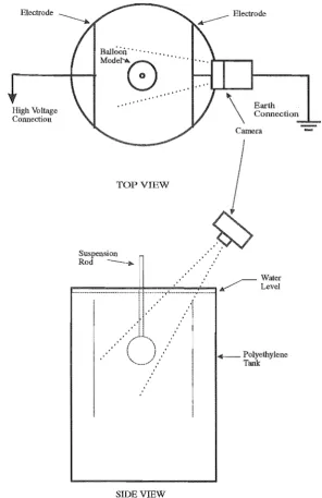
2.3.1 Balloon Model Experimental Setup
A latex rubber balloon was filled with domestic water of resistivity 100nm until the membrane was under tension relative to its unstretched state. Its expanded diameter was approximately 120mm. The balloon was then suspended in a polyethylene tank filled from the same water source. This is shown in Figure 2.2. perspex suspension rod was required to maintain the balloon in a static position and provide electrical insulation.
The tank diameter was 800mm. Two 340mm by 480mm parallel plate electrodes spaced 500mm apart were suspended into the tank such that the balloon model oc-cupied a space between the electrodes. The tank diameter was sufficiently large so that any electric field established between the electrodes was relatively uniform and not significantly influenced by the tank sides. The electrode spacing also allowed for the desired electric field magnitudes with the apparatus available.
One of the electrodes was connected to a ground plane while the other wa..s con-nected to the third stage output of a 14 stage, variable 1.4MV, inverted Marx impulse generator as used in a high voltage testing laboratory for impulse testing of electric power transformers [Khalifa 1990]. An impulse was then applied to the electrode, tank and balloon system with the generator.
2,3 PHYSICAL DIELECTRIC SPHEROID MODEL
Balloon' .. ',' .. "
r - - - -... --t
-"'"8:
" ,
" ,
" , " , " ,
TOP VIEW
Suspension " Rod ---..
:
SIDEvrnw
..
\
Earth Connection
Camera
[image:35.595.158.455.191.649.2]-+ -Polyethylene Tank
Figure 2.2 Balloon model experimental setup.
18 CHAPTER 2 ELECTROPORATION
of weakest dielectric strength.
The peak voltage and electric field magnitudes and
II
e decay time constants wereall measured to an accuracy of ±5%.
2.3.2 Balloon Model Experimental Results
The applied impulses had a peak magnitude ranging between 90 and 200kV and its shape was determined as having a IJ.ls rise time and an 8J.ls
II
e decay time. Figure2.8 shows an oscillograph of an actual capacitive discharge waveform with a peak volt-age of 98kV. The magnitude ofthe effective uniform electric field between the 500mm electrodes was between 1.8k V
I
cm to 4.0k VI
cm which is of the same magnitude as that used in the electroporation of many eukaryotic cell types [Neumann et aZ. 1989, Changet aZ. 1992b]. For the experimental impulse tests the electric field across the membrane
equated to be between 4.8MV
Icm
and 24MVIcm.
These are of the order of the dielec-tric strength of solid dielecdielec-trics. Typically the very best intrinsic breakdown strength of solid dielectrics as determined in physics laboratories [Alston 1968] is 20MVI
cm. How-ever, the practical design field strengths of solid dielectrics used in the electric power industry are very much lower. They are of the order of 2MVIcm
which recognizes the reduction of dielectric strength due to surface imperfections. With a latex rubber balloon, the surface is unmachined and design strengths are most likely to apply. [image:36.595.138.461.469.716.2]2.3 PHYSICAL DIELECTRIC SPHEROID MODEL 19
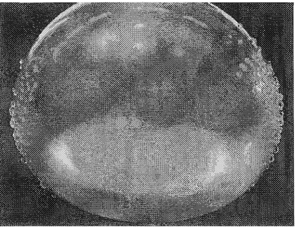
2.3.3 Water Droplet Formations
When the impulsed balloon was removed from the tank it was apparent that a number of holes had been formed in the rubber membrane as shown in Pigure 2.4. Droplets
[image:37.595.155.445.255.478.2]of water formed on the outside of the balloon membrane after it had been towel dried. Repeated drying produced the same droplet formation. It was concluded that the mem-brane was perforated in a large number of places, but each perforation was insufficient in to cause the balloon to undergo spontaneous mechanical fracture.
Figure 2.4 Water droplet formation on the pre-dried surface of an impulsed balloon model.
The perforations produced did so on the poles of the balloon that were facing the electrodes in the tank setup. There was also evidence, by the relative amount of droplet formation, that there were more holes formed on one side relative to the other. In other
words, asymmetrical breakdown was observed. This is due to the surface charge that is transferred to the balloon model during setup. The surface charge produces an inherent transmembrane potential much the same as a biological cell.
A voltage threshold effect was observed for the production of holes. Below about 135kV very little or no droplet formation was observed, even after multiple impulses. Above this threshold, multiple holes were apparent after a single impulse. Also, above the threshold voltage, multiple impulsing on a single balloon produced successively increasing hole numbers on the pole faces as indicated by increasing droplet density.
20 CHAPTER 2 ELECTROPORATION
2.3.4 Photographic Photon Capture
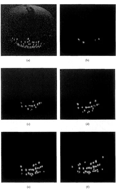
If the breakdown of the balloon membrane was an ionisation dielectric breakdown then such a breakdown would be accompanied by an emission of light from the arc so formed. Photographic emulsions are available which can detect very small emissions. The banoon apparatus was photographed in a completely darkened environment, so determined by a nil exposure of Konica 3200ASA colour film after 30 seconds. The camera position is shown in Figure 2.2. It was placed over the earthed electrode for electromagnetic shielding and safety purposes. Due to this position only the low voltage pole face can be seen or photographed.
A series of impulses was applied to the balloon whilst the camera shutter w&') open. The resulting photographs show a faint but distinct capture of light confirming an ion-isation or arc source. These images were digitally scanned and enhanced to increase their visual contrast relative to the background. The enhancement is required to elimi-nate previously experienced problems of losing definition in reproducing the images on paper (see [Gaynor and Bodger 1994c]). 2.5 shows the light emitted from a se-ries of tests. The image blurring is caused by the impulse generated balloon expansion and elongation parallel to the field direction which is also observed in biological cells during impulsing [Itoh et al. 1990]. The expansion is transient and only lasts about as
long as the impulse.
Figure 2.5(a) is an image showing the ionisation in the membrane due to a single, 3.6kV
jcm
peak electric field, impulse. A low level background illumination was used to indicate the balloon position. Figure 2.5(b) to 2.5(f) show a series of sequential im-ages of a single balloon undergoing subsequent, 3.0kVjcm
peak electric field, impulses. There is an increase in the number of breakdown points for each successive impulse. Also, once a point in the membrane has broken down, it continues to breakdown un-der further impulsing. The peak electric field threshold w&') observed to be at about 2.7kVjcm.
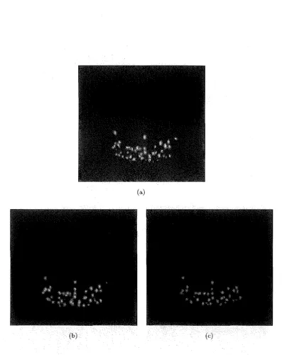
However, Figure 2.6 shows a sequence of three impulses on a single balloon which starts at 3.6kVjcm
and ends with a 2.4kV jcrn impulse. The 2.4 kVjcm
impulse is well below the previously observed threshold, but now shows a high degree of break-down. This indicates that once perforated, the breakdown threshold of a particular point is lowered.2.3.5 Microscope Evaluation
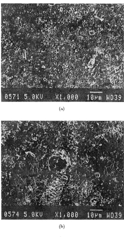
2.3 PHYSICAL DIELECTRIC SPHEROID MODEL 21
(a) (b)
(c) (d)
(e) (f)
[image:39.597.114.493.110.723.2]22 CHAPTER 2 ELECTRO PO RATION
(a)
[image:40.595.97.495.133.633.2](c)
2.4 DISCUSSION 23
the surface appears to be creatored and porous. The electron micrograph in Figure 2. 7 (b) shows a typical impulse generated hole at a peak electric field of 3.3k V
I
cm. The surrounding membrane appears to be more heavily creatored and porous relative to the control.2.4 DISCUSSION
Although not directly observable, it is generally assumed that the initiation of dielectric breakdown in biological membranes is primarily a mechanical molecular rearrangement brought on by electrostatic forces [Kinosita et al. 1992]. The electrical similarities of the balloon model to a biological cell indicate that the balloon membrane should experience similar mechanical forces [Gaynor and Bodger 1994c]. Thus, one might expect that the model would exhibit evidence of electromechanical dielectric breakdown. Experimental data does not corroborate this reasoning.
Some general conditions exi'3t which suggest dominant breakdown mechanisms. Of these conditions, photonic emission is a very strong indicator for ionisation or excita-tion of molecules to a conductive state [Llewellen Jones 1967, Fothergill 1991, Kojima
et at. 1992]. The absence of photon emission would suggest the breakdown was primar-ily electromechanical in nature [Fothergill 1991].
Under similar electrical conditions, different dielectric materials may emit photons which vary in energy depending on the respective material excitation energies. Since the energy of a photon is directly proportional to its wavelength, the emission may produce a light spectrum anywhere between infra red (IR) and ultra violet (UV). The photon emission from the balloon model suggests that the primary mechanism of its membrane dielectric breakdown is molecular ionisation. It is possible that mechanical tearing of the dielectric in the presence of a high electric field could produce the arcing [Donaldson et ~al. 1990]. This is considered unlikely as the reported materials and surrounding media are very different than those used in either the balloon model or biological cell electropermeabilization.
It has been stated [Coster and Zimmermann 1975b] that rubber behaves in a dif-ferent way to biological membranes. The difference has to do with the relative changes in Young's modulus with temperature. Biological membranes can be modelled as bi-layer phospholipid structures which have hydrophillic surfaces and a hydroscopic centre , [Hoppe et al. 1983] and contain various forms of proteins and ion channels. Although the proteins and ion channels do affect the dielectric properties of the membrane, the transmembrane potential required for dielectric breakdown is relatively the same for many different cell types [Chang et al. 1992bJ. This suggests that it is probably the lipid bi-Iayer that mainly determines the dielectric strength of the membrane.
24 CHAPTER 2 ELECTRO PO RATION
(a)
(b)
[image:42.595.169.424.180.667.2]2.4 DISCUSSION 25
appears to be arranged of layers of intertwined strands which are porous enough to let air pass through it over time. However, this may allow the balloon membrane to act more like the biological membrane than would be expected. If water were to 'seep' a little way in on both the outer and inner surfaces due to capillary action, this would mimic the hydrophillic - hydroscopic - hydrophillic characteristics of the biological cell. As in a biological membrane, the balloon membrane has numerous local deformities. In dielectric materials, deformities may cause localised electric field enhancement by up to three or four orders of magnitude [Alston 1968, Fothergill 1991, Khalifa 1990] which aids in the initiation of dielectric breakdown. Although the balloon membrane is a solid, it is also an elastomer which in many ways mechanically models the biological membrane. Whatever the detailed structure of the actual membranes of biological cells and balloons, the results suggest that their behaviour under electric impulse stimulation is similar.
It remains to repeat the experiments which show definite ionisation characteristics, on actual biological cells. As previously stated, the wavelength of any possible photon emission due to ionisation could lie anywhere between IR to UV. Hence this entire spectrum must be investigated before a more definitive conclusion can be made on the mechanisms of biological membrane breakdown. Preliminary experimentation indicates that there is very little or no photon emission from biological cell membranes in the visible region of the spectrum.
In Section 2.3.4, it was shown that once a point in the membrane has exhib-ited dielectric breakdown, it will continue to breakdown at previously sub-breakdown threshold electric field magnitudes. This is an interesting aspect of the general electro-poration system which does not appear to have been considered for multiple impulse electroporation applications.
At present, to obtain hole duration of minutes so that long chain molecules can transverse cell membranes, the temperature of the cells must be lowered and/or multiple impulses must be applied over time [Zimmermann 1986, Neumann et al. 1989, Chang et al. 1992b]. As the temperature lowers, the corresponding peak electric field impulse