warwick.ac.uk/lib-publications
Original citation:
Nichols, Thomas E., Das, Samir, Eickhoff, Simon B., Evans, Alan C., Glatard, Tristan, Hanke, Michael, Kriegeskorte, Nikolaus, Milham, Michael P., Poldrack, Russell A., Poline, Jean-Baptiste, Proal, Erika, Thirion, Bertrand, Van Essen, David C., White, Tonya and Yeo, B.T. Thomas. (2017) Best practices in data analysis and sharing in neuroimaging using MRI. Nature Neuroscience, 20 (3). pp. 299-303.
Permanent WRAP URL:
http://wrap.warwick.ac.uk/85042
Copyright and reuse:
The Warwick Research Archive Portal (WRAP) makes this work by researchers of the University of Warwick available open access under the following conditions. Copyright © and all moral rights to the version of the paper presented here belong to the individual author(s) and/or other copyright owners. To the extent reasonable and practicable the material made available in WRAP has been checked for eligibility before being made available.
Copies of full items can be used for personal research or study, educational, or not-for-profit purposes without prior permission or charge. Provided that the authors, title and full
bibliographic details are credited, a hyperlink and/or URL is given for the original metadata page and the content is not changed in any way.
Publisher’s statement:
https://doi.org/10.1101/054262
A note on versions:
The version presented here may differ from the published version or, version of record, if you wish to cite this item you are advised to consult the publisher’s version. Please see the ‘permanent WRAP URL’ above for details on accessing the published version and note that access may require a subscription.
Best Practices in Data Analysis and
1
Sharing in Neuroimaging using MRI
2
Thomas E. Nichols1,*, Samir Das2,15, Simon B. Eickhoff3, Alan C. Evans2,15, Tristan Glatard2,14, 3
Michael Hanke4,16, Nikolaus Kriegeskorte5, Michael P. Milham6,17, Russell A. Poldrack7, Jean-4
Baptiste Poline8, Erika Proal9, Bertrand Thirion10, David C. Van Essen11, Tonya White12, B.T. 5
Thomas Yeo13 6
7
1University of Warwick, Coventry, UK; 2McGill University, Montreal, Canada; 3Heinrich-Heine
8
University Düsseldorf, Düsseldorf, Germany; 4Otto-von-Guericke-University, Magdeburg, 9
Germany; 5MRC Cognition and Brain Sciences Unit, Cambridge, UK; 6Child Mind Institute, New 10
York, USA; 7Stanford University, Stanford, USA; 8University of California, Berkeley, USA; 11
9NEUROingenia Clinical and Research Center, Mexico City, Mexico; 10Inria, Paris-Saclay
12
University, Paris, France; 11Washington University in St. Louis, St. Louis, USA; 12Erasmus 13
University Medical Center, Rotterdam, The Netherlands; 13National University of Singapore, 14
Singapore; 14Department of Computer Science and Software Engineering, Concordia University, 15
Montreal, Canada; 15Montreal Neurological Institute, Montreal, Canada; 16Center for Behavioral 16
Brain Sciences, Magdeburg, Germany; 17Nathan S. Kline Institute for Psychiatric Research, 17
New York, USA; 18
*Correspondence: [email protected]. 19
20
Words: 3250 (excluding abstract) 21
Figures: 0 22
Tables: 1 23
References: 24 24
25 26
Abstract
27
Given concerns about the reproducibility of scientific findings, neuroimaging must define best 28
practices for data analysis, results reporting, and algorithm and data sharing to promote 29
transparency, reliability and collaboration. We describe insights from developing a set of 30
recommendations on behalf of the Organization for Human Brain Mapping, and identify barriers 31
that impede these practices, including how the discipline must change to fully exploit the 32
potential of the world’s neuroimaging data. 33
[Start of body text]
34The advancement of science requires continuous examination of the principles and practices by 35
which the research community operates. In recent years, this ongoing evaluative process has 36
Ioannidis in 2005 that “most published research findings are false”1 to the recent work by the 38
Open Science Collaboration, which attempted to replicate 100 psychology studies and 39
succeeded in only 39 cases2, there is mounting evidence that scientific results are less reliable 40
than widely assumed. 41
42
Efforts promoting open science principles across fields (e.g.3) as a means of fostering 43
transparency and reproducibility are valuable, but we also need efforts focusing specifically on 44
human neuroimaging. To address this need the Organisation for Human Brain Mapping (OHBM) 45
created the Committee on Best Practices in Data Analysis and Sharing (COBIDAS4, 46
http://www.humanbrainmapping.org/cobidas). This group was charged with creating a report 47
that would compile best practices for open science in neuroimaging and distill these principles 48
into specific research practices. The report was developed in collaboration with the OHBM 49
community, which provided feedback on a draft and ratification of the final version. 50
51
In this commentary, we review the challenging issues that arose in the formation of the report, 52
and identify initial success and the key remaining shortcomings in current practice. 53
What is Reproducibility?
54Open science comprises a number of different goals and principles. The COBIDAS was 55
specifically concerned with ‘Open Data’ and ‘Open Methodology’, both of which are in service 56
of ‘Open Reproducible Research.’ An immediate challenge was to obtain a working definition of 57
reproducibility. We considered a hierarchy of reproducibility concepts ranging from 58
measurement and analytical stability, to broader notions of generalisability (Table 1). A very 59
narrow notion of generalizability would be test-retest reliability on the same scanner, same 60
subject, within 30 minutes, while a more extended notion would be using different scanners on 61
the same subject with re-imaging occurring within 7 days. Generalization over analyses 62
corresponds to re-analysis of the same data using identical or similar tools. One variant of this is 63
“computational reproducibility”5, where independent researchers re-analyse the data and 64
compare their results. We also considered versions of generalizability corresponding to 65
traditional scientific notions of “replication”, such as whether a result is stable over different 66
samples of subjects or populations of subjects. The most challenging, and arguably most 67
important form of generalizability is whether a finding additionally holds under variation in the 68
stimuli and experimental methods. Underlying all of these concerns about reproducibility is how 69
theory-building requires reproducible empirical phenomena, and thus a theory will only be as 70
accurate and generalizable as the data that are used to inspire and/or test it. 71
72
Regardless of the precise scope of generalization, operationalising any of these versions of 73
reproducibility requires explicit definitions of the outcome of interest, which in itself is a 74
challenge. Previous efforts have found generally good measures of test-retest reliability of MRI 75
for both voxel-wise and region of interest measures (e.g. 6-8), but this is the most narrow notion 76
of reproducibility. A large scale project to measure the generalisability of MRI findings across 77
studies, akin to the Open Science Collaboration’s efforts in Psychology2, has not been 78
behavior correlations found only 1 of 17 findings were replicated9, though this work is limited by 80
small replication sample sizes. More work is needed in this area to better quantify the 81
generalisability of MRI findings. 82
83
In short, quantifying “reproducibility” requires precisely defining the scope of variation being 84
considered, the exact outcome that is being measured, and a metric of the stability of that 85
outcome. The COBIDAS did not set out to estimate reproducibility, but was motivated to identify 86
practices that can maximise analytical stability and generalizability of individual studies. 87
[Table 1 about here] 88
Prescribing best practice
89Neuroimaging is a broad field, encompassing a range of approaches across a growing number 90
of modalities. We restricted the scope of the COBIDAS report to include the range of all human 91
neuroimaging using Magnetic Resonance Imaging (MRI), though most of the principles 92
discussed can be applied to other modalities. We established 7 domains of practice, from 93
experimental design and acquisition, through results reporting and data sharing. We quickly 94
realised that it is neither feasible nor desirable to prescribe exactly how any one type of 95
experiment should be conducted. For example, when looking at task fMRI, the optimal 96
experimental design to use will depend on whether one is just trying to detect the presence of 97
an effect or rather estimate the shape of the hemodynamic response function. 98
99
The one “practice” that can be universally commended is the transparent and complete 100
reporting of all facets of a study, allowing a critical reader to evaluate the work and fully 101
understand its strengths and limitations. This also facilitates subsequent research efforts by 102
other investigators, who can exactly follow (or carefully manipulate) each aspect of a study. This 103
includes conveying the “researcher degrees of freedom”, by reporting other analytical paths 104
applied unsuccessfully on the present data before arriving at the published results. Although 105
formidable, the reporting checklists provided in the COBIDAS MRI report reflects the breadth 106
and depth of information needed to ensure another researcher could replicate the work. 107
108
To further facilitate reproducibility, the COBIDAS report includes specific recommendations for 109
statistical modelling, where specific (and common) bad practices have been identified10,11. We 110
have also made concrete recommendations for data sharing, where practice is still evolving. 111
112
From solicited community input, we were struck by the emphatic and diverse views on the types 113
of data to share. Some strongly felt it was essential to share the rawest form of the data from 114
the scanner (DICOM format), while others felt that preprocessed, ready-to-analyze data should 115
be shared; still others emphasized the utility of sharing extensively processed data linked to 116
published figures. We evaluated the pros and cons of each form of data sharing; for example, 117
while sharing preprocessed data can minimize the effort needed for reanalysis and speed 118
advances based on new uses of the data, it may preclude alternate preprocessing options that 119
degree of spatial smoothing used). In the end, we endorsed the sharing of data in as many 121
forms as is feasible. 122
123
Are we ready for open science in neuroimaging?
124Brain imaging research is complicated, not only at the level of the conducting a study, but also 125
at the level of sharing its results and data. The importance of thorough reporting of results is 126
uncontroversial, and practices are improving, and the sharing of data to facilitate replication is 127
increasingly viewed as essential. However, data sharing poses new challenges. Here we 128
consider a number of concerns that investigators have with data sharing that impede adoption 129
of open practices. 130
131
First, some individual researchers may assert ownership of their data and thus may not feel 132
compelled to share. Counter to this is the drive for publically funded research to produce widely 133
accessible data that can be reused and integrated into further research. Researchers may feel 134
that sharing of data will result in a loss of competitive advantage, with other researchers 135
swooping in to publish their planned studies based on the same data. The actual risk of this will 136
depend on the data and hypotheses, but it should be weighed against the opportunity of new 137
collaborations resulting from the sharing. These concerns can be alleviated by delaying the 138
sharing or using a data-sharing repository with an embargo period. 139
140
Another fear is that, upon sharing data, other researchers will discover errors in an analysis or 141
previously undiscovered problems with the data. As scientists, we are supposed to be objective 142
arbiters of evidence and theory, but we are not infallible and must be ready to accept criticism 143
and revise our claims when errors are discovered. Even when no errors are found, a re-144
analyses may support conclusions inconsistent with the original study. For controversial topics, 145
there may also be adversarial reanalyses. We see no better way to advance understanding on a 146
contested finding than to have as many researchers as possible puzzling over the data at hand. 147
However, we need to develop a culture of constructive criticism that recognizes that errors are 148
an inevitable part of scientific progress and protects individual researchers from inappropriately 149
harsh consequences when honest mistakes are discovered. 150
151
A very practical concern, especially for junior investigators, is what is perceived as an 152
unjustifiable cost of data sharing. Current incentives do not justify spending large amounts of 153
time preparing data for sharing, as institutional promotion panels or grant reviewers currently do 154
not adequately reward such efforts. Counter to this is the greater potential impact of a work 155
when it may be cited not just for its scientific findings, but also when its data is reused in other 156
works. Data description papers can document and provide credit for high-quality data 157
acquisition efforts for the open community. We assert that if data sharing and open science 158
priorities in general are to take hold, academic institutions, journals, and granting agencies are 159
crucial for improving the incentives for open practices and developing ways to give appropriate 160
credit for efforts in data sharing. 161
Finally there is the very real worry of failing to comply with human ethics provisions for 163
protecting subject privacy. It can be argued that, once file headers are scrubbed of personally 164
identifiable information and structural images have facial features obscured, that the data are 165
completely anonymised and thus freely sharable. However individual ethics boards have varying 166
views on this and it is best to write ethics consent documents explicitly with data sharing in 167
mind. This topic would greatly benefit from leadership from national research organisations to 168
seek consensus and then establish exactly what comprises anonymized brain imaging data. In 169
particular, ethics boards often only try to minimize the risk to subjects when we are also obliged 170
to maximize the benefit of our research to science and society, so as to honor the contribution of 171
our subjects.12 The future value of shared data must be considered in ethical decision making. 172
173
While studies lacking shared data and having opaque methodological detail are typical, some 174
authors have embraced the challenges of sharing data and analysis methodology. Some recent 175
examples that are particularly thorough and elegant include Waskom et al.13 and Whitaker et 176
al.14, that published a complete array of analysis scripts for generating all figures and results in 177
the paper (https://github.com/mwaskom/Waskom_JNeurosci_2014 and 178
https://github.com/KirstieJane/NSPN_WhitakerVertes_PNAS2016, respectively), and Pernet et 179
al.15 that likewise shared raw data and analysis scripts, as well as all results maps in electronic 180
form. From an organisational perspective, some labs are simply making open science a policy. 181
Most recently the Montreal Neurological Institute announced that their work would be open, with 182
all results and data made freely available at the time of publication16. 183
184
These few examples demonstrate that some researchers are embracing open science 185
principles, but do the tools exist to make it practical on a widespread basis? 186
Existing tools for open neuroimaging
187There is an emerging ecosystem of open science tools for neuroimaging research. Before any 188
data is collected, there are tools to assist in creating human ethics documents that maximise the 189
ease of later data sharing, and for everything from experimental paradigm presentation, 190
preprocessing to statistical modelling, neuroimaging benefits from numerous, free and well-191
supported software tools (see Supplementary Table 1 for an incomplete list). This constellation 192
of tools could be seen as fuel for limitless researcher degrees-of-freedom, and indeed there is a 193
need for the community to identify a set of ‘reference pipelines’ for common analyses. However, 194
since each tool makes particular assumptions about neuroanatomical and neurophysiological 195
processes, it is not possible to recommend the optimal analyses for every possible type of data 196
and analysis objective. Only with user experience and reproducibility comparisons, will the field 197
be able to identify what are the preferred analytical approaches. 198
199
There is a particular embrace of data sharing in the resting-state fMRI community. Since 200
resting-state analyses methods remain in flux, sharing of this data has particular value as it 201
allows future improvements in methods to be assessed and benchmarked relative to previous 202
analyses. For resting and task fMRI and structural MRI, there are a number of projects that have 203
Alzheimer's Disease Neuroimaging Initiative (http://www.adni-info.org). These have become 205
invaluable tools for methodologists to apply novel image processing algorithms, not to mention 206
the primary scientific outputs from these projects. 207
208
One promising new standard is the Brain Imaging Data Structure (BIDS)18, a simple system for 209
organising MRI data after conversion to the NIFTI format. BIDS provides a common, consistent 210
directory hierarchy and naming system for files, as well as supporting ‘side car’ files for key 211
associated data (like stimulus timing information for task fMRI). With a fixed standard for 212
representing data, this has supported the creation of a number of “BIDS Apps”, self-contained 213
programs that can automatically process data arranged according to BIDS. Simple, widely used 214
standards such as this have the potential to dramatically reduce the effort required to exchange 215
and share data. 216
217
New tools are set to dramatically advance computational reproducibility. A challenge to even 218
something as simple as re-running the same data with the same code is the ever-changing 219
versions of software and libraries that software depends on. The last five years has seen the 220
growth of virtual machines and containers to share not just data but a complete environment for 221
processing data. A virtual machine (VM) is an emulator of a computer, including its hardware, 222
operating system and file system. It can be shared as a single file and when run, an entire 223
computer system comes into existence based on a snapshot of the libraries and software 224
interdependencies of one particular system. From within this VM, data can be run through a 225
complete processing pipeline; with the original data of a study this will reproduce the results 226
exactly, while new data can also be imported to evaluate the unique aspects of a pipeline. A 227
downside to VMs is their gross size, as they are as large as any operating system. Containers 228
are miniature VMs, lacking the full operating system but providing the specialised software and 229
libraries required to execute a given task. The BIDS Apps mentioned above rely on such 230
containers, encapsulating software packages large and small that alleviate installation of a 231
myriad of software dependencies. 232
233
Open science tools are gaining traction. For example, the CBRAIN web-based analysis service 234
supports over 260 collaborators in 20 countries; the COINS service currently hosts data on over 235
40,000 subjects for 643 studies; the LONI Pipeline has an average of 100,000 daily jobs from 236
200 different analysis workflows; the Neurovault repository hosts 450 public collections; and the 237
FCP/INDI is openly sharing over 15,000 resting fMRI and structural MRI datasets. 238
Continuous improvement of research practices
239Despite a seeming wealth of tools, there remain specific areas in the field of neuroimaging that 240
need to be embraced to increase reproducibility. Aside from the importance of carefully 241
reporting the study design, methods, and results mentioned above, we also identified priorities 242
including archiving of statistical results, software engineering for reproducibility, and optimizing 243
projects for generalizability. 244
In genetics, the routine sharing of “summary data” (SNP-level statistical results) has facilitated 246
meta-analyses and methodological developments. For example, LD-score regression is a tool 247
that can estimate genetic correlation using just Z-score summary data, and has had dramatic 248
impact in a short timespan due to the availability of such results19. In brain imaging, we have no 249
tradition of sharing summary statistics (i.e. images of T- or Z-scores, or images of percent 250
change effect and standard errors). As a result the quality of meta-analyses are currently limited 251
by their reliance on reported tables of maximum location coordinates, for which there is a 252
substantial loss of information relative to the original statistic images20. In the current age, the 253
costs of sharing such images of summary statistics (~1MB compressed), either through generic 254
or dedicated repositories (e.g., NeuroVault.org, or BALSA, http://balsa.wustl.edu), are relatively 255
minimal. As such, COBIDAS recommends the deposition of unthresholded statistical images 256
into archival resources for all studies. Widespread adoption of this practice will dramatically 257
increase our capacity for more precise meta-analyses, and allow more critical assessment of 258
study results through exploration of the complete 3D image. 259
260
One foundation of computational reproducibility is modern software engineering practice. 261
Whether a small set of scripts or a comprehensive end-to-end pipeline, neuroimaging data 262
analysis depends on coding. Modern software engineering includes practices like version 263
control and unit testing. Version control ensures that revisions of the code are identifiable and 264
archived, and ideally is based on an open platform that allows wide inspection and input; unit 265
tests verify the correctness of individual facets of the code, and can be set to automatically run 266
each time the code is updated. This is not to say that every group should hire a programmer, 267
but rather that every researcher writing scripts or code should obtain proficiency with basic 268
software engineering skills and practices21 (see Software Carpentry for basics instruction for 269
non-programmers, http://software-carpentry.org/). With routine research grounded in well-270
written, less fragile code, it will be much easier to establish analysis pipelines that can both be 271
reused within a lab and facilitate computational reproducibility verified by others. 272
273
Study designs have traditionally been optimised to maximise statistical power to detect 274
differences between groups. With a growing emphasis on prediction, whether (e.g.) identifying 275
early risk for psychosis or progression of a neurodegenerative disease, studies should be 276
optimised for building predictive models that will generalise to the population of interest in yet-277
unseen data. Large multi-site studies that capture wide variation in human populations, as well 278
as site-specific technical idiosyncrasies, are essential to build classifiers with good performance 279
on new data. Whether obtained with prospectively optimized homogeneous acquisition and 280
preprocessing strategies (e.g. Human Connectome Project and its successors22) or via larger 281
but more heterogeneous, aggregate multisite approaches (e.g., FCON1000/INDI; ADNI, PING, 282
and the upcoming ABCD Study) that have optimized image processing strategies determined 283
retrospectively23, generalisability of predictive models will be a key design objective and 284
Beyond the investigator
286Many of the practices advocated here and in the full COBIDAS MRI report require individuals to 287
change the way they conduct research. Almost every such change requires an investment of 288
time and resources. While we argue these have implicit rewards (e.g. shared data will never be 289
lost when the post doc moves on), the advance of open science will require leadership at the 290
institutional level. To provide appropriate incentives, universities and research centers need to 291
explicitly consider the value of sharing of data and code as an unique research output in 292
promotion and review exercises. Journals should require that papers’ statistic images are 293
archived, and promote papers with shared data, provide full analytical detail, and ideally share 294
code or even executable containers or VMs. Foundations and granting agencies need to make 295
data sharing a priority, recognizing and funding the explicit costs of data management required 296
to make this happen. And finally professional organisations like OHBM should prioritize efforts in 297
education to make open science practices accessible to all. 298
299
With the coordinated efforts of individual researchers, academic institutions, journals, granting 300
agencies, and professional organisations, we can accelerate the drive towards open science 301
and maximise the reproducibility of neuroimaging findings going forward. 302
303 304 305 306
Acknowledgements.
307
TEN is supported by the Wellcome Trust (100309/Z/12/Z) and NIH (R01 NS075066-01A1, R01 308
EB015611-01). BTTY is supported by Singapore MOE Tier 2 (MOE2014-T2-2-016), NUS 309
(DPRT/944/09/14, R185000271720), NMRC (CBRG14nov007) and the NUS YIA. SBE is 310
supported by the Deutsche Forschungsgemeinschaft (DFG, EI 816/4-1, LA 3071/3-1; EI 816/6-311
1), the National Institute of Mental Health (R01-MH074457), the Helmholtz Portfolio Theme 312
"Supercomputing and Modeling for the Human Brain" and the European Union Seventh 313
Framework Programme (FP7/2007-2013) under grant agreement no. 604102). MH was 314
supported by funds from the German federal state of Saxony-Anhalt and the European Regional 315
Development Fund (ERDF), project: Center for Behavioral Brain Sciences, and CRCNS 316
BMBF/NSF (01GQ1411/1429999). NK was supported by the UK Medical Research Council 317
and a European Research Council Starting Grant (261352). The authors declare no competing 318
financial interests. ACE, SD and TG are supported by the Irving Ludmer Family Foundation and 319
the Ludmer Centre for Neuroinformatics and Mental Health. TW was supported by a ZonMw 320
TOP grant (91211021). RAP is supported by the Laura and John Arnold Foundation. MPM is a 321
Phyllis Green and Randolph Cowen Scholar and is supported in part by the NIH (U01 322
MH099059; R01 AG047596) 323
324
REFERENCES 325
1. Ioannidis, J. P. A. (2005). Why most published research findings are false. PLoS
327
Medicine, 2(8), e124. doi:10.1371/journal.pmed.0020124 328
2. Open Science Collaboration. (2015). PSYCHOLOGY. Estimating the reproducibility of 329
psychological science. Science (New York, N.Y.), 349(6251), aac4716. 330
doi:10.1126/science.aac4716 331
3. Journals unite for reproducibility. (2014). Nature, 515(7525), 7. doi:10.1038/515007a 332
4. Nichols, T. E., Das, S., Eickhoff, S. B., Evans, A. C., Glatard, T., Hanke, M., Yeo, B. 333
T. T. (2016). Best Practices in Data Analysis and Sharing in Neuroimaging using MRI. 334
bioRxiv, 54262. http://doi.org/10.1101/054262
335
5. Peng, R. D. (2011). Reproducible research in computational science. Science, 336
334(6060), 1226–7. doi:10.1126/science.1213847 337
6. Bennett, C. M., & Miller, M. B. (2013). fMRI reliability: influences of task and 338
experimental design. Cognitive, Affective & Behavioral Neuroscience, 13(4), 690–702. 339
http://doi.org/10.3758/s13415-013-0195-1
340
7. Schnack, H. G., van Haren, N. E. M., Brouwer, R. M., van Baal, G. C. M., Picchioni, M., 341
Weisbrod, M., Hulshoff Pol, H. E. (2010). Mapping reliability in multicenter MRI: voxel-342
based morphometry and cortical thickness. Human Brain Mapping, 31(12), 1967–82. 343
http://doi.org/10.1002/hbm.20991
344
8. Noble, S., Scheinost, D., Finn, E. S., Shen, X., Papademetris, X., McEwen, S. C., 345
Constable, R. T. (2016). Multisite Reliability of MR-Based Functional Connectivity. 346
NeuroImage. http://doi.org/10.1016/j.neuroimage.2016.10.020
347
9. Boekel, W., Wagenmakers, E.-J., Belay, L., Verhagen, J., Brown, S., & Forstmann, B. U. 348
(2015). A purely confirmatory replication study of structural brain-behavior correlations. 349
Cortex, 1–19. http://doi.org/10.1016/j.cortex.2014.11.019
350
10. Kriegeskorte, N., Simmons, W. K., Bellgowan, P. S. F., & Baker, C. I. (2009). Circular 351
analysis in systems neuroscience: the dangers of double dipping. Nature Neuroscience, 352
12(5), 535–40. doi:10.1038/nn.2303 353
11. Poldrack, R. A., Fletcher, P. C., Henson, R. N., Worsley, K. J., Brett, M., & Nichols, T. E. 354
(2008). Guidelines for reporting an fMRI study. NeuroImage, 40(2), 409–14. 355
http://doi.org/10.1016/j.neuroimage.2007.11.048
356
12. Brakewood, B., & Poldrack, R. A. (2013). The ethics of secondary data analysis: 357
Considering the application of Belmont principles to the sharing of neuroimaging data. 358
NeuroImage, 82, 671–676. http://doi.org/10.1016/j.neuroimage.2013.02.040
359
13. Waskom, M. L., Kumaran, D., Gordon, A. M., Rissman, J., & Wagner, A. D. (2014). 360
Frontoparietal Representations of Task Context Support the Flexible Control of Goal-361
Directed Cognition. Journal of Neuroscience, 34(32), 10743–10755. JOUR. 362
http://doi.org/10.1523/JNEUROSCI.5282-13.2014
363
14. Whitaker, K. J., Vértes, P. E., Romero-Garcia, R., Váša, F., Moutoussis, M., Prabhu, G., 364
Bullmore, E. T. (2016). Adolescence is associated with genomically patterned 365
consolidation of the hubs of the human brain connectome. Proceedings of the National 366
Academy of Sciences, 113(32), 201601745. http://doi.org/10.1073/pnas.1601745113 367
15. Pernet, C. R., McAleer, P., Latinus, M., Gorgolewski, K. J., Charest, I., Bestelmeyer, P. 368
individual variability in temporal and extra-temporal cortices. NeuroImage, 119, 164–74. 370
http://doi.org/10.1016/j.neuroimage.2015.06.050 371
16. Owens, B. (2016). Montreal institute going “open” to accelerate science. Science. 372
http://doi.org/10.1126/science.aae0265
373
17. Mennes, M., Biswal, B. B., Castellanos, F. X., & Milham, M. P. (2013). Making data 374
sharing work: The FCP/INDI experience. NeuroImage, 82, 683–691. 375
http://doi.org/10.1016/j.neuroimage.2012.10.064
376
18. Gorgolewski, K. J., Auer, T., Calhoun, V. D., Craddock, R. C., Das, S., Duff, E. P., 377
Poldrack, R. A. (2016). The brain imaging data structure, a format for organizing and 378
describing outputs of neuroimaging experiments. Scientific Data, 3, 160044. 379
http://doi.org/10.1038/sdata.2016.44
380
19. Bulik-Sullivan, B. K., Loh, P.-R., Finucane, H. K., Ripke, S., Yang, J., Patterson, N., 381
Neale, B. M. (2015). LD Score regression distinguishes confounding from polygenicity in 382
genome-wide association studies. Nature Genetics, 47(3), 291–295. 383
http://doi.org/10.1038/ng.3211
384
20. Salimi-Khorshidi, G., Smith, S. M., Keltner, J. R., Wager, T. D., & Nichols, T. E. (2009). 385
Meta-analysis of neuroimaging data: A comparison of image-based and coordinate-386
based pooling of studies. NeuroImage, 45(3), 810–823. 387
http://doi.org/10.1016/j.neuroimage.2008.12.039
388
21. Goble, C. (2014). Better software, better research. IEEE Internet Computing, 18(5), 4–8. 389
http://doi.org/10.1109/MIC.2014.88
390
22. Glasser, M. F., Smith, S. M., Marcus, D. S., Andersson, J. L. R., Auerbach, E. J., 391
Behrens, T. E. J., Van Essen, D. C. (2016). The Human Connectome Project’s 392
neuroimaging approach. Nature Neuroscience, 19(9), 1175–87. 393
http://doi.org/10.1038/nn.4361 394
23. Abraham, A., Milham, M., Martino, A. D., Craddock, R. C., Samaras, D., Thirion, B., & 395
Varoquaux, G. (2016). Deriving reproducible biomarkers from multi-site resting-state 396
data: An Autism-based example. Neuroimage. 397
http://doi.org/10.1016/j.neuroimage.2016.10.045
398
24. ISO. (2006). Statistics - Vocabulary and symbols. Part 2: Applied statistics. ISO 3534–2 399
(Second.). Geneva: ISO. 400
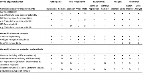
Levels of generalization
Generalization over measurements Population Sample Scanner Visit Data
Stimulus Population
Stimulus
Sample Method Code Experi-menter
Data Analyst
ISO Repeatability
e.g. 30-minute intra-scanner reliability • • • • D • • • • • • ISO Intermediate Reproducibility
e.g. 7-day intra-scanner reliability • • • D D • • • • • • ISO Reproducibility
e.g. 7-day inter-scanner reliability • • D D D • • • • • •
Generalization over analyses
Analysis Replicability • • • • • • • • • • • Collegial Analysis Replicability • • • • • • • • • • D Peng5 Reproducibility • • • • • • • • D D D
Generalization over materials and methods
Near Replicability (different subjects) • D • - - • • • • • • Intermediate Replicability (different labs) • D D - - • • • • D D Far Replicability (different experimental &
analytical methods) • D D - - • D D D D D Hypothesis Generalisability (different subject
[image:12.612.55.558.210.466.2]populations & types of stimuli) D D D - - D D D D D D Table 1. A partial taxonomy of reproducibility in neuroimaging. For each type of reproducibility (row), the variable (column) that is held constant (•, bullet) or allowed to vary (D=different) is indicated; minus (-) indicates not applicable. Variations in the participant studied can be described in terms of the population they belong to (e.g. different patient groups or people from different cultures), or whether the same sample or a distinct sample of individuals is used. The MRI scanner used can be the same or not, and if the same participant sample is considered, the very same data can be used or new data can be acquired on the same or different days (visits) to the scanner. Experimental variation has many forms including the particular experimental design, but here we only consider stimuli. The type of stimulus used (stimulus population) may change, for example in a working memory experiment, letter stimuli might be replaced with shape stimuli; a more subtle change would be to use a different sample of stimuli of the same type, e.g. different particular shapes. The analysis method may vary; for example, with structural MRI for prediction of patient disease status, a linear discriminant might be used instead of a nonlinear support vector machine. Analysis code more narrowly reflects the particular implementation of a given method. Personnel conducting the research is another important source of variation, whether this is the experimenter or data analyst. Finally, note that the International Standards Organisation (ISO) has precise definitions of reproducibility24 as indicated in the first three rows, but these capture only the minimal levels of generalizability.
Personnel Analysis
MRI Acquisition
Resource Type Short Description Free Open Source Link
Open Brain Consent Consent Ethics template oriented for neuroimaging data sharing x x http://open-brain-consent.readthedocs.io
OpenSesame Paradigm software Graphical experiment builder x x http://osdoc.cogsci.nl
PsychoPy Paradigm software Psychophysics software in Python x x http://www.psychopy.org
Psychtoolbox Paradigm software Psychophysics Toolbox x x http://psychtoolbox.org/
aa Pipeline Automatic Analysis, Matlab-based workflow tool x (Matlab) x http://automaticanalysis.org
C-BRAIN Pipeline Web-based software for computationally intensive analyses x x http://cbrain.mcgill.ca
CCS Pipeline Connectome Computation System, a pipline primarily for resting data x x http://github.com/zuoxinian/CCS
C-PAC Pipeline Configurable Pipeline for the Analysis of Connectomes x x http://fcp-indi.github.io
DPARSF/DPABI Pipeline Data Processing & Analysis for Brain Imaging, inlcuding resting-state fMRI x x http://rfmri.org/dpabi
DTIPrep Pipeline Pipeline for diffusion weighted / diffusion tensor image data x x http://www.nitrc.org/projects/dtiprep/
HCP Pipeline Pipeline Human Connectome Project Pipeline x x http://github.com/Washington-University/Pipelines
LONI Pipeline Pipeline Cross-platform workflow tool for neuroimaging, genomics, bioinformatics NC http://pipeline.loni.usc.edu
LORIS Pipeline Web-based data and project management software for neuroimaging x x http://loris.ca
NIAK Pipeline Llibrary of modules and pipelines for fMRI processing in Matlab/Octave x x http://www.nitrc.org/projects/niak
NiDB Pipeline Neuroimaging database software that includes pipeline tools x x http://github.com/gbook/nidb
NiPype Pipeline Neuroimaging in Python Pipelines and Interfaces x x http://nipy.org/nipype
PANDA Pipeline Pipeline for Analyzing braiN Diffusion imAges x x http://www.nitrc.org/projects/panda
SimNIBS Pipeline Simulation of Non-invasive Brain Stimulation x x http://simnibs.de
AFNI Scriptable Analysis Neuroimaging analysis software for functional MRI x x http://afni.nimh.nih.gov/afni
CONN Scriptable Analysis Functional connectivity toolbox, Matlab-based pipeline tool x (Matlab) x http://www.nitrc.org/projects/conn
Connectir Scriptable Analysis Analysis software for Connectome-Wide Association Studies, based in R x x http://czarrar.github.io/connectir
DiPy Scriptable Analysis Diffusion analysis pipeline using Python x x http://nipy.org/dipy
Freesurfer Scriptable Analysis Neuroimaging analysis software for MRI, empahsis on surface-based analysis x x http://surfer.nmr.mgh.harvard.edu
FSL Scriptable Analysis Neuroimaging analysis software for MRI NC x http://www.fmrib.ox.ac.uk/fsl
MindBoggle Scriptable Analysis Automated labeling and shape analysis of brain images x x http://www.mindboggle.info
SPM Scriptable Analysis Neuroimaging analysis software based in Matlab, for MRI, M/EEG, PET. x (Matlab) x http://www.fil.ion.ucl.ac.uk/spm
Voxel Scriptable Analysis Mass-Univariate Voxelwise Analysis of Medical Imaging Data, based in R x x http://cran.r-project.org/web/packages/voxel
BIDS Data Sharing Standard for organising MRI data and associated supporting data http://bids.neuroimaging.io
COINS Data Sharing Web-based data management and analysis tool http://coins.mrn.org
FCP/INDI Data Sharing Repository for resting state fMRI data http://fcon_1000.projects.nitrc.org
Figshare Data Sharing Generic data sharing repository http://figshare.com
LONI IDA Data Sharing Image data archive, repository for primarily neuroimaging data http://ida.loni.usc.edu
LORIS Data Sharing Database for longitudinal imaging studies http://bigbrain.loris.ca
NDA Data Sharing NIMH Data Archive, repository for data from NIMH-funded studies http://data-archive.nimh.nih.gov
NITRC-IR Data Sharing Image repository for neuroimaging data http://www.nitrc.org/ir
OpenfMRI Data Sharing Repository for task fMRI data, inlcuding all image and task paradam data https://openfmri.org
PCP Data Sharing Preprocessed connectome project - pipelines for resting state data http://preprocessed-connectomes-project.org
XNAT-Central Data Sharing Repository for raw MRI data http://central.xnat.org
BALSA Results Sharing Sharing of surface-based statistical results x http://balsa.wustl.edu
NeuroVault Results Sharing Sharing tool for statistical maps x x http://neurovault.org
NIDM Results Sharing Standard for exporting statistical results independent of the analysis tool x x http://nidm.nidash.org
Docker Reproducibility tool Containerisation tool x x http://www.docker.com
Resource Type Short Description Free Open Source Link