Address for correspondence
Dr. Dr. Vinma Shetty, Associate Professor, Department of Dermatology, Venereology and Leprosy,
A.J Institute of Medical Sciences, Mangalore, Karnataka, India
Email: [email protected]
Original Article
Dermoscopic study of hair loss in females and its
correlation with serum ferritin levels
Introduction
Alopecia can result in loss of self-esteem and lack of self-confidence1,2 and is often difficult to treat.3 Some relationship has been documented between iron deficiency and hair loss, however it is yet to be confirmed.3
Dermoscopy is a non-invasive technique used to visualise pigmented skin lesions.4 “Trichoscopy” is the term used for dermoscopy of hair and the
scalp.5 Trichoscopy is used to distinguish the different types of alopecia and identify diagnostic signs.6
In this study, we examined the dermoscopic pattern of different types of hair loss in females and its correlation with serum ferritin levels.
Materials and Methods
This was a hospital based, cross sectional observational study. Female subjects in the age group of 15-55 years with complaints of alopecia attending the Dermatology OPD from April 2018 to August 2018, were studied after obtaining ethical clearance from the Institutional Ethics Committee. A convenient sample size of 70 was adopted. For this study, we defined
Vinma Shetty, Hafsa Eram, Saumya Goel, Amita Murali Babu
Department of Dermatology, Venereology and Leprosy, A.J. Institute of Medical Sciences, Mangalore, Karnataka, India.
Abstract
Background Alopecia can be inherited or acquired, cicatricial or non-cicatricial. Sometimes the diagnosis can pose difficulties, and in such cases, dermoscopy can aid in the diagnosis. The treatment can also be difficult due to its multiple etiologies. Iron deficiency is an important cause of alopecia but its relationship with alopecia remains controversial.Methods This was a hospital based, cross sectional observational study with 70 female patients. After taking consent, dermoscopic evaluation of the scalp was done and photographs were taken. Serum ferritin levels were done for all the subjects. Software (SPSS version 16.0 statistical package) was used throughout. Mean and standard deviation were used for continuous variables.
Results Most common cause of alopecia was telogen effluvium. Most common dermoscopic finding was thin hair. In our study, we found a correlation between serum ferritin levels and certain patterns of alopecia like telogen effluvium, female pattern hair loss (FPHL), alopecia areata.
Conclusion Dermoscopy of the scalp can aid in the diagnosis of difficult conditions. Serum ferritin and haemoglobin levels are reduced in certain types of alopecia like telogen effluvium, FPHL and alopecia areata.
Key words
alopecia as visible thinning or loss of hair from the scalp. Subjects who were unwilling to participate in the study, those with active secondary bacterial infection at the site of alopecia, and those with other co morbidities like diabetes and hypertension were excluded from the study.
For all the subjects enrolled in the study, clinical examination was done, digital photographs were taken, dermoscopic findings were recorded and blood was drawn for haemoglobin and ferritin levels. A manual dermoscope with a 10x10 magnification was used.
Software (SPSS version 16.0 statistical package) was used throughout. Mean and standard deviation were used for continuous variables.
Results
70 female subjects were included in the study. All the subjects completed the study.
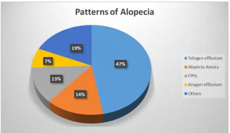
Majority of the subjects (33 subjects, 47.1%) had telogen effluvium, out of which most of them (13 subjects, 39.4%) were in the age group 26-35 years. The second most common type of alopecia was alopecia areata with 10 (14.3%) subjects, out of which majority (4 subjects, 40%) were in the 15-25 years age group. 9 (12.8%) subjects had female pattern hair loss (FPHL) with 5 (55.5%) subjects in the 46-55 years age group. 5 (7.1%) subjects had anagen effluvium with 4 (80%) subjects in the 46-55 years age group (Table 1, Figure 1).
There was a lot of overlap in the dermoscopic findings of the different patterns of alopecia. The most common dermoscopic finding was thin hair, found in 47 (67.1%) subjects, followed by yellow dots in 39 (55.7%) subjects, diameter diversity >20% in 32 (45.7%) subjects and vellus hair in 23 (32.8%) subjects (Table 2). Other findings like black dots, honeycomb pigment network, peripilar sign and loss of follicles were seen in a lesser proportion of subjects.
Table 1 Distribution of patterns of alopecia in the different age groups
Age(years) Patterns of alopecia
Telogen effluvium
Anagen effluvium
FPHL Alopecia areata
Pseudopelade of Brocq
DLE Pediculosis capitis
Pyoderma
15-25 8 0 0 4 1 0 2 2
26-35 13 0 1 3 2 1 0 1
36-45 6 1 3 2 0 1 2 1
46-55 6 4 5 1 0 0 0 0
Total n (%) 33 (47.1%) 5 (7.1%) 9 (12.8%) 10 (14.3%) 3 (4.2%) 2 (2.8%) 4 (5.7%) 4 (5.7%) FPHL: Female pattern hair loss; DLE: Discoid lupus erythematosus
Table 2 Dermoscopic findings observed among study subjects Dermoscopic findings No. of subjects*
n (%)
Thin hair 47 (67.1%)
Yellow dots 39 (55.7%)
Diameter diversity >20% 32 (45.7%)
Vellus hair 23 (32.8%)
Black dots 14 (20%)
Honeycomb pigment network 8 (11.4%)
Peripilar sign 4 (5.7%)
Figure 1 Patterns of alopecia among study subjects
Table 3 Prominent dermoscopic features in different patterns of alopecia Type of alopecia Dermoscopic findings
Telogen effluvium Thin hair (81.8%), yellow dots (54.5%), peripilar erythema (18.2%)
Alopecia areata Yellow dots (70%), black dots (60%), thin hair (40%), exclamation mark hair (30%) FPHL Diameter diversity >20% (66.7%), thin hair (66.7%), yellow dots (55.6%)
DLE Loss of follicles (100%), Honeycomb pigment network (50%) FPHL: Female pattern hair loss; DLE: Discoid lupus erythematosus
Table 4 Correlation of pattern of hair loss with serum ferritin and haemoglobin levels
Investigation Telogen effluvium FPHL Alopecia areata Serum ferritin (µg/L) Mean ± SD 19.3 ± 4.7 32.8 ± 19.6 22.7 ± 5.6
Hb (g/dL) Mean ± SD 10.7 ± 1.2 11.2 ± 1.4 11.5 ± 1.2
Hb: Haemoglobin; FPHL: Female pattern hair loss
The most common finding in telogen effluvium was thin hair (81.8%) followed by yellow dots (54.5%). In alopecia areata, yellow dots (70%) was the most common finding, followed by black dots (60%) and thin hair (40%), exclamation mark hair was found in 30% of the subjects. Diameter density >20% and thin hair (66.7%) was the most common finding in FPHL, followed by yellow dots (55.6%) (Table 3).
The serum ferritin (19.3±4.7) and haemoglobin (10.7±1.2) were lowest in subjects with telogen effluvium, followed by alopecia areata (Serum ferritin=22.7±5.6, haemoglobin = 11.5±1.2) and FPHL (serum ferritin = 32.8±19.6, haemoglobin =11.2±1.4). The other patterns of alopecia did not show any correlation with serum ferritin and haemoglobin levels (Table 4).
Clinical and dermoscopic pictures are shown in
Figures 2-7.
Discussion
Telogen effluvium is usually the most common pattern of alopecia in females, followed by FPHL and alopecia areata.7 In our study too, the most common type of alopecia was telogen effluvium, followed by alopecia areata and FPHL.
Figure 2a, 2b Clinical photographs of telogen effluvium
Figure 3a, 3b Dermoscopic pictures of telogen effluvium
Figure 4a, 4b Clinical photographs of FPHL
Figure 6a, 6b Clinical photographs of alopecia areata
Figure 7a, 7b Dermoscopic pictures of alopecia areata
degenerating keratinocytes and sebum.8,9 Black dots are seen within the yellow dots and they characterize cadaverised hair, that break before emergence from the scalp.4
On dermoscopy, telogen effluvium shows decreased hair density and empty hair follicles, and it is a diagnosis of exclusion.4 In our study, thin hair was the most common finding. In alopecia areata, the trichoscopic findings differ based on stage, site and severity of disease.8,9 Black dots, tapering and cadaverised hair are the most specific findings and associate with disease activity.10 Yellow dots are seen in all phases and associate with disease severity.11 Acute stage shows exclamation hairs and black dots.4 In our study, yellow dots and thin hair were the most common findings in alopecia areata. Reduced terminal hair, increased vellus hair and empty follicles in the frontal area in female pattern hair loss were found in a study by Kowalska-Oledzka et al.12 Diameter diversity >20% and thin hair
were the most common findings in our study. DLE of the scalp is infrequent and dermoscopy helps to discern it from other cicatricial alopecias. Atrophy with complete loss of follicular openings is seen.4 Loss of follicles was seen in all DLE subjects in our study.
telogen effluvium who received oral iron therapy. However, Olsen et al.17 reported no difference in the prevalence of iron deficiency between patients with or without alopecia. There was no association between serum ferritin levels and alopecia in women in a study by Bregy and Trueb.18 In our study, we found that the serum ferritin and haemoglobin levels were low in subjects with telogen effluvium, FPHL and alopecia areata. Other patterns of alopecia showed no correlation with the anaemia status.
Conclusion
Dermoscopy can be used to study different patterns of hair loss and can aid in the diagnosis of difficult conditions. Serum ferritin and haemoglobin levels are reduced in certain types of alopecia like telogen effluvium, FPHL and alopecia areata, while it has no correlation with the other patterns of alopecia.
References
1. N. Otberg and J. Shapiro, “Hair growth disorders,” in Fitzpatrick’s Dermatology in General Medicine, L.A. Goldsmith, S.I. Katz, B.A. Gilcherst, A.S. Paller, D.J. Lefeell, and K. Wolff, Eds., pp. 979–1008, McGraw-Hill, New York, NY, USA, 8th edition, 2012.
2. T.F. Cash, “The psychological effects of androgenetic alopecia in men,” J Am Acad Dermato 1992; 26(6): 926–31.
3. Park S, Na S, Kim J, Cho S, Lee J. Iron Plays a Certain Role in Patterned Hair Loss. J Korean Med Sci 2013; 28(6): 934.
4. Jain N, Doshi B, Khopkar U. Trichoscopy in alopecias: Diagnosis simplified. Int J Trichol 2013; 5: 170-8.
5. Rudnicka L, Olszewska M, Rakowska A, Kowalska-Oledzka E, Slowinska M. Trichoscopy: A new method for diagnosing hair loss. J Drugs Dermatol 2008; 7: 651-4. 6. Chiramel MJ, Sharma VK, Khandpur S,
Sreenivas V. Relevance of trichoscopy in the differential diagnosis of alopecia: A cross‑sectional study from North India.
Indian J Dermatol Venereol Leprol 2016; 82: 651‑8
7. S.B. Shrivastava, Diffuse hair loss in an adult female: approach to diagnosis and management. Indian J Dermatol Venereol Leprol 2009; 75(1): 20-28.
8. Tosti A. Hair shaft disorders. In: Tosti A, editor. Dermoscopy of Hair and Scalp: Pathological and Clinical Correlation. Illustrated ed. USA: CRC Press; 2007. p. 51-3.
9. Tosti A, Duque-Estrada B. Dermoscopy in hair disorders. J Egypt Womens Dermatol Soc 2010; 7: 1-4
10. Kharkar V. Overview of trichoscopy. In: Khopkar U, editor. Dermoscopy and Trichoscopy in Diseases of the Brown Skin. 1st ed. New Delhi: Jaypee; 2012. p. 169-81. 11. Rudnicka L, Olszewska M, Rakowska A,
Slowinska M. Trichoscopy update 2011. J Dermatol Case Rep 2011; 5: 82-8.
12. Kowalska‑Oledzka E, Slowinska M, Rakowska A. Sensitivity and specificity of the trichoscopy. Indian J Dermatol Venereol Leprol 2012; 78: 636‑7.
13. Brittenham GM. Disorders of iron metabolism: iron deficiency and overload. In: Hoffman R, Benz EJ Jr, Shattil SJ, Furie B, Cohen HJ, Silberstein LE, McGlave P, editors. Hematology: basic principles and practice. 3rd ed. New York: Churchill-Livingstone, 2000, p 397-428.
14. Kantor J, Kessler LJ, Brooks DG, Cotsarelis G. Decreased serum ferritin is associated with alopecia in women. J Invest Dermatol 2003; 121: 985-8.
15. Moeinvaziri M, Mansoori P, Holakooee K, Safaee Naraghi Z, Abbasi A. Iron status in diffuse telogen hair loss among women. Acta Dermatovenerol Croat 2009; 17: 279-84.
16. Rushton DH, Norris MJ, Dover R, Busuttil N. Causes of hair loss and the developments in hair rejuvenation. Int J Cosmet Sci 2002; 24: 17-23.
17. Olsen EA, Reed KB, Cacchio PB, Caudill L. Iron deficiency in female pattern hair loss, chronic telogen effluvium and control groups. J Am Acad Dermatol 2010; 63: 991-9.