Altaf Hussain Chalkoo et al JMSCR Volume 06 Issue 09 September 2018 Page 766
Morphometric Analysis of the Mental Foramen using Cone-Beam
Computed Tomography
Authors
Altaf Hussain Chalkoo
1,
Bashir Ahmad Wani
2*, Rayees Ahmad Sheikh
3,
Shazia Maqbool
41
Professor, Department of Oral Medicine and Radiology, Government Dental College Srinagar
2
Postgraduate student, Department of Oral Medicine and Radiology, Government Dental College Srinagar
3
Postgraduate student, Department of Oral Medicine and Radiology, Government Dental College Srinagar
4
Postgraduate student, Department of Oral Medicine and Radiology, Government Dental College Srinagar *Corresponding Author
Bashir Ahmad Wani
Email: [email protected]
Abstract
Aim: To study the morphology and morphometric analysis of the mental foramen using cone-beam computed tomography (CBCT) in dentate adult patients.
Materials and Methods: 108 CBCT’s were analyzed and assessment of the distance between the upper and lower cortical areas of the mental foramen to the alveolar crest and the inferior border of the mandibular bone respectively, as well as the location, shape and size of the mental foramen.
Results: The mean age of patients with right mandibular measurements was 39.9 years (SD = 15.93) and with left mandibular measurements was 41.0 years (SD= 16.91). Oval was the most frequent shape and most frequent position of the mental foramen was position IV followed by positions III, V and VI. The mean distance from the upper cortical area of the mental foramen to the alveolar crest was 13.54mm (SD = 2.11) on right side and 12.85mm (SD = 1.80) on left side, and the lower cortical area to the mandibular basal bone was 13.77mm (SD = 1.66) on right side and 13.75mm (SD = 1.65) on left side respectively. The size of the mental foramen as per right and left side of the mandible was 2.67mm (SD = 0.75) and 2.85mm (SD = 1.06) respectively with a range of 0.8mm to 6.0mm.
Conclusion: The knowledge of the exact location of the mental foramen and its variations helps to properly plan surgical procedures and to administer anesthesia effectively without damaging the neurovascular bundle.
Keywords: Cone-beam computed tomography, Mental foramen, Mandibular basal bone, Alveolar crest.
Introduction
The mental foramen is an opening present on the lateral part of the mandible, where the inferior alveolar nerve branches into the mental nerve and the incisive nerve. These terminal branches supply the sensory innervations of the soft tissues in the
vestibular area, the lower lip and the chin, up to the midline.1 The mental foramen location varies in different age groups. It is located closer to the alveolar crest in children before the eruption of teeth and midway between the alveolar crest and lower border of mandible in adults. With bone
www.jmscr.igmpublication.org Impact Factor (SJIF): 6.379
Index Copernicus Value: 79.54 ISSN (e)-2347-176x ISSN (p) 2455-0450
Altaf Hussain Chalkoo et al JMSCR Volume 06 Issue 09 September 2018 Page 767 resorption, it is located closer to the alveolar crest
or even can be found over it.2 Knowledge of the accurate mental foramen position is of utmost important for any surgical procedure like implant surgery in the region of mental foramen.3 Recently due to increased surgical procedures and implant surgeries near mental foramen area, there are increased chances of injury to the mental nerve, thereby causing temporary or permanent sensorial, tactile or thermal change.4
Cone beam computed tomography (CBCT) is nowadays being used extensively in dentistry because it has ability to reconstruct three dimensional images. In comparison to computed tomography, CBCT offers accurate 3-dimensio-nal scanning with radiation doses that are lower, thereby enables its use in a normal clinical dental set-up.5
Cone-beam CT can be used to accurately determine the location, shape, and size of the mental foramen and the presence of accessory canals.6 In order to conduct invasive procedures without damaging the mental foramen, CBCT allows an accurate morphometric analysis, thereby enabling us to develop a suitable treatment plan and to administer anesthesia effectively.7
As the mental foramen is an important anatomical landmark to facilitate surgical, local anesthetic, and other invasive procedures, the present study was aimed to assess morphological and morphometric analysis of mental foramen with reference to surrounding landmarks.
Materials and Methods
The CBCT images of 108 patients were collected from a CBCT centre. The patients with age of more than 15 years were included in this study. Patients with impacted teeth, radiolucent or radio-opaque bony lesions, any surgical procedure or graft placed in the mandible, related syndromes or orthodontic treatments were excluded from the study. CBCT images were obtained using New Tom VGi scanner (QR srl; Verona, Italy) in standard resolution mode [palatal plane parallel to the horizontal plane, allowing the axial cuts
parallel to the palatal plane with voxel size of 0.3mm], exposure parameters include kVp=110, exposure time of 3.6 s and FOV 8×8cm, or 8×12cm. Axial, Coronal and sagittal cross sections with 1mm thickness at an interval of 0.5mm were prepared. Under the direct super-vision of a radiologist, images were evaluated by a trained post graduate student.
The different parameters recorded were shape of mental foramina whether oval or round, number of mental for-amina, position of mental foramen in relation to the roots of mandibular teeth and the distance between the upper and lower cortical areas of the mental foramen to the alveolar crest and the inferior border of the mandible respectively (fig-1 & 2). The position of the mental foramen was expressed in relation to the lower teeth, in accordance with Tebo and Telford8 as:
I – mesial to the first premolar II – beneath the first premolar III – between the premolars IV – beneath the second premolar
Altaf Hussain Chalkoo et al JMSCR Volume 06 Issue 09 September 2018 Page 768 Statistical software SPSS (version 20.0) and
Microsoft Excel (version 5.00) were used to carry out the statistical analysis of data. Data was analysed by means of descriptive statistics viz, means, standard deviations and ranges. Student’s independent t-test was employed for comparison of various parameters between right and left mandible. A P-value of less than 0.05 was considered statistically significant.
Results
The mean age of patients with right mandibular measurements was 39.9 years (SD = 15.93) and with left mandibular measurements was 41.0 years (SD= 16.91) with no statistically significant (p> 0.05) difference between right and left mandible as shown in table-1.
Oval was the most frequent shape found in this study with no significant difference observed between right and left as shown in table-2.
Table-2
Shape No. Percentage
Oval 62 57.40
Round 46 42.60
Total 108 100
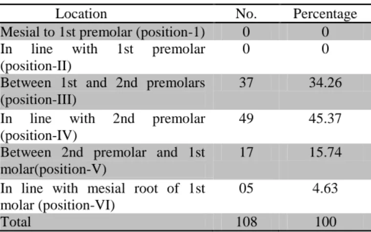
The most frequent position of the mental foramen was position IV followed by positions III, V and VI. No mental foramen was found in position I and II.
Table-3
Location No. Percentage
Mesial to 1st premolar (position-1) 0 0
In line with 1st premolar
(position-II)
0 0
Between 1st and 2nd premolars (position-III)
37 34.26
In line with 2nd premolar
(position-IV)
49 45.37
Between 2nd premolar and 1st molar(position-V)
17 15.74
In line with mesial root of 1st molar (position-VI)
05 4.63
Total 108 100
The distance from the upper cortical area of the mental foramen to the alveolar crest and the lower cortical area to the mandibular basal bone respectively was measured as shown in table-4. There was no statistically significant differences found between the right and left sides in either measurement (p = 0.071) and (p = 0.941) respectively.
Table 4: Showing distance from upper cortical area to alveolar crest (mm) and distance from lower cortical area to mandibular basal bone (mm) as per right and left mandible
Parameter
Right Mandible
Left
Mandible P-value Mean SD Mean SD
Distance from upper cortical area to
alveo-lar crest (mm) 13.54 2.11 12.85 1.80 0.071
Distance from lower cortical area to
mandibular basal bone (mm) 13.77 1.66 13.75 1.65 0.941
39.9 41.0
15 18 21 24 27 30 33 36 39 42 45
Right Mandible Left Mandible
Ag
e
(y
ea
rs)
Showing mean age (years) as per right and left mandible
Table 1: Showing mean age (years) as per right and left mandible in study patients
Age
(years) N Mean SD Range P-value Right
Mandible 54 39.9 15.93 16-68
0.739 Left
Altaf Hussain Chalkoo et al JMSCR Volume 06 Issue 09 September 2018 Page 769 The size of the mental foramen as per right and
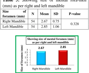
left side of the mandible was measured with a range of 0.8mm to 6.0mm. There was no significant difference observed between the two sides (p = 0.328) as shown in table-5.
Discussion
The location of the mental foramen varies in different age groups with location closer to the alveolar crest in children before the eruption of teeth and in old age due to resorption of bone, and midway between the alveolar crest and lower border of mandible in adults.2 Knowledge of the accurate mental foramen position is of utmost important for any surgical procedure like implant surgery in the region of mental foramen. Cone-beam CT can be used to evaluate accurately the location, shape, and size of the mental foramen and the presence of accessory canals.6
Oval was the most frequent shape observed in our study that is similar with other studies who take the oval and round shapes as evaluation criteria.9,10,11 However, Gupta S. et al and Córdova L. et al disagree with our results as they reported the round shape as the most frequent one.12,13
The most frequent position of the mental foramen was position-IV followed by positions-III, V and VI. This study showed that mental foramen was found in all the samples between the roots of the first premolar and of the first molar. Similar
results were observed by Igbigbi PS. et al1 and Amorin M. et al14 while asHaghanifar S. et al15 and Oliveira J. et al11 found that the most frequent location of the mental foramen was position III. These variations in the position of the mental foramen could be due to ethnic differences.
The mean distance from the upper cortical area of the mental foramen to the alveolar crest was 13.54mm (SD = 2.11) on right side and 12.85mm (SD = 1.80) on left side, and the lower cortical area to the mandibular basal bone was 13.77mm (SD = 1.66) on right side and 13.75mm (SD = 1.65) on left side respectively. Igbigbi PS. et al1 and Agarwal D. et al had found similar results in their studies. Oliveira J. et al11 and Budhiraja V. et al 6 observed measurements on dry mandibles and reported shorter distances as compared to this study.
The size of the mental foramen as per right and left side of the mandible was 2.67mm (SD = 0.75) and 2.85mm (SD = 1.06) respectively with a range of 0.8mm to 6.0mm. Ilayperuma et al17 has found mental foramen size as 2.50 mm ± 0.61 and 2.50 mm ± 0.61 on right and left respectively.
Conclusion
The localization of mental foramen is difficult as it can’t be visualized or palpated clinically, hence can be localized in relation to lower teeth. The knowledge of the position and size of the mental foramen provide valuable information to dental surgeon, thus avoiding complications from local anesthetic, surgical and otherinvasive procedures. It is concluded that the mental foramen is located on an average 13.19mm below the alveolar crest in dentate adult population. The most frequent position of the mental foramen was position-IV (along the longitudinal axis of the second premolar).
References
1. Igbigbi PS, Lesbona S. The Position and Dimensions of the Mental Foramen in Adult Malawian Mandibles. West African J Med. 2005; 24 (3): 184–189.
2.67 2.85
0 0.5 1 1.5 2 2.5 3
Right Mandible Left Mandible
S
iz
e
o
f
fo
ra
m
en
(
m
m
)
Showing size of mental foramen (mm) as per right and left mandible
Table 5: Showing size of mental fora-men (mm) as per right and left mandible
Size of
foramen (mm) N Mean SD P-value
Right Mandible 54 2.67 0.75
0.328
Altaf Hussain Chalkoo et al JMSCR Volume 06 Issue 09 September 2018 Page 770 2. Hassan T, Fauzi M, Hassan D. Bilateral
Absence of Mental Foramen: A Rare Variation. Int J Anatomic Variations 2010; 3: 187–189. [cited: 2014 Jan 8].
3. Al-Juboori MJ, Al-Wakeel HA, Yun CM and Wen FS. Location of mental foramen among Malaysia populations: Retrospective study by using orthopanto-mogram. World J Med Sci Res 1: 85-90, 2013.
4. Hwang K, Lee WJ, Song YB and Chung IH. Vulnerability of the inferior alveolar nerve and mental nerve during genioplasty: An anatomic study. J Craniofac Surg 16: 10- 14, 2005
5. Bashir A, Altaf H.Chalkoo, Rizwan H. morphometric analysis of nasopalatine canal using cone beam computed tomography:
6. Budhiraja V, Rastogi R, Lalwani R, Goel P, Chandra S. Study of Position, Shape, and Size of Mental Foramen Utilizing Various Parameters in Dry Adult Human Mandibles From North India. Int Scholarly Res Notices [Internet] 2012; 1–5. [cited: 2013 Nov 27]. Available from: http://dx.doi.org/10.5402/2013/961429 7. Sekerci A, Sahman H, Sisman Y, Aksu Y.
Morphometric analysis of the mental foramen in a Turkish population based on multi-slice computed tomography. J Oral and Maxillofacial Radio [Internet] 2013; 1: 1–7. [cited: 2014 Apr-28]. Available from: http://www.joomr.org/temp/JOralMaxillof acRadiol112- 8554038_234540.pdf
8. Tebo HG, Telford IR. An Analysis of the Variations in Position of the Mental Foramen. Anat Rec. 1950;107:61-66. 9. Hasan T. Mental foramen morphology: a
must know in clinical dentistry. J Pak Dent Assoc. [Internet] 2012; 21 (03): 167–172. [cited: 2014 Apr 21]. Available from: http://www.researchgate.net/
publication/233790049_Morphology_
of_the_mental_foramena_ must_know_ in_clinical_dentistry
10.Budhiraja V, Rastogi R, Lalwani R, Goel P, Chandra S. Study of Position, Shape, and Size of Mental Foramen Utilizing Various Parameters in Dry Adult Human Mandibles From North India. Int Scholarly Res Notices [Internet] 2012; 1–5. [cited: 2013 Nov 27]. Available from: http://dx.doi.org/10.5402/2013/961429 11.Oliveira J, Araujo A, Da Silva C, Sousa R,
Lima F. Morphological and Morphometric Study of the Mental Foramen on the M-CP.18 Jianchenjiang Point. Int J Morpho [Internet] 2009; 27 (1): 231–238. [cited: 2013 Dec 12]. Available from: http://www.scielo.cl/ scielo.php?script= sci_pdf&pid=S0717-
95022009000100039&lng=es&nrm=iso &tlng=en.
12.Gupta S, Soni JS. Study of Anatomical Variations and Incidence of Mental Foramen and Accessory Mental Foramen in Dry Human Mandibles. Natl J Med Res 2012; 2 (1): 28–30.
13.Córdova L. Características Radiográficas del Foramen Mentoniano en Pacientes del Instituto de Salud Oral de la FAP del 2000 al 2008 [thesis] Lima, Perú. Universidad Nacional Federico Villarreal: 2009.
14.Amorin M, Prado F, Borini C, Bittar T, Volpato M, Groppo F, et al. The Mental Foramen Position in Dentate and Edentulous Brazilian’s Mandible. Int J Morpho [Internet] 2008; 26 (4): 981–987. [cited: 2014 Jan 21]. Available from: http://www.scielo.cl/scielo.
php?script=sci_pdf& pid=S0717- 95022008000400033&lng=es&nrm=iso &tlng=en
Altaf Hussain Chalkoo et al JMSCR Volume 06 Issue 09 September 2018 Page 771 http://www.ijdr.in/article.asp?issn=0970-
9290;year=2009;volume=20; issue=2; spag e=150;epage=152;aulast=Haghanifar 16.Agarwal D, Gupta S. Morphometric
Analysis of Mental Foramen in Human Mandibles of South Gujarat. People’s Journal of Scientific Research 2011; 15.4(1).