ISSN(Online): 2320-9801
ISSN (Print): 2320-9798
International Journal of Innovative Research in Computer
and Communication Engineering
(A High Impact Factor, Monthly, Peer Reviewed Journal) Website: www.ijircce.com
Vol. 5, Issue 11, November 2017
A Comparative Study on Image Isolation and
Classification Techniques in Microscopic
Blood Smear Images
Renuka V Tali
1, Siddalingesh Bandi
2Assistant Professor, Department of Electronics and Communication Engineering, K S School of Engineering and
Management, Bengaluru, Karnataka, India
1Associate Professor, Department of Electronics and Communication Engineering, Global Academy of Technology,
Bengaluru, Karnataka, India
2ABSTRACT:
In relevance to the scope of Biomedical Engineering, Blood Cell Analysis plays a significant role in
assisting Medical Practitioner to diagnose the presence or level of severity of diseases. Blood tests usually carried out
by pathologists to determine number of blood cell components is very tiring, monotonous, time consuming and requires
highly experienced personnel’s. The switching of manual methods to completely automized requires image processing
and computer vision techniques. This paper reveals most of the segmentation and classification methods of White
blood cells (Leukocytes) in microscopic blood smear images. The selection of suitable segmentation technique is a
challenging task. The classification technique also depends on success of segmentation. We mainly focus on methods
used in segmenting White blood cells and its major types from its RBC’s, Platelets and background.
KEYWORDS:
Leukocytes, segmentation, classification, Machine learning, blood smear images, Medical tests
I. INTRODUCTION
When a doctor suspects any disease, firstly suggests a complete blood test or differential blood test based on
severity of symptoms. Complete blood test results in complete count of red blood cells platelets, leukocytes (white
blood cells), and erythrocytes (red blood cells). Differential blood test classifies and counts the five major types of
leukocytes. Two broad classifications of WBC’s are Granulocytes and Agranulocytes. Granulocytes are Eosinophil’s,
Basophils and Neutrophils. Agranulocytes are Lymphocytes and Monocytes. The WBC count range for a normal adult
is 4,500 to 10,000. If the count is above or below this range, it may detect the presence of some diseases. Leukopenia is
the low WBC count and may detect the presence of HIV, autoimmune disorders, liver & spleen diseases, and radio
therapy and so on. Leucocytosis is the high WBC count that may predict the presence of anaemia, allergies, pregnancy,
tissue damages, asthma, blood cancer and many more. Even during the medical treatment continuous monitoring of
WBC count has to be done through blood tests to analyse the effect of treatment on the patients. Hence blood tests are
very crucial and helpful in correctly diagnosing and treating the diseases
.
ISSN(Online): 2320-9801
ISSN (Print): 2320-9798
International Journal of Innovative Research in Computer
and Communication Engineering
(A High Impact Factor, Monthly, Peer Reviewed Journal) Website: www.ijircce.com
Vol. 5, Issue 11, November 2017
presents a review of some existing techniques available in segmentation and classification of leukocytes (WBC) in microscopic blood smear images.
A powerful alternate to automatic haematology analysers is systems based on image processing and machine learning techniques. Here images of microscopic blood smear are acquired using microscope and camera. Later, these images are analysed using different robust segmentation and classification algorithms which can give accurate count results.
A successful Segmentation highly depends on type of application and algorithm selected which in turn leads to efficient recognition and classification. Over the decades, many research scholars are continuously working on different traditional and hybrid techniques to find optimized solution. Unfortunately failed to prove the best one since no single technique is applicable to all applications. The following paragraphs discuss various segmentation and classification techniques analysed by many researchers in segmenting White blood cells and its types from background and other components of blood.
Rong Chu, et al. proposed a method to segment WBC using contour sub image Cosegmentation method. All types of white blood cells: Eosinophil, Basophil, Neutrophil, Monocyte, Lymphocyte can be segmented using this hybrid method [7]. Huey Nee Lim, et al. proposed a combination of k-means clustering for color based clustering and watershed transform based morphological segmentation for segmenting only WBC’s [8]. Khaled A. Abuhasel, et al. [9] suggested a technique to isolate WBC nucleus using modified gram Schmidt method and region growing method to segment cytoplasm of WBC. Jyoti Rawat et.al [6] reviewed on different techniques like global thresholding, color space transformation, morphological methods for segmenting and classifying all types of White blood cells: Eosinophil, Basophil, Neutrophil, Monocyte, Lymphocyte and concludes that blob analysis method proves better.
Biplab Kanti Das, et al. [11] proposed a technique to detect cytoplasm and nucleus of blood cells using thresholding, morphological analysis and contouring on 20 images of color sample blood image and reports 85% of accuracy. Chaitali Raje, et al. [10] proposed a method of segmenting nucleus in WBC using Otsu’s thresholding and statistical parameters like Standard Deviation and Mean. Mostafa Mohamed et al. [13] tested on 365 blood images using Gray scale contrast enhancement and filtering method for automatic WBC nuclei segmentation and reports 79.7% of accuracy but did not overcome the problem of non-homogeneous lighting and over staining [15].
R. Adollah et al. [5] Segmented leukocyte from background using multilevel thresholding technique but this method may not be applicable for dark or bright images. N H Abd Halim et al. [17] worked on image quality and Extraction of nucleus from WBC using a global contrast stretching and HSI color models. Jun Duan et al. [18] proposed a color image segmentation technique Integrating color histogram and region growing and merging-based for segmenting nucleus and cytoplasm from WBC and states that results vary if there is cell adhesion. Subrajeet Mohapatra et al. [2][4] also worked on Leukocyte color based segmentation using a rough set based clustering approach for Nucleus and cytoplasm of WBC extraction. P Sukumar et al. [19] proposed a technique to identify abnormal WBC nucleus in cervical cancer cells using Fast particle swarm optimization with ELM method (extreme learning machine, standard particle swarm optimization) also calculates Execution time of the techniques. F Boray Tek et al. [20] worked on segmentation of various components of blood by computing Cell area using area granulometry. Minimum area watershed transform, circle radon transform are used for segmentation. Transform used is not completely capable of detecting under or over segmentation. Pramit Ghosh, et.al. Makes use of HSI color model, shape and color characteristics, Fuzzy logic functions to identify the 5 types of leukocytes. It finds that nucleus of monocyte is oval and that of lymphocyte is circular. Considering height and width of cell nucleus, identification of monocyte and lymphocytes can be done. Average color and saturation helps in detecting neutrophils. Fuzzy logic assigns two colors red and blue which is used to detect eosinophil and basophil [3].
Vanika Singhal, et.al. Proposes a method to detect abnormal lymphocyte from a given blood image. Local Binary Pattern, a classifying texture feature is used to identify blast cell. It encounters various features of shape like perimeter, area, convex area, solidity, eccentricity, major axis length, compactness and orientation to segment nucleus from cytoplasm. Data set are trained using Support vector machine. System reports 89.72% of accuracy [12].
Sheng-Fuu Lin, et.al. Focusses on un-uniform stain caused due to methods involved in preparation of blood smear and dying agents. It presents a feature of nuclei lobes to differentiate nucleus granulocyte from agranulocytes. To segment the nucleus, Region growth algorithm is used. Redescription algorithm is used to identify bi or multilobed nucleus. Shape features used to classify cells are relative area, variance of boundary curvature and distance of center to boundary, roundness, solidity and number of lobes. Texture features used are energy, contract, correlation, entropy andhomogeneity. Multiple SVM is used to train the images. Approximately 85% of accuracy in detecting all types of WBC’s is reported [14].
S. H. Rezatofighi S. H, et.al. Proposed a method to isolate WBC nuclei using Gram Schmidt Orthogonalization. This technique is used to strengthen desired vectors of colors and weaken the undesired vectors of colors. Three vectors are selected due to large variation in colors. Reports 93% of accuracy. It imposes restrictions in application on new data in which it requires calculation of new vectors. Suggests that methods not based on color features are complex and require more time [21]. A.S.Abdul Nasir, et.al. Presents a combined methods of linear contrast technique used to enhance the quality of image and unsupervised k-means clustering to obtain fully segmented abnormal leukaemia images. By application of median filtering and region growing segmented nucleus is obtained. This work reveals that most of the WBC information is hidden in H component and structure of nucleus can be analysed better using S component of HSI Model [22].
ISSN(Online): 2320-9801
ISSN (Print): 2320-9798
International Journal of Innovative Research in Computer
and Communication Engineering
(A High Impact Factor, Monthly, Peer Reviewed Journal) Website: www.ijircce.com
Vol. 5, Issue 11, November 2017
watershed transform. The work is carried out in three steps like identifying WBC’s and blast cells as objects of interest
and deleting the background, applying Otsu’s global thresholding and finally applying watershed algorithm to segment
objects of interest. Author reports drawback of technique as localization of nucleus and cytoplasm are incomplete [23].
Hemant Tulsani, et.al. Presents a method to count all blood cell components. Watershed transform and
regional maxima computations are used to segment the cells that solves the problem of overlapping cells. Work begins
with smoothening of obtained image and converting to YCbCr color model and binary images. Later different masks
are applied on WBC and platelets using morphological and thresholding to compute regional maximal points. Finally
performs watershed algorithm on masks to count number of cells. Author suggests that marker based techniques solves
the problem of over segmentation associated with watershed transform [24].
Table 1 shows comparison of various segmentation and classification techniques with respect to classifiers used,
accuracy and no. of images used.
II. PROPOSED BLOCK DIAGRAM
The figure 1 below shows basic block diagram of Machine learning based classification of WBC’s system
which consists of a high magnification microscope to view cells on stained glass slides of blood droplets. Later a digital
camera if mounted to microscope can be capable of capturing digital images. Using various image processing
segmentation techniques, individual cells can be extracted. Classification of these cells into WBC types is carried out
by applying machine learning models.
Figure 1: Block Diagram [25]
III. PERFORMANCE PARAMETERS
Medical treatment prescribed by doctors mainly rely on medical test reports. The accuracy of these tests help
doctors in predicting presence or absence of diseases. Fortunately tests parameters can be measured using sensitivity,
specificity and accuracy. These parameters evaluate how reliably better a test is. Sensitivity measures performance of
the test in detecting the presence of a sickness. Specificity decides how best the test is in detecting the absence of a
disease. Accuracy determines overall performance of system in correctly detecting presence or absence of a disease.
There are other parameters also that assist in evaluating specificity, sensitivity and accuracy. They are True
Positive (TP), True Negative (TN), False Positive (FP), and False Negative (FN). If there is a presence of disease, and
test also proves the presence of sickness, then test result is said to be TP. Similarly if there is absence of disease, and
test also proves the absence of disease, then test result is said to be TN. If medical test indicates presence of disease, but
actually patient is healthy, then result of test is said to be FP. If medical test indicates absence of disease, but actually
patient suffers from such disease then test result is said to be FN. Sensitivity measures true positives that are identified
correctly by a medical test. Specificity measures true negatives that are detected correctly by medical tests. Accuracy is
a measure of both true positives and true negatives.
1. True positive (TP) is no of WBC’s (or components of WBC) correctly classified as WBC’s.
2. True negative (TN) is the number of other cells correctly classified as other cells.
ISSN(Online): 2320-9801
ISSN (Print): 2320-9798
International Journal of Innovative Research in Computer
and Communication Engineering
(A High Impact Factor, Monthly, Peer Reviewed Journal) Website: www.ijircce.com
Vol. 5, Issue 11, November 2017
Author, Year Work Classifiers Accuracy Remarks No of sample
Images Dipti Patra, et
al. 2010 [2]
A fuzzy clustering based two stage color
segmentation strategy is employed
Discriminative features: nucleus shape, texture are used for detection. Shape features: Hausdorph Dimension & contour signature. Support vector machine (SVM) for classification
95% Advantage over existing schemes is images considered are smear images with many lymphocytes 108 blood smear images Pramit Ghosh, et al. 2011 [3]
Work is to detect different WBC’s
Image enhancement is done using Laplacian filter. Thresholding is used to segment, HIS color model and fuzzy logic functions are used
97.33% Specifically not mentioned techniques only algorithms are explained. 150samples of blood image are taken. Subrajeet Mohapatra, et al. 2011 [4] Work on Leukocyte color based segmentation
A rough set based clustering approach Not mentioned Improved rough k-means clustering is highlighted 100 images R.Adollah, M Y Mashor, 2011. [5] Work on segmentation of microscopic bone marrow images
Multilevel Thresholding Not mentioned
Works well on normal images but not on too dark or bright images 2 images Normal bone marrow and ALL Jyoti Rawat, et al. 2015 [6] Review on different techniques by different researchers
Global Thresholding, color space transformation, Morphological methods.
Approximatel y 80%
States that blob analysis method proves better
Not mentioned
Rong Chu, et al.
2015 [7]
A new method to obtain entire WBC contour
Sub image Cosegmentation method based on region-based active contour model.
92.3% Successfully solves Adhesion problem
103 blood cell images
Huey Nee Lim, et al. 2015 [8]
A combination of Color and Morphological based techniques
k-means clustering for Color based clustering and Watershed transform based morphological
segmentation
Claims to be 100% accurate but no record of it. Overcomes the problem of over segmentation Not mentioned Khaled A. Abuhasel, et al. 2015 [9] Method to segment Nucleus and Cytoplasm of White Blood Cells. Modified Gram-Schmidt method for segmenting nucleus of WBC and Region growing method to segment cytoplasm of WBC. 95.21% for Lymphocyte cell type. 92.31% for Neutrophil cell type: with comparison to manual segmenting. Not mentioned how feature extraction and classification is carried out.
100 samples of microscopic WBC images Chaitali Raje, et al. 2014 [10] Method of Segmenting nucleus in WBC
Statistical parameters like Mean and Standard Deviation and Otsu’s thresholding based segmentation. Good performance Fully segmented nucleus is not obtained 128 microscopic blood slide images Biplab Kanti Das, et al
To detect nucleus and
Thresholding,
morphological analysis and
85% of accuracy
Not clearly mentioned
ISSN(Online): 2320-9801
ISSN (Print): 2320-9798
International Journal of Innovative Research in Computer
and Communication Engineering
(A High Impact Factor, Monthly, Peer Reviewed Journal) Website: www.ijircce.com
Vol. 5, Issue 11, November 2017
2014 [11]
cytoplasm of blood cells
contouring about
techniques used Vanika
Singhal, et al. 2014 [12] Methodology for automatic detection of Acute Lymphoblastic Leukemia
Segmentation is done using HIS color model,
thresholding and morphological operation like dilation is used.
89.72% of accuracy Methods for segmentation, feature extraction and classification are clearly mentioned
75 blast cell images and 65 normal cell images. Mostafa Mohamed, et al. 2012 [13] Method for automatic WBC nuclei segmentation
Gray scale contrast enhancement and filtering
79.7% Major problem is non-homogeneous lighting and over staining. 365 blood images Sheng-Fuu Lin, et al. 2011 [14]
A method to ease the influence of non-uniform stain and extract the nucleus of WBC
Region growing algorithm for segmentation, shape feature is extracted using distance transformation and mean-shift to analyze no of lobes of nucleus,
classification is done using support vector machine
Min of 76.47%
An attempt to solve problem of non-uniform stain is considered by modification of RGB channels. 400 images Behrouz Far, et al. 2012 [15] Technique for automatic blood cell nuclei segmentation Gram-Schmidt Orthogonalization technique for segmentation and morphological operations used to enhance the segmentation.
85.4% Non-homogeneous lighting and over staining is the main reason of segmentation inaccuracy 367 blood images Cecilia Di Ruberto, et.al. 2016 [16] Technique to segment and count leukocytes using learning by sampling
Nearest Neighbor and SVM for Segmentation. Counting by Hough Transform
99.2% Considered only normal cells. Future work may segment blast cells. 367 blood smear images of size 640X 480
N H Abd Halim, et al. 2011 [17]
To improve image quality
A global contrast stretching and segmentation WBC based on HIS color space
Not mentioned Details about image acquisition is missing Not mentioned
Jun Duan, et al. 2011 [18] Color image segmentation technique
Integrate color histogram based method and region growing and merging-based segmentation method
Not mentioned
Segmentation results vary if there is cell adhesion.
Not mentioned
P Sukumar, et al. 2015 [19] To detect abnormal WBC nucleus in cervical cancer cells
Fast particle swarm optimization with ELM method (extreme learning machine, standard particle swarm optimization)
90% Execution time of the
techniques are mentioned.
50 images
F Boray Tek, et al. 2005 (Springer) [20] To segment various components of blood.
Cell area is computed using area granulometry. Minimum area watershed transform, circle radon transform.
ISSN(Online): 2320-9801
ISSN (Print): 2320-9798
International Journal of Innovative Research in Computer
and Communication Engineering
(A High Impact Factor, Monthly, Peer Reviewed Journal) Website: www.ijircce.com
Vol. 5, Issue 11, November 2017
Table 1: Comparison of various segmentation and classification techniques
Range of sensitivity lies between 0 and 1. As the value approaches 1, system is highly sensitive in identifying correct
WBC.
1
Sensitivity is the probability of correctly identifying a Leukocyte (WBC) cell.
=
( + )
2
Specificity determines the probability of correctly classifying other cell
.=
( + )
Range of Specificity lies between 0 and 1. As the value approaches 1, system is highly specific in detecting other cells.
3
Misclassification is the total number of cells classified incorrectly and is given by
Misclassification= FP+FN
The range of misclassification lies between 0 and 1. 0 indicates absence of wrong classification i.e. all WBC’s and
other cells are classified respectively. 1 indicates systems incapability in identifying correctly WBC’s and others.
4
Accuracy
= ( )( )
segmentation.
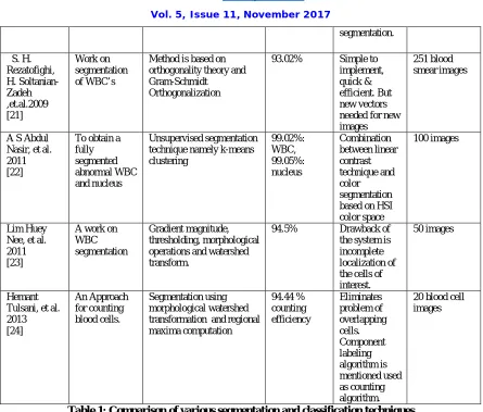
S. H. Rezatofighi, H. Soltanian-Zadeh ,et.al.2009 [21]
Work on segmentation of WBC’s
Method is based on orthogonality theory and Gram-Schmidt
Orthogonalization
93.02% Simple to implement, quick & efficient. But new vectors needed for new images
251 blood smear images
A S Abdul Nasir, et al. 2011 [22]
To obtain a fully segmented abnormal WBC and nucleus
Unsupervised segmentation technique namely k-means clustering
99.02%: WBC, 99.05%: nucleus
Combination between linear contrast technique and color segmentation based on HSI color space
100 images
Lim Huey Nee, et al. 2011 [23]
A work on WBC segmentation
Gradient magnitude, thresholding, morphological operations and watershed transform.
94.5% Drawback of the system is incomplete localization of the cells of interest.
50 images
Hemant Tulsani, et al. 2013 [24]
An Approach for counting blood cells.
Segmentation using morphological watershed transformation and regional maxima computation
94.44 % counting efficiency
Eliminates problem of overlapping cells. Component labeling algorithm is mentioned used as counting algorithm.
ISSN(Online): 2320-9801
ISSN (Print): 2320-9798
International Journal of Innovative Research in Computer
and Communication Engineering
(A High Impact Factor, Monthly, Peer Reviewed Journal) Website: www.ijircce.com
Vol. 5, Issue 11, November 2017
Accuracy is systems performance measurement parameter. Its range lies between 0 and 1. 1 indicates 100% accurate
system.
Execution Time is the time taken by the system from segmenting, correctly classifying and counting a normal
or abnormal WBC’s. Execution time should be as minimum as possible. Usually in terms of milli seconds to micro
seconds or even less.
The system sensitivity and specificity should approach 100%. Misclassification should approach 0% claiming highly
accurate system with value approaching 100%. Execution time should be in terms of micro seconds.
IV. CONCLUSION
This work reveals most of segmentation and classification methods used in isolating and counting WBC’s in
microscopic blood smear images. Main causes of inaccurate or failure in segmentation step are contract of nucleus,
overexposed images, clustering of leukocytes, similar leukocyte erythrocyte color, Some neutrophils not resembling
typical multilobed nucleus and some image articrafts.. Before selecting any method thorough study of structure, area,
development stages, staining effects, illumination conditions, and contextual information of cells is utmost importance.
Most important is to select moderately illuminated and uniformly stained smear slide images. For improved
classification accuracy, typical images should be considered in the training set of samples. Contextual information,
marker based, color features based segmentation techniques blend completely into such application.
Acknowledgment
We extend our gratitude to our esteemed organizations KSSEM and GAT for encouraging and supporting our research
throughout the period which led to the successful bring out of this work. We also thank our family members for their
kind cooperation.
REFERENCES
1. Chair and Department of laboratory diagnostics, Medical University of Lublin, “The automated hematology analyzers”, Annales Universitatis Mariae Curle, vol XXIV, N 3,7 Section DDD 2011
2. Subrajeet Mohapatra, Dipti Patra, et.al. “Image Analysis of blood microscopic images for acute leukemia detection”, International Conference on Industrial Electronics, Control and Robotics, IEEE, 2010.
3. Pramit Ghosh, et.al. “Automatic white blood cell measuring aid for medical diagnosis”, IEEE, 2011.
4. Subrajeet Mohapatra, Dipti Patra, Kundan Kumar , “Blood Microscopic Image Segmentation using Rough Sets”, IEEE international Conference on Image Information Processing, 2011.
5. R.Adollah, M Y Mashor, “Bone Marrow Image Segmentation Based on Multilevel Thresholding”, IEEE International Conference on Biomedical Engineering, pp 457-461, February 2011.
6. Jyoti Rawat, H S Bhadauria, et.al, “Review of Leukocyte Classification Techniques for Microscopic Blood Images”, 2nd IEEE International Conference on Computing for Sustainable Global Development, pp 1948-54, 2015.
7. Rong Chu, Xiaoqin Zeng, et.al, “Subimage Cosegmentation in a Single White Blood Cell Image”, 7th IEEE International Conference on Computational Systems and Networks (CICSyN), pp 152-157, 2015.
8. Huey Nee Lim, et al, “Color and Morphological Based Techniques on White Blood Cells Segmentation”, 2nd IEEE International Conference on Biomedical Engineering, 2015.
9. Khaled A Abuhasel, Chastine Fatichah, et.al, “A Commixed Modified Gram-Schmidt and Region Growing Mechanism for White Blood Cell Image Segmentation”, IEEE, 2015.
10. Chaitali Raje, Jyoti Rangole, “Detection of Leukemia in Microscopic Images Using Image Processing”, IEEE International Conference on Communication and Signal Processing, pp 255-259, April 2014.
11. Biplab Kanti Das, Krishna Kumar Jha, “A New Approach for Segmentation and Identification of Disease Affected Blood Cells”, IEEE International Conference on Intelligent Computing Applications, pp 208-212, 2014.
12. Vanika Singhal, Preety Singh, “Local Binary Pattern for Automatic Detection of Acute Lymphoblastic Leukemia”, IEEE, 2014.
13. Mostafa Mohammed, Behrouz Far, “An Efficient Technique for White Blood Cells Nuclei Automatic Segmentation”, IEEE International Conference on Systems, Man and Cybernetics, pp 220-225, 2012.
14. Sheng-Fuu Lin, Yu-Bi Hong, “Differential Count of White Blood Cell in Noisy Normal Blood Smear”, 7th IEEE International Conference on Industrial Electronics and Applications, pp 1784-1789, 2011 .
15. Mostafa Mohamed, Behrouz Far, “A Fast Technique for White Blood Cells Nuclei Automatic Segmentation Based on Gram-Schmidt Orthogonalization”, 24th IEEE International Conference on Tools with Artificial Intelligence, pp 947-952, 2012.
ISSN(Online): 2320-9801
ISSN (Print): 2320-9798
International Journal of Innovative Research in Computer
and Communication Engineering
(A High Impact Factor, Monthly, Peer Reviewed Journal) Website: www.ijircce.com
Vol. 5, Issue 11, November 2017
17. N H Abd Halim, M Y Mashor, “Nucleus Segmentation Technique for Acute Leukemia”, 7th IEEE International Colloquium on Signal processing and its Applications, pp 192-197, 2011.
18. Jun Duan, Le Yu, “A WBC Segmentation Method Based on HIS Color Space”, Proceedings of IEEE IC-BNMT, pp 629-632, 2011. 19. P Sukumar, R K Gnanamurthy, “Segmentation and Abnormality Detection of Cervical Cancer Cells using Fast ELM with Particle Swarm
Optimization”, GENETIKA, Vol 47, No 3, 863-876, 2015.
20. F Boray Tek, Andrew G Dempster, “Blood Cell Segmentation using Minimum Area Watershed and Circle Radon Transformations”, Springer Mathematical Morphology: 40 years On, 441-454,2005.
21. S. H. Rezatofighi, H. Soltanian-Zadeh, R. Sharifian and R. Zoroofi, "A New Approach to White Blood Cell Nucleus Segmentation Based on Gram-Schmidt Orthogonalization," Proceeding of IEEE International Conference on Digital Image Processing (ICDIP), Iran, 2009. 22. A.S.Abdul Nasir, M.Y.Mashor, H.Rosline, “Unsupervised Colour Segmentation of White Blood Cell for Acute Leukaemia Images”,
IEEE, 978-1-61284-896-9/11/$26.00 ©2011.
23. Lim Huey Nee, Mohd Yusoff Mashor, Rosline Hassan, “White Blood Cell Segmentation for Acute Leukemia Bone Marrow Images”, 2012 International Conference on Biomedical Engineering (ICoBE),Penang,Malaysia,27-28 February 2012.
24. Hemant Tulsani, Rashmi Gupta, Rajiv Kapoor, “An Improved Methodology for Blood Cell Counting”, IMPACT-2013, IEEE, 978-1-4799-1205-6/13/$31.00 ©2013.
25. http://cdn.shopclues.net/images/detailed/5397/100x1500xEducationMicroscope_1403604000.jpg. http://www.microscopesblog.com/2012/08/differential-white-blood-cell-count.html,
![Figure 1: Block Diagram [25]](https://thumb-us.123doks.com/thumbv2/123dok_us/1388578.1171559/3.595.74.536.410.510/figure-block-diagram.webp)