Review Paper
The Structure-to-Function Relationships of
Gammaherpesvirus-Encoded Long Non-Coding RNAs and their
Contributions to Viral Pathogenesis
Gabriela Chavez-Calvillo†, Sarah Martin†, Chad Hamm, Joanna Sztuba-Solinska*
*Auburn University, Department of Biological Sciences, 120 W. Samford Ave, Rouse Life Sciences
Building, Auburn, AL, 36849; [email protected], Tel.: 334-844-1650
† These joint-first authors contributed equally to this review.
* Author to whom correspondence should be addressed.
Abstract:
Advances in next-generation sequencing have facilitated the discovery of a multitude of long
non-coding RNAs (lncRNAs) with pleiotropic functions in cellular processes, disease and viral
pathogenesis. It came as no surprise when viruses were also revealed to transcribe their own
lncRNAs. Among them, gammaherpesviruses, one of the three subfamilies of the Herpesviridae, code their largest number. These structurally and functionally intricate non-coding (nc) transcripts
modulate cellular and viral gene expression to maintain viral latency or prompt lytic reactivation.
The lncRNAs allow the virus to escape cytosolic surveillance, sequester and re-localize essential
cellular factors and modulate the cell cycle and proliferation. Some viral lncRNAs act as
‚messenger molecules‛, transferring information about viral infection to neighboring cells. This
broad range of lncRNA functions is achieved through lncRNA structure-mediated interactions
with effector molecules of viral and host origin, including other RNAs, proteins and DNAs. In this
review, we discuss examples of gammaherpesvirus-encoded lncRNAs, emphasize their unique
structural attributes, and link them to viral life cycle, pathogenesis and disease progression. We
will address their potential as novel targets for drug discovery and propose future directions to
explore lncRNA structure and function relationship.
Keywords: gammaherpesviruses, long non-coding RNAs, RNA structure and function, viral
pathogenesis
1. Introduction: defining long non-coding RNAs
Only about 1.2% of the human genome encodes protein-coding genes, however a large
majority is transcribed into non-coding RNAs (ncRNAs); products that seem to lack protein-coding
capacity and are functional upon transcription [1], [2]. This diverse group can be arbitrarily divided
into: (i) small ncRNAs (sncRNA), transcripts shorter than 200 nucleotides (nts), which include
microRNAs (miRNAs), small nucleolar RNAs (snoRNAs), piwiRNAs (piRNAs) and many others,
and (ii) long ncRNAs (lncRNAs), transcripts longer than 200 nts [3]. This classification system is
based solely on RNA length, and as such does not reflect biological properties, biogenesis, stability,
abundance, and/or mechanism of action.
The majority of lncRNAs are generated by RNA polymerase II, have a 5′ terminal
methylguanosine cap and are often spliced and polyadenylated [4]. Alternative pathways contribute
to the generation of non-polyadenylated lncRNAs, likely expressed from RNA polymerase III
promoters [5], and lncRNAs that are excised during splicing and small nucleolar RNA production
[6]. No specific biochemical features can be exclusively ascribed to lncRNAs, but rather the lack of a
defined open reading frame (ORF) suggests that many transcripts function intrinsically as lncRNAs
[7]. Exceptions to these conventions include lncRNAs that have been shown to associate with
polysomes and encode short or non-canonical peptides [8], [9], and bifunctional mRNAs that are
also lncRNAs [10].
LncRNAs have been identified as major players involved in the regulation of almost every
stage of gene expression, the cell cycle, pluripotency and modulation of host-pathogen interactions
[3], [11]–[15]. Despite wide distribution in genomes of complex organisms, only a small fraction of
lncRNAs have been functionally and structurally characterized, and even less is known about
virus-encoded lncRNAs [16], [17]. In this review, we will highlight the most prominent examples of
gammaherpesvirus-encoded lncRNAs, emphasize their multifunctionality in the viral life cycle and
pathogenesis, and finally, propose a path for prospective studies.
2. The role of lncRNAs in gammaherpesviruses
Gammaherpesviruses form one of the three subfamilies of the Herpesviridae. They are characterized by their cellular tropism for lymphocytes and are distinct from alpha- and
betaherpesviruses in molecular phylogenetic analyses [18]. Similar to host cells, herpesviruses
produce lncRNAs and intriguingly, gammaherpesviruses genomes encode the greatest number
(Table 1). This viral lncRNA production allows precise regulation of an unusual life cycle [13], [14],
few viral genes (latent genes) and no production of infectious virions, and (ii) lytic, during which the
virus expresses most of its genes, viral DNA is amplified, and progeny virions are assembled and
released from the cells [21], [22].
The gammaherpesviruses establish latency as a strategy for avoiding host immune surveillance
and fusing symbiotically with the host for persistent lifetime infection. However, the transition to
the lytic phase of infection is critical for viral dissemination within and between hosts. Timing of
both phases must be finely tuned, and that daunting task can be achieved only by molecules that can
either slip under the radar of host immune response and/or modulate cellular immune response. In
most cases, lncRNAs appear to be more immune inert, perhaps due to a complex structure that
sequesters cellular factors, preventing their detection by host surveillance system. In support of that
notion, the EBV-encoded RNA1 (EBER1) interaction with the lupus antigen (La) has been shown to
protect the 5’pppEBER1 from being recognized by cytoplasmic RNA sensors [23]. In addition,
tampering with interferon signaling and cellular response genes establishes lncRNAs as essential
modules of escape strategies used by viruses to avoid antiviral pathways. For example, Kaposi’s
sarcoma-associated herpesvirus (KSHV) polyadenylated nuclear (PAN) RNA expression has been
shown to interfere with the ability of transcription factors to activate the interleukin-4 (IL-4)
promoter, and to knockdown the expression of RNase L, an essential interferon effector [24]. Further
instances of extensive immunomodulatory lncRNA functions will be discussed in more detail
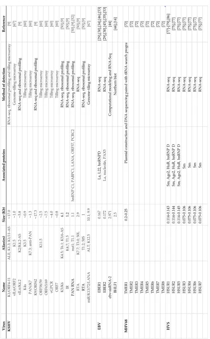
Table 1. LncRNAs in gammaherpesvirus. The table includes: aliases ascribed to the aforementioned lncRNAs, the molecular size expressed in kilobases, proteins associated with lncRNAs, the detection method and the original references.
3. Kaposi's sarcoma herpesvirus (KSHV)-encoded lncRNAs
Kaposi's sarcoma herpesvirus (KSHV) is the etiologic agent of Kaposi’s sarcoma, as well as
certain B-cell lymphomas, including primary effusion lymphoma (PEL) and multicentric
Castleman’s disease (MCD) [47]–[49].KSHV encodes several lncRNAs [9], [50]–[53] (Table 1), and
PAN RNA is the most abundantly produced transcript during lytic reactivation [54]. PAN RNA has
been shown to fold into three branched domains, each of which contains well-defined motifs
connected by less structurally constrained regions (Figure 1) [55]. Domain I includes 9-nt element
termed the Mta-responsive element (MRE) that binds mRNA transcript accumulation protein (Mta,
also ORF57), which modulates PAN RNA stability and function [56]. Domain IIIoccludes a 79-nt
long nuclear retention element (ENE) that sequesters the poly(A) tail of PAN RNA by formation of a
triple helix [57]. This structural motif contributes to intracellular stability and allows PAN RNA to
‚escape‛ decay mechanisms. Domain II is characterized by a flexible conformation, likely to
accommodate long-range tertiary interactions (e.g., formation of the ENE triple helix), support more
compact folding in adjacent regions and provide an accessible ‚landing pad‛ for protein interaction
[55].
PAN RNA has been identified as a key player involved in regulation of almost every stage of
viral gene expression, cell cycle, pluripotency, modulation of host-pathogens interactions and
production of infectious virus [58]–[60]. It localizes mainly to the nucleus, yet, deep-sequencing
studies also indicate its presence in the cytoplasm and in latently infected cells [61]. Arias and
colleagues have suggested that the presence of PAN RNA in the cytoplasm might be explained by a
potential protein coding capacity. They observed initiating ribosomes at the PAN start codon,
elongating ribosomes throughout the body of the transcript, and accumulation of releasing
ribosomes at the stop codon[9].
PAN RNA knockdown experiments demonstrated compromised viral lytic gene expression
and virion production [19], [58], [63], likely due to essential epigenetic regulatory roles. PAN
actively participates in chromatin remodeling by recruiting the protein components of polycomb
repressive complex 2 (PRC2) (Figure 1A), as well as the histone methyltransferase and the
demethylases (Figure 1B) [58], [61], [64], [65]. Using chromatin isolation by RNA purification
(ChIRP-Seq), Rossetto et al. (2013) demonstrated the great extent to which PAN RNA manipulates
viral and host gene expression programs. Eighty-four cellular gene promoters involved in regulation
of the inflammatory and antiviral responses (IFNγ, IL-18, IFNA16, and RNase L), cell death
(TRIM68, RAD52, INPP5E, EPHB2, PAX2) and development (HIST1H4A, HIST3H3, PAX6, PAX5,
CDKN2B), and thirty-five viral gene promoters involved in direct regulation of KSHV lytic gene
expression (i.e. PAN, orLyt-L, K14, ORF4, ORF64, ORF50, ORF74), were shown to be directly
protein C1 (PABPC1), which relocalizes to the nucleus during the lytic phase of KSHV infection [56],
[63]. This relocalization is directly caused by the shutoff exonuclease (SOX) protein, which
downregulates expression of host mRNAs and upregulates levels of PAN RNA [63]. Therefore, PAN
RNA acts downstream of SOX, further contributing to viral manipulation of gene expression.
In addition, multiple viral proteins have been shown to associate with PAN RNA (Figure 1C).
The interaction with ORF26 likely facilitates PAN packaging into virions [54], [55], while the ORF59
likely facilitates the recruitment of PAN RNA to the viral episome [24], [66]. PAN RNA has been also
shown to regulate the function of the latency-associated nuclear antigen (LANA) protein (Figure
1D) [12]. During latency, LANA wraps around the KSHV episome and silences the expression of
lytic genes. Lytic reactivation is marked by an abundance of PAN RNA, which sequesters LANA
away from the episome, thereby relieving the repressive activity and facilitating the expression of
lytic genes.
Other KSHV-encoded lncRNAs, including T1.2, T3.0, T6.1 and antisense-to-latency transcript
(ALT) have been discovered, but only a few have been proven to be functional [51], [52]. T1.5 is
expressed from a region near one of the two origins of lytic replication (ori-Lyt). It is produced
during the early stages of infection and is required for viral replication. T1.5 accumulates in the
cytoplasm and is packaged into virions. T3.0 and T6.1 have the same transcription start site (TSS)
and are antisense to the replication and transcription activator (RTA/ORF50), but do not inhibit RTA
function. Although these three lncRNAs do not have canonical ORFs, all have been reported to be
ribosome-associated, similar to PAN RNA [9]. ALT is a 10 Kb polyadenylated early lytic transcript
expressed antisense to the major viral latency transcripts encoding LANA and the viral microRNAs
[50]. In addition, ALT is on the same strand and is co-terminal with a bicistronic lytic transcript
containing ORF K14 (v-OX2, a homolog of cellular surface receptor OX2) and ORF74 (vGPCR, viral
G protein-coupled receptor) [67]. It has been suggested that the 3’ UTR common to ALT and the
K14-ORF74 mRNA is likely regulated by microRNAs. This overlapping arrangement of viral
transcripts represents a strategy by which KSHV maximizes its coding capacity and level of gene
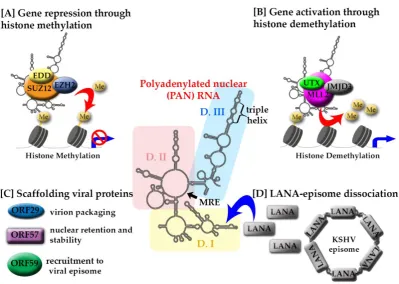
Figure 1: Structure-mediated multifunctionality of PAN RNA. The secondary structure of KSHV PAN RNA is represented in the middle with color-coded domains: I (yellow), II (pink)
and III (light blue). The position of two cis-acting motifs involved in PAN RNA stability and functionality, MRE and triple helix, are indicated. [A] PAN RNA interaction with PRC2
components: EZH2 (blue), SUZ12 (orange), EDD (yellow), leads to histone methylation and gene repression.[B] The interaction of PAN RNA with UTX (lime green)/MLL2 (purple)/JMJD3 (light grey) targets histones for demethylation to increase gene expression. [C] PAN RNA
interacts with viral proteins ORF29 (blue), ORF57 (purple), ORF59 (green) and LANA (grey). [D] The interaction of PAN RNA with LANA is partially responsible for the LANA-episome
dissociation leading to KSHV lytic reactivation.
4. Epstein-Barr virus (EBV)-encoded lncRNAs
EBV, also known as Human Herpesvirus-4 (HHV4), is the causative agent of infectious
mononucleosis [25] and it can lead to the development of Hodgkin’s lymphoma [26], endemic
Burkitt’s lymphoma [25] and nasopharyngeal cancer [27]. This lymphotropic gammaherpesvirus
undergoes five distinct phases of latency, and each can be characterized by the production of specific
viral products, including lncRNAs. For example, EBV-encoded RNA1 and 2 (EBER1 and EBER2) are
transcribed during all phases of latency, but EBV stable intronic-sequence RNA-2 (ebv-sisRNA-2) is
detected only during latency III, while BHLF1 is expressed during latency I and III [28]–[30].
The EBERs are the most abundantly expressed nuclear ncRNAs in EBV-infected cells. More
than 30 years after their discovery [29], they remain a functional puzzle to the study of EBV latency,
of viral latency establishment or tumorigenic potential [31]. When it comes to their classification,
they fall in an arbitrary gap, as both EBERs are shorter than a typical lncRNA at ~180 nts each (Table
1). Yet, their biogenesis is distinct from that of miRNAs, prompting us to include them in the context
of this review. EBERs are synthesized by RNA polymerase III [29] at approximately equal rates
(Table 1), but they differ in half-lives, EBER1 at 8–9 h and EBER2 at 0.75 h [32]. While there is a low
sequence homology between the EBERs (~54%), interestingly they share a highly conserved
secondary structure [33].
The composition and functionality of all EBER ribonucleoprotein (RNP) complexes essential for
EBV pathogenesis are as of yet undefined. It is known, however, that EBER1 folds into four
stem-loops [33], [34], and that stem-loops I, III, and IV each bind an L22 ribosomal protein (Figure
2A). L22 is normally present in the nucleoli and cytoplasm during latency, however, following EBV
lytic activation, the complex relocates to the nucleoplasm [35], [36]. Additional factors regulate the
EBER1:L22 complex, including binding of the interferon-inducible protein kinase R (PKR) and the
association of La antigen with the EBER 3’ polyuridylate stretch [35]. The latter interaction has been
proposed to dampen the recognition of a small fraction of cytoplasmic EBER1 by RNA sensors, and
facilitate sorting of 5’pppEBER1 into exosomes in order to relay information about viral infection
into neighboring cells [23]. Other known EBER1-mediated interactions reported include AU-rich
element binding factor (AUF1)/ heterogeneous nuclear ribonucleoprotein D (hnRNP D), a protein
involved in destabilization of mRNA upon binding to AU-rich elements (AREs) [37]. The interplay
between AUF1 and EBER1 potentially disrupts normal mRNA homeostasis and contributes to
EBV-associated oncogenesis [38].
EBER2 has been shown to localize to the terminal repeats (TRs) of the latent EBV genome, likely
to regulate EBV lytic reactivation (Figure 2B). Binding of the EBER2 stem-loop (nts 32 – 53) by both
an RNA transcript expressed from TR locus and the B-cell transcription factor paired box protein 5
(PAX5) facilitates this localization. In addition, intermediary host proteins, including splicing factor
proline and glutamine rich (SFPQ), non-POU domain-containing octamer-binding protein (NONO),
and RNA binding motif protein 14 (RBM14), have been reported to mediated the PAX5-EBER2
interaction [39]. EBER2 and PAX5 have been proposed to act in concert, clearing adjacent chromatin
of interfering factors to induce lytic gene expression. Disruption of the PAX5-EBER2 complex
impacts genome packaging and depletion of either PAX5 or EBER2 has been shown to decrease EBV
lytic reactivation [36].
EBERs structural and functional similarity to the adenoviral ncRNAs VAI and VAII, suggests
that like VAI and VAII, the EBERs could interact with PKR. It has been confirmed in vitro that PKR
dimerization, a requirement for activation, is inhibited in the presence of either EBERs or VAI/II [40]
activation within cells in response to interferon, challenge that hypothesis demonstrating that the
EBERs are unable to inhibit phosphorylation of either cytoplasmic or nuclear PKR [41]. EBERs also
regulate a variety of host cell genes, including those active in deamination, cell adhesion, apoptosis,
and receptor signaling [42]. They have been shown to elicit transcription of cytokines, such as IL-9,
IL-10 and IGF1 (insulin-like growth factor-1), that act as autocrine growth factors within
EBV-infected cancer cells.
BHLF1, another EBV-encoded lncRNA, is 2.5 kb in length and has an unusually high GC
content (~78%) (Table 1). This lncRNA is transcribed from the LF3/BHLF1 promoter and it is the
most abundantly expressed poly(A) viral transcript in chemically induced cells [14]. BHLF1
associates with one of the two origins of viral lytic replication to induce lytic reactivation, aiding
initial strand separation and loading of core replication proteins [14]. Interestingly, around 10% of
RNA-seq reads mapping to the BHLF1 locus contain G at position 40080 instead of an expected A
residue encoded in the template, likely due to double-stranded RNA-specific adenosine deaminase
(ADAR)-mediated deamination [43], [44]. Because BHLF1 RNA actively influences initiation of
viral replication, ADAR-mediated RNA editing may also impact this process [45].
The long W repeat intron of EBV is responsible for expression of ebv-sisRNA-2, a structurally
conserved, stable transcript. Ebv-sisRNA-2 is produced during the most oncogenic phase of EBV
latency (III), and folds into a thermodynamically stable 586 nt hairpin that contains intermittent
bulges between canonically paired regions (Table 1) [46]. Similar thermodynamically stable hairpins
have also been found in viral transcripts of other EBV strains, murine herpesvirus 4, and in the 3’
untranslated region (UTR) of some human transcripts [46]. Presently, the function of ebv-sisRNA-2
remains undefined, however, conservation between ebv-sisRNA-2 sequence and secondary
structure and similar hairpins of other herpesviruses suggest a shared functionality in latent
Figure 2: The structure-to-function relationship of EBER 1 and 2. [A] EBER 1 stem-loops I
(violet), III (pink), and IV (green) create a scaffold for interaction with L22 (orange) resulting in the re-localization of EBER1:L22 into the nucleoplasm. EBER1 binds to the La protein (green)
via the 3’ polyuridylate stretch (purple), shielding EBERs from recognition by host proteins. [B] EBER2 participates in the formation of a ternary complex with PAX5 (red), which involves host proteins (orange and green) and a nascent transcript (blue) expressed from the terminal repeats
(TR) of the EBV genome. This interaction influences genome packaging and induces lytic gene expression, resulting in EBV reactivation.
5. Murine herpesvirus-68-encoded lncRNAs
While Murine herpesvirus-68 (MHV-68) is a natural pathogen of rodents, the pathogenesis of
MHV-68 infection in mice mimics that of EBV/KSHV infection in humans with acute lytic viral
replication, followed by dissemination and establishment of persistent latency [68].
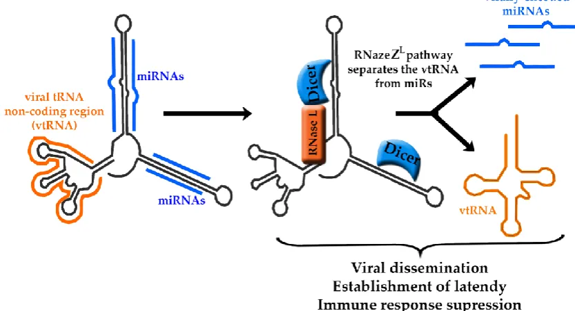
During latency, MHV-68 produces eight tRNA-microRNA-encoded RNAs, referred to as
TMERs, which are highly-abundant during asymptomatic infection and in proliferating B cells in the
context of lymphoproliferative disease [69]. TMERs include a viral tRNA non-coding region,
referred to as vtRNA followed by two to four miRNAs sequences localized downstream of the
vtRNA (Figure 3). Because of their tight structural arrangement, it has been a challenging task to
separate potential functions of the vtRNAs and miRNAs from each other [70].
When it comes to biogenesis of TMERs, the transcription utilizes the second type of RNA
(TRGYNNARNNG) and a B box (RGTTCRANTCC), which are separated from one another by
~30-60 nts [71]. The promoter sequences recruit transcription factors, while simultaneously
containing the D- and T- loops sequences that make up the vtRNA. The A and B boxes are required
for the structural development of the pseudo-tRNA, which in turn facilitates production of
miRNA-producing stem-loops. Elimination of any of the box elements inhibits miRNA production.
The tRNA and the miRNA portions of TMERs are co-transcribed and subsequently cleaved into
separate components via the tRNA maturation pathway, referred to as RNaseZL. On the other hand,
the miRNAs are processed through association with Dicer, and they are subsequently involved in
regulation of viral gene expression [72]. It has been proposed that the pol III transcription of tRNA
stem-loops could also result in production of siRNAs through the RNaseZL pathway [73] (Figure 3).
Figure 3: Maturation pathway of TMERs. Each TMER transcript contains a viral tRNA non-coding region (vtRNA, orange) and miRNA hairpins (blue). Through the RNaseZL
pathway, the vtRNA is separated from the hairpins that are then processed by Dicer into miRNAs. TMERs are essential for the establishment of latency and viral dissemination,
however, due to their close structural relationship their individual functions are not well defined.
The in vivo contribution of TMERs to MHV68 biology has been established based by a panel of
individual TMER mutant viruses [69], [72]. It has been shown that most TMER mutants had little to
no influence over viral latency, with the exception of TMER4, which has been established as a key
mediator in MHV68 hematogenous dissemination and latency. Interestingly, TMER4 vtRNA4
stem-loops, but not miRNAs, were shown to be essential for wild-type TMER4 activity, as they likely
Also, a TMER4 transcript retaining a stem-loop would be expected to interact with components of
the RNA-induced silencing complex (RISC) machinery independent of sequence specificity. A
multitude of additional functions have been ascribed to TMER4, including acting as a pro-survival
signal for the cell, blocking apoptosis of infected cells, suppressing an antiviral immune response
and initiating an immune response responsible for movement of the infection into peripheral
circulation. Each of these functions is essential for MHV68 pathogenesis and the establishment of
latency.
Another study established that one or all TMERs as essential molecular factors triggering the
development of viral pneumonia in an immunocompromised host, as TMER-deficient MHV68
showed reduced virulence, despite having an enhanced frequency of virus-infected cells [74].
Strikingly, expression of a single viral tRNA-like molecule, in the absence of all other virus-encoded
TMERs and miRNAs, reverses both attenuation in virulence and enhanced frequency of infected
cells. These data show that TMERs play essential functions in acute infection and virulence in
immunocompromised hosts and identify them as a new target to modulate MHV68 infection and
pathogenesis.
7. Herpesvirus saimiri (HVS)-encoded lncRNAs
HVS establishes latency in the T cells of New World primates and has the ability to cause
aggressive leukemias and lymphomas [75], [76]. During latency HVS expresses seven small
nuclear uracil-rich non-coding RNAs, called HSURs (Figure 4) [77]–[80]. The HSURs have
common features with Sm-class small nuclear RNAs (snRNAs) and share the same biogenesis
pathway. They are transcribed by RNA pol II in the nucleus, and subsequently exported to the
cytoplasm, where they associate with Sm core proteins and acquire a trimethylguanosine 5’ -end
cap before being imported back to the nucleus.
The 5’ termini of HSURs 1, 2 and 5 contain a highly conserved the AUUUA pentamer
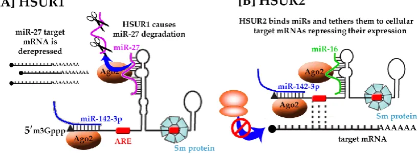
characteristic of AREs that regulate the stability of many host mRNAs [81]. In the case of HSUR1,
the ARE-like elements associate with cellular proteins hnRNPD and HuR, to mediate
ARE-dependent degradation pathway, leading to HSUR1 decay [82].
HSUR1 and HSUR2 have been shown to modulate expression of transcription factors involved
in the apoptotic response, cell cycle checkpoints, and cellular metabolism, i.e. fork-head box1,
FOXO1, PAX3 and RUNX1 [83]–[85]. Cazalla et al., 2010, observed complementarity between
HSUR1 and HSUR2 sequences and the seed regions of three host miRNAs, namely, miR-161, miR-27
and miR-42 (Figure 4). Both HSURs have been also found in Argonaute 2 (Ago2) complexes from
HSUR1 reduces miR-27 levels in infected marmoset T cells through target RNA-directed
miRNA degradation (TDMD), thereby derepressing miR-27 cellular target mRNA production and
promoting T cell activation (Figure 4A). TDMD of miR-27 is required for efficient HSV replication,
as viral strains with HUSR1 bearing a mutated miR-27 binding site have reduced titers [87].
Knockdown of HSUR1 confirmed the negative effects of HSUR1 on miR-27 accumulation and
mutation of the miR-27 complementary sequence in HSUR1 abolished the interaction and the
reduction of miR-27 levels.
Conversely, HSUR2 does not deplete the miRNAs it binds but instead acts as a tether that
recruits the Ago–miR-142-3p and Ago–miR-16 complexes to cellular mRNAs that encode
pro-apoptosis factors. These complexes then induce the silencing of these tethered mRNAs and thus
prevent apoptosis (Figure 4B) [88]. In addition, the in vivo crosslinking analysis indicated that also
HSUR2 base-pairs with mRNAs encoding retinoblastoma and factors involved in p53 signaling and
apoptosis.
Figure 4. Putative functions of HSUR RNAs. HSURs have been found to have highly conserved regions responsible for binding of host miRNAs, i.e., miR-16 (green), miR-27 (pink), miR-142
(blue) and host proteins, i.e. Ago2 (orange), involved in RISC complex formation, spliceosomal Sm proteins (blue). While the exact mechanism and function of HSURs are not yet understood
the recruitment of host miRNAs and proteins likely regulates gene expression of the target messenger RNA.
8. Gammaherpesvirus-encoded lncRNAs as therapeutic targets
As discussed above, gammaherpesvirus-encoded lncRNAs are key modulators of viral
pathogenesis and replication. As such, they represent an as of yet unexplored opportunity for
pharmacological intervention as specific targets in the context of structure-function relationships.
The currently available therapeutic options targeting gammaherpesviruses rely mostly on the
antiviral strategies are limited by the continual emergence of resistant strains [90], and the fact that
the latent viral reservoir is not eliminated.
RNA therapeutics can take advantage of the unusually high abundance of viral lncRNAs, the
presence of these molecules at various stages of viral life cycle, often including latency, and the parts
played in crucial functions in cellular processes and pathogenesis. Frequently, a single lncRNA
scaffolds or targets several molecular factors, therefore manipulation of the levels of this RNA may
serve to modulate the functions of multiple genes. However, siRNA- or antisense oligonucleotide
(ASO)-mediated knockdown strategies designed to disrupt lncRNAs function are often cytotoxic,
and this application is often linked with off-target effects. In addition, single-stranded ASOs have
often reduced stability, are frequently subjected to cellular degradation, and sometimes have low
target affinity and potency. To overcome these issues, chemical modifications to ASOs, including
addition of 2’-O-methyl and/or locked nucleic acid (LNA) bases, have been shown to increase
affinity, improve cellular uptake and decrease toxicity [91]. Off-target hybridization effects can also
be minimized by cautious bioinformatic selection of ASO sequences.
Recently, small molecule targeting of RNA structures has emerged as a promising avenue
against viral disease [92]. Small molecules offer the advantage of having desirable properties such as
good absorption, distribution, and oral bioavailability. They bind RNA by virtue of secondary or
tertiary structure, as opposed to sequence, and as such, they provide an orthogonal means to target
unique motifs [93], [94]. Ligands binding to lncRNA architectures would be able to affect RNA:RNA,
RNA:DNA, RNA:protein interactions, structural stability or conformational changes, and thereby
block processes essential for viral replication. Proof-of-concept for targeting functional RNAs by
small molecules has been demonstrated for multiple viruses, including HIV, HCV[94], SARS CoV
[95], and Influenza [96].
Another challenge in developing an effective antiviral strategy against gammaherpesviruses is
that lytic reactivation is needed before antiviral agents targeting virus can be employed. Most of the
current therapeutics target viral products present only during a productive infection, and the latent
virus reservoir is impervious to these treatments. Thus, latency represents an attractive target for
viral eradication, and indeed, recent studies using latency-reversing agents showed effectiveness
during treatment of HIV infection [97]–[99]. Currently available latency-reversing agents against
EBV and KSHV infections [98], [100]–[102], manipulate an epigenetic pathway i.e. histone epigenetic
modifications to achieve viral reactivation, and none of them target viral lncRNAs. In this review,
we have educed multiple examples of gammeherpesvirus-encoded lncRNA directly involved in
viral latent-to-lytic switch. Targeting them with therapeutics may lead to viral reactivation, which in
combination with other antiviral agents can create a platform for effective eradication of
9. Gammaherpesvirus-encoded lncRNAs: future directions
Targeting gammaherpesvirus-encoded lncRNAs requires a comprehensive understanding of
their structure to create an effective approach against viral infection and associated diseases. Some
lncRNA regions can be structurally flexible, and as such targeting them may not be successful.
Others may be occupied by strong intermolecular contacts, which would make them inaccessible for
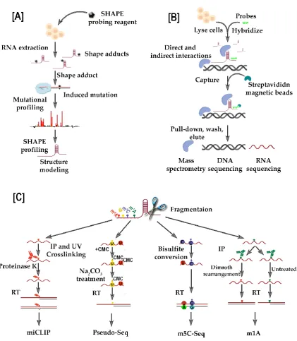
such a therapeutic approach. The application of deep-sequencing RNA structure probing
techniques, including the selective 2’-hydroxyl acylation analyzed by primer extension and
mutational profiling, (SHAPE-MaP [55]), as well as SHAPE-seq [103] and DMS treatment coupled
with massively parallel sequencing readout (DMS-seq)[104], can offer an insight into the
conformation of targeted lncRNAs, aiding the choice of a functional motifs against which one can
successfully design a therapeutic approach (Figure 5A). Also, RNA-centric biochemical affinity
techniques, such as RNA antisense purification (RAP) (Figure 5B) [105], chromatin isolation by RNA
purification (ChIRP) [106], and capture hybridization analysis of RNA targets (CHART) [107] can be
useful in revealing the comprehensive lncRNA interactome network, including contacts with
chromatin, proteins and other RNAs.
Finally, another layer of RNA structure and function regulation has captured the attention of
the scientific field, namely, epitranscriptomic modifications. Several of them, including
N6-methyladenosine (m6A), 5-methylcytidine (m5C), inosine (I), pseudouridine (Ψ), and
N1-methyladenosine (m1A), are present in lncRNAs and influence metabolism, stability, structure
and function [108]. Advances in the development of high-throughput and site-specific sequencing
technologies to identify distinct epitranscriptomic markers provide us with new tools to identify the
location and dynamic distribution of RNA modifications, and to reveal how these modifications
affect lncRNAs (Figure 5C). For example, lncRNA metastasis-associated lung adenocarcinoma
transcript 1 (MALAT1), has been shown to contain m5C [109] and Ψ [110] modifications, however an
influence on RNA structure-to-function relationship has yet to be proven. In addition, at least two
m6A signatures have been localized to the MALAT1 stem-loop, which reduce its stability and
facilitate the binding of heterogeneous nuclear ribonucleoprotein C [111]. While
the MALAT1 hairpin is the first example of so called m6A-switch, structural changes induced by the
presence of m6A modification, this phenomenon likely applies to other lncRNAs. It would be
interesting to address whether gammaherpesvirus-encoded lncRNAs also undergo
epitranscriptomic modifications and how these changes might affect RNA structure and function
during viral replication and pathogenesis. This information would contribute greatly to the
unraveling of novel modes of action by lncRNAs, as well as reveal potential ‚weak spots‛ in the
N6-methyladenosine (m6A, red) is detected with individual-nucleotide-resolution cross-linking
and immunoprecipitation (miCLIP) methodology. Here, immunoprecipitation (IP) and UV crosslinking with m6A-specific antibodies is coupled with reverse transcription and
deep-sequencing, and the sites of modification are detected as either misincorporation of base-pairs or truncation. Pseudouridine (Ψ, yellow) is detected by CMC-derivatization, where sodium carbonate removes the CMC derivative from non-pseudouridine modifications. The 5-methylcytosine (m5C, purple) uses bisulfite conversion that causes non-methylated cytosines
to be converted to guanine. The N1-methyladenosine (m1A, green), similar to miCLIP, relies on
using m1A-specific antibodies.
Acknowledgments: JSS, SEM, GCC are funded by Alabama Agricultural Experiment Station, Hatch
Funding Program and start-up funds from the Department of Biological Sciences, College of Science
and Mathematics, and Office of the Vice President for Research, Auburn University.
Author Contributions: JSS, GCC and SEM contributed to the manuscript writing and preparation.
CHH edited the manuscript and prepared the illustrations. Authors would like to thank Jennifer T.
Miller (NIH/NCI) and Jacek Wower (Auburn University) for constructive criticism of the
manuscript.
Conflicts of Interest: The authors declare no conflict of interest.
References:
[1] A. F. Palazzo and E. S. Lee, ‚Non-coding RNA: What is functional and what is junk?,‛ Front. Genet., vol. 5, no. JAN, 2015.
[2] B. Uszczynska-Ratajczak, J. Lagarde, A. Frankish, R. Guigó, and R. Johnson, ‚Towards a
complete map of the human long non-coding RNA transcriptome,‛ Nature Reviews Genetics, pp. 1–14, 2018.
[3] H. Wu, L. Yang, and L. L. Chen, ‚The Diversity of Long Noncoding RNAs and Their
Generation,‛ Trends in Genetics, vol. 33, no. 8. pp. 540–552, 2017.
[4] M. Guttman et al., ‚Chromatin signature reveals over a thousand highly conserved large non-coding RNAs in mammals,‛ Nature, vol. 458, no. 7235, pp. 223–227, 2009.
[5] P. Kapranov et al., ‚RNA maps reveal new RNA classes and a possible function for pervasive transcription,‛ Science (80-. )., vol. 316, no. 5830, pp. 1484–1488, 2007.
[7] M. E. Dinger, K. C. Pang, T. R. Mercer, and J. S. Mattick, ‚Differentiating protein-coding and
noncoding RNA: Challenges and ambiguities,‛ PLoS Computational Biology, vol. 4, no. 11. 2008.
[8] M. I. Galindo, J. I. Pueyo, S. Fouix, S. A. Bishop, and J. P. Couso, ‚Peptides encoded by short
ORFs control development and define a new eukaryotic gene family,‛ PLoS Biol., vol. 5, no. 5, pp. 1052–1062, 2007.
[9] C. Arias et al., ‚KSHV 2.0: A Comprehensive Annotation of the Kaposi’s Sarcoma-Associated Herpesvirus Genome Using Next-Generation Sequencing Reveals Novel Genomic and
Functional Features,‛ PLoS Pathog., vol. 10, no. 1, 2014.
[10] M. E. Dinger, D. K. Gascoigne, and J. S. Mattick, ‚The evolution of RNAs with multiple
functions,‛ Biochimie, vol. 93, no. 11. pp. 2013–2018, 2011.
[11] S. Cao et al., ‚New Noncoding Lytic Transcripts Derived from the Epstein-Barr Virus Latency Origin of Replication, oriP, Are Hyperedited, Bind the Paraspeckle Protein, NONO/p54nrb,
and Support Viral Lytic Transcription,‛ J Virol, vol. 89, no. 14, pp. 7120–7132, 2015.
[12] M. Campbell et al., ‚A Lytic Viral Long Noncoding RNA Modulates the Function of a Latent Protein,‛ J. Virol., vol. 88, no. 3, pp. 1843–1848, 2014.
[13] L. T. Krug, ‚Complexities of gammaherpesvirus transcription revealed by microarrays and
RNAseq,‛ Curr. Opin. Virol., vol. 3, no. 3, pp. 276–284, 2013.
[14] A. J. Rennekamp and P. M. Lieberman, ‚Initiation of Epstein-Barr Virus Lytic Replication
Requires Transcription and the Formation of a Stable RNA-DNA Hybrid Molecule at
OriLyt,‛ J. Virol., vol. 85, no. 6, pp. 2837–2850, 2011.
[15] F. Kopp and J. T. Mendell, ‚Functional Classification and Experimental Dissection of Long
Noncoding RNAs,‛ Cell, vol. 172, no. 3. pp. 393–407, 2018.
[16] I. V. Novikova, S. P. Hennelly, and K. Y. Sanbonmatsu, ‚Structural architecture of the human
long non-coding RNA, steroid receptor RNA activator,‛ Nucleic Acids Res., vol. 40, no. 11, pp. 5034–5051, 2012.
[17] J. A. Brown et al., ‚Structural insights into the stabilization of MALAT1 noncoding RNA by a bipartite triple helix,‛ Nat. Struct. Mol. Biol., vol. 21, no. 7, pp. 633–640, 2014.
Health/Lippincott Williams & Wilkins, 2007.
[19] M. Campbell, H.-J. Kung, and Y. Izumiya, ‚Long Non-Coding RNA and Epigenetic Gene
Regulation of KSHV,‛ Viruses, vol. 6, no. 11, pp. 4165–4177, 2014.
[20] S. Cao et al., ‚New Noncoding Lytic Transcripts Derived from the Epstein-Barr Virus Latency Origin of Replication, oriP , Are Hyperedited, Bind the Paraspeckle Protein, NONO/p54nrb, and Support Viral Lytic Transcription,‛ J. Virol., vol. 89, no. 14, pp. 7120–7132, 2015.
[21] P. Purushothaman, P. Dabral, N. Gupta, R. Sarkar, and S. C. Verma, ‚KSHV genome
replication and maintenance,‛ Frontiers in Microbiology, vol. 7, no. FEB. 2016.
[22] T. Tsurumi, M. Fujita, and A. Kudoh, ‚Latent and lytic Epstein-Barr virus replication
strategies,‛ Reviews in Medical Virology, vol. 15, no. 1. pp. 3–15, 2005.
[23] S. R. Baglio et al., ‚Sensing of latent EBV infection through exosomal transfer of 5′pppRNA,‛ Proc. Natl. Acad. Sci., vol. 113, no. 5, pp. E587–E596, 2016.
[24] C. C. Rossetto and G. S. Pari, ‚Kaposi’s Sarcoma-Associated Herpesvirus Noncoding
Polyadenylated Nuclear RNA Interacts with Virus- and Host Cell-Encoded Proteins and
Suppresses Expression of Genes Involved in Immune Modulation,‛ J. Virol., vol. 85, no. 24, pp. 13290–13297, 2011.
[25] G. Henle et al., ‚Viral lncRNA: A regulatory molecule for controlling virus life cycle,‛ J. Virol., vol. 11, no. 1, pp. 5894–5904, Mar. 2016.
[26] B. Johansson, G. Klein, W. Henle, and G. Henle, ‚Epstein-Barr virus (EBV)-associated
antibody patterns in malignant lymphoma and leukemia. I. Hodgkin’s disease,‛ Int. J. Cancer, vol. 6, no. 3, pp. 450–462, Nov. 1970.
[27] L. S. Young and C. W. Dawson, ‚Epstein-Barr virus and nasopharyngeal carcinoma.,‛ Chin. J. Cancer, vol. 33, no. 12, pp. 581–590, Dec. 2014.
[28] A. T. Phan, S. G. Fernandez, J. J. Somberg, K. M. Keck, and J. J. L. Miranda, ‚Epstein–Barr
virus latency type and spontaneous reactivation predict lytic induction levels,‛ Biochem. Biophys. Res. Commun., vol. 474, no. 1, pp. 71–75, May 2016.
[29] M. R. Lerner, N. C. Andrews, G. Miller, and J. A. Steitz, ‚Two small RNAs encoded by
Epstein-Barr virus and complexed with protein are precipitated by antibodies from patients
1981.
[30] D. P. Toczyski, A. G. Matera, D. C. Ward, and J. A. Steitz, ‚The Epstein-Barr virus (EBV) small
RNA EBER1 binds and relocalizes ribosomal protein L22 in EBV-infected human B
lymphocytes.,‛ Proc. Natl. Acad. Sci., vol. 91, no. 8, pp. 3463–3467, 1994.
[31] Y. Wu, S. Maruo, M. Yajima, T. Kanda, and K. Takada, ‚Epstein-Barr Virus (EBV)-Encoded
RNA 2 (EBER2) but Not EBER1 Plays a Critical Role in EBV-Induced B-Cell Growth
Transformation,‛ J. Virol., vol. 81, no. 20, pp. 11236–11245, Oct. 2007.
[32] P. A. Clarke, N. A. Sharp, and M. J. Clemens, ‚Expression of genes for the Epstein--Barr virus
small RNAs EBER-1 and EBER-2 in Daudi Burkitt’s lymphoma cells: effects of interferon
treatment,‛ J. Gen. Virol., vol. 73, no. 12, pp. 3169–3175, Dec. 1992.
[33] D. Iwakiri, ‚Epstein-Barr Virus-Encoded RNAs: Key Molecules in Viral Pathogenesis,‛
Cancers (Basel)., vol. 6, no. 3, pp. 1615–1630, Aug. 2014.
[34] V. Fok, R. M. Mitton-Fry, A. Grech, and J. A. Steitz, ‚Multiple domains of EBER 1, an
Epstein-Barr virus noncoding RNA, recruit human ribosomal protein L22,‛ RNA, 2006. [35] T. V Sharp et al., ‚Comparative analysis of the regulation of the interferon-inducible protein
kinase PKR by Epstein-Barr virus RNAs EBER-1 and EBER-2 and adenovirus VAI RNA.,‛
Nucleic Acids Res., vol. 21, no. 19, pp. 4483–4490, Sep. 1993.
[36] E. K. Lee et al., ‚Effects of lymphocyte profile on development of EBV-induced lymphoma subtypes in humanized mice.,‛ Proc. Natl. Acad. Sci. U. S. A., vol. 112, no. 42, pp. 13081–13086, Oct. 2015.
[37] N. Lee, G. Pimienta, and J. A. Steitz, ‚AUF1/hnRNP D is a novel protein partner of the EBER1
noncoding RNA of Epstein-Barr virus.,‛ RNA, vol. 18, no. 11, pp. 2073–2082, Nov. 2012. [38] A. Gouble, S. Grazide, F. Meggetto, P. Mercier, G. Delsol, and D. Morello, ‚A New Player in
Oncogenesis: AUF1/hnRNPD Overexpression Leads to Tumorigenesis in Transgenic Mice 1,‛
2002.
[39] N. Lee, T. A. Yario, J. S. Gao, and J. A. Steitz, ‚EBV noncoding RNA EBER2 interacts with host
RNA-binding proteins to regulate viral gene expression,‛ Proc. Natl. Acad. Sci., vol. 113, no. 12, pp. 3221–3226, 2016.
apoptosis by Epstein-Barr virus small RNAs is not mediated by inhibition of PKR.,‛ J. Virol., vol. 79, no. 23, pp. 14562–14569, 2005.
[41] S. A. McKenna, D. A. Lindhout, T. Shimoike, C. E. Aitken, and J. D. Puglisi, ‚Viral dsRNA
Inhibitors Prevent Self-association and Autophosphorylation of PKR,‛ J. Mol. Biol., vol. 372, no. 1, pp. 103–113, 2007.
[42] Z. Wang, Y. Zhao, and Y. Zhang, ‚Viral lncRNA: A regulatory molecule for controlling virus
life cycle,‛ Non-coding RNA Res., vol. 2, no. 1, pp. 38–44, 2017.
[43] A. Arvey et al., ‚An atlas of the Epstein-Barr virus transcriptome and epigenome reveals host-virus regulatory interactions,‛ Cell Host Microbe, vol. 12, no. 2, pp. 233–245, 2012. [44] K. T. Jeang and S. D. Hayward, ‚Organization of the Epstein-Barr virus DNA molecule. III.
Location of the P3HR-1 deletion junction and characterization of the NotI repeat units that
form part of the template for an abundant 12-O-tetradecanoylphorbol-13-acetate-induced
mRNA transcript.,‛ J. Virol., vol. 48, no. 1, pp. 135–148, Oct. 1983.
[45] H. Iizasa and K. Nishikura, ‚A new function for the RNA-editing enzyme ADAR1,‛ Nat. Immunol., vol. 10, no. 1, pp. 16–18, Jan. 2009.
[46] W. N. Moss, N. Lee, G. Pimienta, and J. A. Steitz, ‚RNA families in Epstein-Barr virus.,‛ RNA Biol., vol. 11, no. 1, pp. 10–17, 2014.
[47] E. Oksenhendler et al., ‚High incidence of Kaposi sarcoma-associated herpesvirus-related non-Hodgkin lymphoma in patients with HIV infection and multicentric Castleman disease,‛
Blood, vol. 99, no. 7, pp. 2331–2336, 2002.
[48] N. Dupin et al., ‚HHV-8 is associated with a plasmablastic variant of Castleman disease that is linked to HHV-8-positive plasmablastic lymphoma.,‛ Blood, vol. 95, no. 4, pp. 1406–1412, 2000.
[49] M. Q. Du et al., ‚Kaposi sarcoma-associated herpesvirus infects monotypic (IgMλ) but polyclonal naive B cells in Castleman disease and associated lymphoproliferative disorders,‛
Blood, vol. 97, no. 7, pp. 2130–2136, 2001.
[50] J. M. Schifano, K. Corcoran, H. Kelkar, and D. P. Dittmer, ‚Expression of the
Antisense-to-Latency Transcript Long Noncoding RNA in Kaposi’s Sarcoma-Associated
[51] Y. Xu and D. Ganem, ‚Making sense of antisense: seemingly noncoding RNAs antisense to
the master regulator of Kaposi’s sarcoma-associated herpesvirus lytic replication do not
regulate that transcript but serve as mRNAs encoding small peptides.,‛ J. Virol., vol. 84, no. 11, pp. 5465–75, Jun. 2010.
[52] V. Majerciak, T. Ni, W. Yang, B. Meng, J. Zhu, and Z. M. Zheng, ‚A Viral Genome Landscape
of RNA Polyadenylation from KSHV Latent to Lytic Infection,‛ PLoS Pathog., vol. 9, no. 11, 2013.
[53] K. T. Tycowski et al., ‚Viral noncoding RNAs: More surprises,‛ Genes Dev., vol. 29, no. 6, pp. 567–584, 2015.
[54] J. Bechtel, A. Grundhoff, and D. Ganem, ‚RNAs in the Virion of Kaposi’s
Sarcoma-Associated Herpesvirus,‛ J. Virol., vol. 79, no. 16, pp. 10138–10146, 2005. [55] J. Sztuba-Solinska, J. W. Rausch, R. Smith, J. T. Miller, D. Whitby, and S. F. J. Le Grice,
‚Kaposi’s sarcoma-associated herpesvirus polyadenylated nuclear RNA: A structural
scaffold for nuclear, cytoplasmic and viral proteins,‛ Nucleic Acids Res., vol. 45, no. 11, pp. 6805–6821, 2017.
[56] M. J. Massimelli et al., ‚Stability of a long noncoding viral RNA depends on a 9-nt core element at the RNA 5’ end to interact with viral ORF57 and cellular PABPC1.,‛ Int. J. Biol. Sci., vol. 7, no. 8, pp. 1145–60, 2011.
[57] N. K. Conrad and J. A. Steitz, ‚A Kaposi’s sarcoma virus RNA element that increases the
nuclear abundance of intronless transcripts,‛ EMBO J., vol. 24, no. 10, pp. 1831–1841, 2005. [58] C. C. Rossetto and G. Pari, ‚KSHV PAN RNA associates with demethylases UTX and JMJD3
to activate lytic replication through a physical interaction with the virus genome,‛ PLoS Pathog., vol. 8, no. 5, 2012.
[59] C. C. Rossetto and G. S. Pari, ‚PAN’s Labyrinth: Molecular biology of Kaposi’s
sarcoma-associated herpesvirus (KSHV) PAN RNA, a multifunctional long noncoding
RNA.,‛ Viruses, vol. 6, no. 11, pp. 4212–26, Nov. 2014.
Viral and Cellular Gene Expression by Kaposi’s Sarcoma-Associated Herpesvirus
Polyadenylated Nuclear RNA,‛ J. Virol., vol. 87, no. 10, pp. 5540–5553, 2013.
[62] J. Carlevaro-Fita, A. Rahim, R. Guigó, L. A. Vardy, and R. Johnson, ‚Cytoplasmic long
noncoding RNAs are frequently bound to and degraded at ribosomes in human cells,‛ RNA, vol. 22, no. 6, pp. 867–882, 2016.
[63] S. Borah, N. Darricarrère, A. Darnell, J. Myoung, and J. A. Steitz, ‚A viral nuclear noncoding
RNA binds re-localized poly(A) binding protein and is required for late KSHV gene
expression,‛ PLoS Pathog., vol. 7, no. 10, 2011.
[64] M. He et al., ‚Cancer angiogenesis induced by Kaposi sarcoma-associated herpesvirus is mediated by EZH2,‛ Cancer Res., vol. 72, no. 14, pp. 3582–3592, 2012.
[65] A. Forero, K. D. McCormick, F. J. Jenkins, and S. N. Sarkar, ‚Downregulation of IRF4 induces
lytic reactivation of KSHV in primary effusion lymphoma cells,‛ Virology, vol. 458–459, no. 1, pp. 4–10, 2014.
[66] M. E. McDowell, P. Purushothaman, C. C. Rossetto, G. S. Pari, and S. C. Verma,
‚Phosphorylation of Kaposi’s Sarcoma-Associated Herpesvirus Processivity Factor ORF59 by
a Viral Kinase Modulates Its Ability To Associate with RTA and oriLyt,‛ J. Virol., vol. 87, no. 14, pp. 8038–8052, 2013.
[67] S. Chandriani, Y. Xu, and D. Ganem, ‚The lytic transcriptome of Kaposi’s sarcoma-associated
herpesvirus reveals extensive transcription of noncoding regions, including regions antisense
to important genes.,‛ J. Virol., vol. 84, no. 16, pp. 7934–42, Aug. 2010.
[68] J. Aligo, M. Walker, P. Bugelski, and D. Weinstock, ‚Is murine gammaherpesvirus-68
(MHV-68) a suitable immunotoxicological model for examining immunomodulatory
drug-associated viral recrudescence?,‛ J. Immunotoxicol., vol. 12, no. 1, pp. 1–15, Jan. 2015. [69] E. R. Feldman et al., ‚A Gammaherpesvirus Noncoding RNA Is Essential for Hematogenous
Dissemination and Establishment of Peripheral Latency,‛ mSphere, vol. 1, no. 2, pp. e00105-15, Apr. 2016.
[71] K. W. Diebel, D. J. Claypool, and L. F. van Dyk, ‚A conserved RNA polymerase III promoter
required for gammaherpesvirus TMER transcription and microRNA processing,‛ Gene, vol. 544, no. 1, pp. 8–18, 2014.
[72] E. R. Feldman et al., ‚Virus-Encoded MicroRNAs Facilitate Gammaherpesvirus Latency and Pathogenesis In Vivo,‛ MBio, vol. 5, no. 3, pp. e00981--14, May 2014.
[73] L. J. Scherer, R. Frank, and J. J. Rossi, ‚Optimization and characterization of tRNA-shRNA
expression constructs,‛ Nucleic Acids Res., vol. 35, no. 8, pp. 2620–2628, Apr. 2007.
[74] K. W. Diebel et al., ‚Gammaherpesvirus small noncoding RNAs are bifunctional elements that regulate infection and contribute to virulence in vivo.,‛ MBio, vol. 6, no. 1, pp. e01670--14, Feb. 2015.
[75] J. C. Albercht and B. Fleckenstein, ‚Nucleotide sequence of HSUR 6 and HSUR 7, two small
RNAs of herpesvirus saimiri,‛ Nucleic Acids Research, vol. 20, no. 7. p. 1810, 1992.
[76] A. Ensser and B. Fleckenstein, ‚T-cell transformation and oncogenesis by γ2-herpesviruses,‛
Advances in Cancer Research, vol. 93. pp. 91–128, 2005.
[77] S. I. Lee, S. C. S. Murthy, J. J. Trimble, R. C. Desrosiers, and J. A. Steitz, ‚Four novel U RNAs
are encoded by a herpesvirus,‛ Cell, vol. 54, no. 5, pp. 599–607, 1988.
[78] V. E. E. Myer, S. I. I. Lee, and J. A. A. Steitz, ‚Viral small nuclear ribonucleoproteins bind a
protein implicated in messenger RNA destabilization,‛ Proc. Natl. Acad. Sci., vol. 89, no. 4, p. 1296, 1992.
[79] H. L. Cook, H. E. Mischo, and J. A. Steitz, ‚The Herpesvirus saimiri Small Nuclear RNAs
Recruit AU-Rich Element-Binding Proteins but Do Not Alter Host AU-Rich
Element-Containing mRNA Levels in Virally Transformed T Cells,‛ Mol. Cell. Biol., vol. 24, no. 10, pp. 4522–4533, 2004.
[80] J. C. Albrecht et al., ‚Primary structure of the herpesvirus saimiri genome.,‛ J. Virol., vol. 66, no. 8, pp. 5047–58, 1992.
[81] C. Y. A. Chen and A. Bin Shyu, ‚AU-rich elements: characterization and importance in
mRNA degradation,‛ Trends in Biochemical Sciences, vol. 20, no. 11. pp. 465–470, 1995.
[82] X. C. Fan, V. E. Myer, and J. A. Steitz, ‚AU-rich elements target small nuclear RNAs as well as
[83] I. K. Guttilla and B. A. White, ‚Coordinate regulation of FOXO1 by miR-27a, miR-96, and
miR-182 in breast cancer cells,‛ J. Biol. Chem., vol. 284, no. 35, pp. 23204–23216, 2009. [84] R. Ben-Ami et al., ‚A multinational survey of risk factors for infection with
extended-spectrum beta-lactamase-producing enterobacteriaceae in nonhospitalized
patients.,‛ Clin. Infect. Dis., vol. 49, no. 5, pp. 682–90, 2009.
[85] C. G. Crist et al., ‚Muscle stem cell behavior is modified by microRNA-27 regulation of Pax3 expression.,‛ Proc. Natl. Acad. Sci. U. S. A., vol. 106, no. 32, pp. 13383–7, 2009.
[86] D. Cazalla, T. Yario, and J. Steitz, ‚Down-regulation of a host MicroRNA by a herpesvirus
saimiri noncoding RNA,‛ Science (80-. )., vol. 328, no. 5985, pp. 1563–1566, 2010.
[87] L. Marcinowski et al., ‚Degradation of cellular miR-27 by a novel, highly abundant viral transcript is important for efficient virus replication in vivo,‛ PLoS Pathog., vol. 8, no. 2, 2012. [88] C. Gorbea, T. Mosbruger, and D. Cazalla, ‚A viral Sm-class RNA base-pairs with mRNAs &
recruits microRNAs to inhibit apoptosis,‛ Nature, vol. 550, no. 7675, pp. 275–279, 2017. [89] N. Coen, S. Duraffour, D. Topalis, R. Snoeck, and G. Andrei, ‚Spectrum of activity and
mechanisms of resistance of various nucleoside derivatives against gammaherpesviruses,‛
Antimicrob. Agents Chemother., vol. 58, no. 12, pp. 7312–7323, 2014.
[90] C. Gilbert, J. Bestman-Smith, and G. Boivin, ‚Resistance of herpesviruses to antiviral drugs:
Clinical impacts and molecular mechanisms,‛ Drug Resistance Updates, vol. 5, no. 2. pp. 88– 114, 2002.
[91] X. Shen and D. R. Corey, ‚Chemistry, mechanism and clinical status of antisense
oligonucleotides and duplex RNAs,‛ Nucleic Acids Res., vol. 46, no. 4, pp. 1584–1600, 2018. [92] T. Hermann, ‚Small molecules targeting viral RNA,‛ Wiley Interdisciplinary Reviews: RNA,
vol. 7, no. 6. pp. 726–743, 2016.
[93] C. M. Connelly, M. H. Moon, and J. S. Schneekloth, ‚The Emerging Role of RNA as a
Therapeutic Target for Small Molecules,‛ Cell Chemical Biology, vol. 23, no. 9. pp. 1077–1090, 2016.
[94] J. Parsons, M. P. Castaldi, S. Dutta, S. M. Dibrov, D. L. Wyles, and T. Hermann,
[95] S. J. Park, Y. G. Kim, and H. J. Park, ‚Identification of rna pseudoknot-binding ligand that
inhibits the - 1 ribosomal frameshifting of SARS-coronavirus by structure-based virtual
screening,‛ J. Am. Chem. Soc., vol. 133, no. 26, pp. 10094–10100, 2011.
[96] M. K. Lee et al., ‚Correction: A novel small-molecule binds to the influenza A virus RNA promoter and inhibits viral replication,‛ Chem. Commun., vol. 50, no. 83, p. 12578, 2014. [97] S. G. Deeks, ‚HIV: Shock and kill,‛ Nature, vol. 487, no. 7408. pp. 439–440, 2012.
[98] S. K. Ghosh, S. P. Perrine, R. M. Williams, and D. V. Faller, ‚Histone deacetylase inhibitors
are potent inducers of gene expression in latent EBV and sensitize lymphoma cells to
nucleoside antiviral agents,‛ Blood, vol. 119, no. 4, pp. 1008–1017, 2012.
[99] Y. Kim, J. L. Anderson, and S. R. Lewin, ‚Getting the ‘Kill’ into ‘Shock and Kill’: Strategies to
Eliminate Latent HIV,‛ Cell Host and Microbe, vol. 23, no. 1. pp. 14–26, 2018.
[100] W. H. Feng and S. C. Kenney, ‚Valproic acid enhances the efficacy of chemotherapy in
EBV-positive tumors by increasing lytic viral gene expression,‛ Cancer Res., vol. 66, no. 17, pp. 8762–8769, 2006.
[101] F. Zhou et al., ‚Oncolytic reactivation of KSHV as a therapeutic approach for primary effusion lymphoma,‛ Mol. Cancer Ther., vol. 16, no. 11, 2017.
[102] Q. Li, M. He, F. Zhou, F. Ye, and S.-J. Gao, ‚Activation of Kaposi’s Sarcoma-Associated
Herpesvirus (KSHV) by Inhibitors of Class III Histone Deacetylases: Identification of Sirtuin
1 as a Regulator of the KSHV Life Cycle,‛ J. Virol., vol. 88, no. 11, pp. 6355–6367, 2014. [103] R. Diaz-Toledano, G. Lozano, and E. Martinez-Salas, ‚In-cell SHAPE uncovers dynamic
interactions between the untranslated regions of the foot-and-mouth disease virus RNA,‛
Nucleic Acids Res., vol. 45, no. 3, pp. 1416–1432, 2017.
[104] R. Francisco-Velilla, J. Fernandez-Chamorro, G. Lozano, R. Diaz-Toledano, and E.
Martínez-Salas, ‚RNA-protein interaction methods to study viral IRES elements,‛ Methods, vol. 91. pp. 3–12, 2015.
[105] J. Engreitz, E. S. Lander, and M. Guttman, RNA Antisense Purification (RAP) for Mapping RNA Interactions with Chromatin, vol. 1262. New York, NY: Springer New York, 2015.
[106] C. Chu, K. Qu, F. L. Zhong, S. E. Artandi, and H. Y. Chang, ‚Genomic Maps of Long
vol. 44, no. 4, pp. 667–678, 2011.
[107] M. D. Simon, ‚Capture Hybridization Analysis of RNA Targets (CHART),‛ Curr. Protoc. Mol. Biol., vol. 2013, no. January, pp. 1–16, 2013.
[108] R. Jacob, S. Zander, and T. Gutschner, ‚The dark side of the epitranscriptome: Chemical
modifications in long non-coding rnas,‛ International Journal of Molecular Sciences, vol. 18, no. 11. 2017.
[109] J. E. Squires et al., ‚Widespread occurrence of 5-methylcytosine in human coding and non-coding RNA,‛ Nucleic Acids Res., vol. 40, no. 11, pp. 5023–5033, 2012.
[110] X. Li et al., ‚Chemical pulldown reveals dynamic pseudouridylation of the mammalian transcriptome,‛ Nat. Chem. Biol., vol. 11, no. 8, pp. 592–597, 2015.
[111] N. Liu, Q. Dai, G. Zheng, C. He, M. Parisien, and T. Pan, ‚N6-methyladenosine-dependent
![Figure 2: The structure-to-function relationship of EBER 1 and 2. [A] EBER 1 stem-loops I (violet), III (pink), and IV (green) create a scaffold for interaction with L22 (orange) resulting in the re-localization of EBER1:L22 into the nucleoplasm](https://thumb-us.123doks.com/thumbv2/123dok_us/7994133.1327141/10.595.94.465.74.387/structure-function-relationship-scaffold-interaction-resulting-localization-nucleoplasm.webp)