Complementary role of cardiac CT
in the assessment of aortic valve
replacement dysfunction
Alastair J Moss,1Marc R Dweck,1John G Dreisbach,2Michelle C Williams,1 Sze Mun Mak,3Timothy Cartlidge,1Edward D Nicol,4Gareth J Morgan-Hughes5
To cite:Moss AJ, Dweck MR, Dreisbach JG, et al. Complementary role of cardiac CT in the assessment of aortic valve replacement
dysfunction.Open Heart
2016;3:e000494.
doi:10.1136/openhrt-2016-000494
Received 4 July 2016 Accepted 14 October 2016
For numbered affiliations see end of article.
Correspondence to Dr Alastair J Moss; [email protected]
ABSTRACT
Aortic valve replacement is the second most common cardiothoracic procedure in the UK. With an ageing population, there are an increasing number of patients with prosthetic valves that require follow-up. Imaging of prosthetic valves is challenging with conventional echocardiographic techniques making early detection of valve dysfunction or complications difficult. CT has recently emerged as a complementary approach offering excellent spatial resolution and the ability to identify a range of aortic valve replacement complications including structural valve dysfunction, thrombus development, pannus formation and prosthetic valve infective endocarditis. This review discusses each and how CT might be incorporated into a multimodal cardiovascular imaging pathway for the assessment of aortic valve replacements and in guiding clinical management.
INTRODUCTION
The utility of cardiac CT for the assessment of possible aortic valve replacement dysfunc-tion has risen rapidly over the past 10 years following a similar, albeit delayed, trajectory to CT coronary imaging. It can be used to assess mechanical and bioprosthetic valves inserted surgically as well valves inserted using transcutaneous aortic valve implant-ation (TAVI). Clinicians familiar with both cardiac CT and valvular heart disease have
identified a number of specific situations
where CT can help in the assessment of pos-sible aortic valve replacement dysfunction by providing complementary diagnostic infor-mation to transthoracic echocardiography,
transoesophageal echocardiography and
cardiac MR. These include the identification
of pannus formation, thrombus, premature
bioprosthetic leaflet degeneration,
assess-ment of bileaflet mechanical valve leaflet
motion and aortic root abscess formation. While the role of cardiac CT is relatively new in this setting, its use is steadily expanding across the world, with many experienced
centres now using it routinely. This article evaluates the evidence in support of cardiac CT imaging for the detection of possible aortic valve replacement dysfunction and aims to prompt clinicians to consider it in
specific clinical scenarios.
BIOPROSTHETIC AORTIC VALVE REPLACEMENT DYSFUNCTION
Selecting the appropriate prosthetic heart
valve has traditionally been a difficult
deci-sion for many patients undergoing surgical aortic valve replacement. However, recent
advances in bioprosthetic valve design,
coupled with an ageing population, have wit-nessed increasing use of these valves in pref-erence to metallic valves. With the recent addition of TAVI, the number of patients with functioning bioprostheses is only set to expand further. Surveillance of bioprosthesis function, looking for evidence of valve degeneration, forms an integral part of the long-term management of patients with these valves and a substantial healthcare
burden.1 This continues to remain relevant
in the modern era where newer generation bioprosthetic valves offer improved longevity
but still have a limited life span of 10–
15 years. Despite technological advances, bio-prostheses exhibit more frequent structural valve dysfunction than mechanical valves
(2.17%/patient-year vs 0%/patient-year,
p=0.0001), resulting in higher rates of repeat operation (2.32%/patient-year vs 0.62%/
patient-year, p=0.0003).2 The high spatial
and temporal resolution of cardiac CT makes it well suited to visualise many of the complications that can arise following bio-prosthetic valve implantation, namely,
struc-tural valve dysfunction, thrombus
development, pannus formation and
pros-thetic valve infective endocarditis.3 We will
here examine the role of CT in assessing each of these problems.
on September 12, 2020 by guest. Protected by copyright.
Structural valve dysfunction
Structural valve dysfunction can have catastrophic conse-quences, yet its underlying pathophysiology remains incompletely understood. The term structural valve dys-function encompasses intrinsic dys-functional changes to
the valve leaflets including retraction or tearing,
progres-sive stenosis and disruption of the annular housing or sewing ring. The principal pathological driver behind this degeneration and eventual failure appears to be
leaflet calcification. This most commonly results in
valvu-lar regurgitation due to tearing of the leaflets but may
also cause increasing valve stiffness, restenosis and
per-ipheral embolism.4 At the subcellular level, scanning
electron microscopy has revealed that microcrystalline
hydroxyapatite and amorphous calcium phosphate
aggregate into plate-like structures that are incorporated
into the collagen matrix.5 Calcification has been
observed in over half of porcine prosthetic valves implanted 5 years previously, rising to over
three-quarters of valves aged 8 years or more.6 7Interestingly
valve calcification and degeneration appears accelerated
in younger patients, perhaps due to increased mechan-ical stresses in these patients, as highlighted by the fatal complications following implantation of bovine
pericar-dial bioprostheses in children and young adults.8
Indeed, such is the importance of calcification in
struc-tural valve dysfunction that efforts by valve manufac-turers aimed at improving longevity have largely focused
on anticalcific strategies including the application of
topical anticalcific agents to the leaflet surfaces.
The mechanisms driving prosthetic valve calcification
are incompletely understood with several different
pro-cesses having been implicated.9The combination of
glu-taraldehyde pretreatment and an immune rejection response to residual animal antigens in the
bioprosth-eses leads to the accumulation of extracellular
calcium.10–13 However, recent data have suggested a
third potential mechanism with evidence supporting
prosthetic valve calcification as an active disease process
with many similarities to those observed in aortic
sten-osis and atherosclersten-osis.13 14 CT is the technique of
choice for imaging macroscopic deposits of calcium in
the vasculature and so potentially allows leaflet calcifi
ca-tion and degeneraca-tion to be identified at an earlier
sub-clinical stage.
While large-scale clinical trials are currently lacking, the requirement for improved assessment of
biopros-thetic valve calcification was recently illustrated in the
case of severe bioprosthetic valve obstruction in a 13-year-old girl who died suddenly just 23 months after implantation of a bovine pericardial aortic
bioprosth-esis.8 The rapid deterioration in her valve function
occurred despite recent echocardiography demonstrat-ing only mildly stenotic gradients and restricted motion
of a single leaflet. Ex vivo CT was performed on this and
two other explanted bioprostheses demonstrating
exten-sive calcification within the central part of the valve
leaf-lets between the aortic and ventricular surfaces.
Immunohistochemistry showed that these calcium
deposits extended along collagen fibres within the
leaflet thereby increasing leaflet thickness. In principle,
calcium is best imaged on non-contrast CT scans, such as those used for CT calcium scoring of the coronary arteries and native aortic valves. However, using
dedi-cated software, it is also possible to quantify leaflet
calci-fication on contrast-enhanced cardiac CT, allowing
improved localisation of calcific deposits to the
biopros-thetic leaflets rather than surrounding structures.15 The
challenge is deciding on a suitable threshold for identi-fying calcium on these scans, although a detection
threshold of 850 Hounsfield units (HU) has recently
been used to identify calcification in native valves that
ultimately goes on to cause clinical complications
follow-ing TAVI.15 Structural components within the prosthesis
such as the stent frame and sewing ring may generate beam hardening artefact that may obscure or even be
confused with bioprosthetic calcification. An
appreci-ation of the different structural designs of the various bioprosthetic valves is therefore of use when trying to
identify true structural valve dysfunction.16 While TAVI is
currently reserved for high-risk surgical candidates with a reduced life expectancy, this procedure is increasingly used in high-risk younger adults where the long-term durability of these valves has not been evaluated.
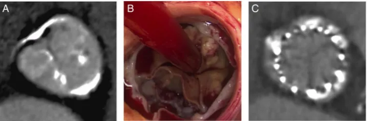
Figure 1 Structural valve degeneration. (A and B) Bioprosthetic aortic valves calcify along the cusp commissures mimicking the pattern found in native aortic valve disease. (C) Calcified leaflets are reflected onto the sinus walls during transcatheter valve implantation.
on September 12, 2020 by guest. Protected by copyright.
Reports of early TAVI valve degeneration have again
implicated calcification as the key pathological driver,
suggesting that CT may once again be off use in detect-ing early degeneration. This is supported by several recent observations. Deutsch and colleagues noted
struc-tural valve dysfunction with several areas of macrocalcifi
-cation on CT in a 4-year-old CoreValve TAVI
bioprosthesis implanted in to a 44-year-old.17 Similar
findings have also been found in elderly patients early
after valve implantation.18 19 Importantly residual
calcium from the original aortic valve around the perim-eter of the TAVI bioprosthesis must be differentiated
from new calcium formation within the leaflets of the
new prosthetic valve (figure 1).
Reduced leaflet mobility and hypoattenuation leaflet thickening
One of the major advantages biological prosthetic heart valves have over mechanical alternatives is the lack of a requirement for long-term anticoagulant use. During
bioprosthesis endothelialisation, in the first 3 months
after implantation, thrombus formation can occur in
0.8–4.0% of cases.20 However, clinical sequelae of
sys-temic thromboembolism and obstructive thrombosis are very rare phenomena occurring in 0.9% and 0.03% of
cases, respectively.21 Recent studies performing
contrast-enhanced cardiac CT in TAVI patients have
identified a new phenomenon termed hypoattenuation
leaflet thickening (HALT; figure 2). This was first
described by Pache et al22 in an 86-year-old man with
hypoattenuation of a single prosthetic cusp 7 days after implantation of a 29 mm SAPIEN XT valve. Subsequent resolution following coumadin anticoagulation
sug-gested that this finding represented cusp thrombosis
rather than pannus formation. Following this initial report, three more recent studies have further
investi-gated the frequency and possible clinical significance of
HALT,23–25and each is discussed below.
Leetmaa et al23 reported the incidence of HALT in a
consecutive cohort of 140 patients undergoing SAPIEN
XT transcatheter aortic valve implantation. Using
prospective-gated cardiac CT, five patients (3.6%) were
found to have hypoattenuating masses on the aortic surface of the transcatheter valve at 3 months postimplan-tation. Combined data from the PORTICO IDE study as well as the RESOLVE and SAVORY registries have
sug-gested a higher incidence of HALT (13–40% of patients)
if scans are performed at earlier time points
postimplan-tation (30 days to 3 months).24In these pooled registries,
reduced leaflet motion and hypoattenuated lesions were
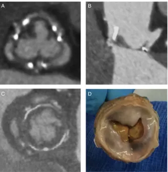
associated with an increased risk of systemic thrombo-embolism (3 of 17 patients with these abnormalities vs 1 of 115 patients without; p=0.007), with both features responding to therapeutic anticoagulation. Ex vivo Figure 2 (A and B)
Hypoattenuation leaflet thickening suggestive of thrombus formation on the supravalvular surface impinging leaflet motion. (C and D) Pannus formation underneath the prosthesis can also restrict leaflet motion necessitating surgical removal of the prosthesis.
on September 12, 2020 by guest. Protected by copyright.
modelling established that changes in mean pressure gra-dients are dependent on the size of valve and the number of cusps occluded. This may explain why these
findings on cardiac CT are commonly observed in the
absence of any increase in velocities or pressure gradient on echocardiography. Indeed at least two cusps must be involved in a 23 mm PORTICO valve for the effective
orifice area to be sufficiently stenosed to meet current
echocardiographic criteria for detection.26 Early CT
assessment following SAPIEN 3 transcatheter valve implantation revealed hypoattenuated thickening in 10.3% of patients with a trend towards a higher incidence
in patients administered single-antiplatelet therapy
com-pared with dual-antiplatelet therapy.25In 13 patients with
hypoattenuated thickening who underwent repeat CT angiography after 3 months of treatment, all those on a
combination of clopidogrel and phenprocoumon
(WOEST regimen27) had complete resolution of CT
abnormalities. Importantly, there were no thrombo-embolic or bleeding complications after 8 months of follow-up.
In light of the asymptomatic presentation of cusp thrombosis that occurs in spite of dual-antiplatelet therapy, important questions have been raised regarding the clinical relevance of hypoattenuated thickening and whether CT may more appropriately stratify antithrom-botic therapy following transcatheter valve implantation.
Importantly, the clinical significance of HALT has not
been validated beyond these observational studies and the recent publication of low 30-day stroke rates
follow-ing TAVI suggests that the presence of this finding does
not always translate into significant thromboembolic
events.28Randomised control trials are warranted before
recommending routine anticoagulation in a group of elderly patients that are at high risk of bleeding
compli-cations, although CT may find a role in pinpointing
those patients with most to gain from this therapy.
MECHANICAL AORTIC VALVE REPLACEMENT DYSFUNCTION
Imaging of mechanical prosthetic valves has traditionally been performed using transthoracic echocardiography,
transoesophageal echocardiography andfluoroscopy.29 30
Table 1 Differentiating pannus from thrombus on cardiac CT
Pannus Thrombus
Timing of presentation
Usually 12 months after surgery
Occurs at any time
Location Below the aortic prosthesis
Above or below the aortic prosthesis Morphology
on CT
Circular mass extending from sewing ring Contrast enhancement Calcification may be present
Irregularly shaped mass attached to leaflet or hingepoint No contrast enhancement
CT attenuation
>145 HU <145 HU
HU, Hounsfield units.
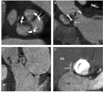
Figure 3 Prosthetic valve infective endocarditis. (A) Complications of prosthetic valve infective endocarditis include the development of perivalvular pseudoaneurysm formation (white asterix). (B) Saccular pseudoaneurysms (white asterix) occur in regions where
vegetations (white arrow) erode through the annulus with a loss of sewing ring integrity. (C) Bacterial spread into the aortic root results in abscess formation (white asterix). (D) The anatomical location of erosive shunts (white arrow) and paravalvular leaks due to suture dehiscence can be readily identified using cardiac CT. LA, left atrium; RA, right atrium.
on September 12, 2020 by guest. Protected by copyright.
However, the anatomic evaluation of the annular
housing, prosthetic valve leaflets and perivalvular
struc-tures by echocardiography has intrinsic limitations, most notably the acoustic shadowing artefacts produced by metallic components of the valve. Although by no means free of technical limitations, complementary roles for cardiac CT are emerging in the assessment of mechanical valves, particularly in acquired mechanical obstruction and endocarditis. While the metallic compo-nents of metallic valves also result in artefact on CT, diagnostic quality images can be obtained in the major-ity of mechanical valves.
Acquired mechanical prosthetic valve obstruction— thrombosis and pannus formation
Acquired mechanical prosthetic valve obstruction (PVO) is an uncommon but serious and potentially fatal
complication of valve replacement.31A definitive
diagno-sis is of paramount importance due to the associated morbidity and mortality and the frequent need for
appropriately timed intervention.32 The two main
mechanisms of acquired mechanical PVO are throm-bosis and pannus formation, both of which restrict
normal leaflet motion.31 Differentiation of these two
causes is of critical importance in guiding appropriate treatment (table 1). While both can be treated with urgent surgical revision, valve thrombosis is also
poten-tially amenable to thrombolysis.33 Moreover accurate
assessments of the degree of valve obstruction and the size of the thrombus will help to determine the urgency with which intervention is required alongside standard
clinical assessments.31 CT is increasingly being used in
this role to complement standard imaging and to help guide patient management, in particular when a clear
aetiology has not been established.30
Cardiac CT in suspected acquired mechanical PVO
enables evaluation of leaflet opening and closing angles,
dynamic leaflet motion and the composition of
perivalv-ular masses valve helping to differentiate between valve
thrombosis and pannus formation.33 A recent
prospect-ive trial compared the imaging results from cardiac CT
with post-thrombolysis imaging and/or surgical findings
in patients with acquired mechanical PVO.33 Of the 39
patients with a periprosthetic mass visible on cardiac CT, thrombus demonstrated a mean attenuation value of 87 (±59 HU) compared with 322 (±122 HU) for pannus. The investigators recommended a cut-off point of >145 HU for differentiating pannus from thrombus,
pro-viding a sensitivity of 88% and specificity of 96%.
Additionally, in the patients who underwent thromboly-sis, those with the lowest attenuation masses demon-strated the best response to thrombolysis with complete dissolution in all the <90 HU masses compared with just 42% in the masses with attenuation of between 90 and
145 HU.33
Table 2 The complementary role of CT in the assessment of aortic valve replacement dysfunction
CT Echocardiography
Prosthetic valve pathology
Calcification +++ +
Pannus ++ +
Thrombus ++ +
Vegetations + +++
Leaflet perforations/tears − +++
Valve dehiscence + +++
Paraprosthetic regurgitation − +++ Aortic root pathology
Perivalvular abscesses +++ ++
Pseudoaneurysm +++ +
Prosthetic valve function + +++ Coronary artery anatomy +++ − Presurgical planning +++ + Prevalve-in-valve planning +++ +
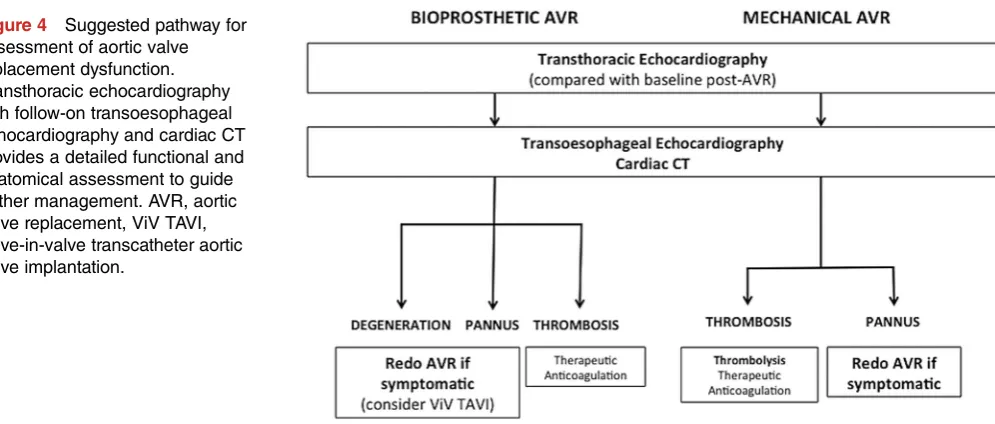
Figure 4 Suggested pathway for assessment of aortic valve replacement dysfunction. Transthoracic echocardiography with follow-on transoesophageal echocardiography and cardiac CT provides a detailed functional and anatomical assessment to guide further management. AVR, aortic valve replacement, ViV TAVI, valve-in-valve transcatheter aortic valve implantation.
on September 12, 2020 by guest. Protected by copyright.
AORTIC VALVE REPLACEMENT INFECTIVE ENDOCARDITIS Prosthetic valve infective endocarditis carries a very high
in-hospital mortality rate of 20–40%.34 Transthoracic
echocardiography and transoesophageal echocardiog-raphy are the front-line investigations in the assessment
of suspected prosthetic valve infection endocarditis;29
however, cardiac CT can prove useful particularly in
cases where echocardiography is inconclusive.32 35
Furthermore, emerging data suggest it can provide superior detection to echocardiography of several
spe-cific complications including perivalvular abscesses,
pseudoaneurysms, valve dehiscence and extracardiac
foci of infection.35–38 While echocardiography is the
modality of choice for detecting vegetations and valve dehiscence, the complementary assessment of aortic root involvement provided by CT improves the
diagnos-tic accuracy for planning surgical intervention (κstatistic
0.66–0.79 for CT or echocardiography alone vsκstatistic
0.88 for combined testing).37
While the temporal resolution of echocardiography often means that highly mobile vegetations are better visualised on echocardiography, large prosthetic valve vegetations can be readily seen on CT as microlobulated,
hypoattenuating lesions attached to the leaflets or
sewing ring.39 If the infection spreads to involve the
sewing ring, suture dehiscence can result in a
paravalvu-lar leak where there is a breach between the inflow and
outflow tract. Loss of the sewing ring integrity leads to
perivalvular pseudoaneurysm formation, which is
evident as a focal, contrast-filled saccular or fusiform
out-pouching arising from the annulus (figure 3).
Extension of infection into the aortic root is more easily detected using CT compared with transoesophageal echocardiography and can further inform surgical man-agement by detecting ancillary features such as pulmon-ary septic emboli, patency of the coronpulmon-ary arteries,
mediastinal gas and collections.38 Importantly, the
add-ition of CT imaging to standard assessments has been shown to change the clinical treatment strategy in a quarter of patients with suspected prosthetic valve
infect-ive endocarditis.35
TECHNICAL CONSIDERATIONS
In 2014, Ghersin et al40 published a suggested protocol
for the CT evaluation of suspected prosthetic valve dys-function. A retrospective contrast-enhanced cardiac CT is performed as the primary diagnostic scan for mechan-ical and bioprosthetic aortic valves. Scans acquired retro-spectively can be reconstructed to provide images of the valve at multiple points across the cardiac cycle, allowing assessment of its open and closed positions, as well as the visualisation of dynamic motion through systole and
diastole.40 The field of view can be limited to cover the
level of the valve alone (reducing radiation exposure) or, depending on the clinical indications, expanded to
Figure 5 An example adjunctive CT imaging in the management of aortic valve replacement dysfunction. (A) Severe prosthesis regurgitation on transoesophageal
echocardiography resulted from calcific structural valve
dysfunction of the bioprosthetic leaflets in a 27 mm Aspire bioprosthesis demonstrated on CT (B). (C and D) Preprocedural planning facilitated implantation of a transcatheter aortic valve in valve.
on September 12, 2020 by guest. Protected by copyright.
cover other areas of interest, including the coronary arteries.
Multiple factors can affect image quality when asses-sing mechanical aortic valve prostheses uasses-sing CT, in par-ticular the presence of cardiac arrhythmia and older
prosthetic valve types.41Anecdotal evidence suggests that
modern low profile bileaflet mechanical valves can be
imaged with diagnostic certainty by cardiac CT with minimal beam hardening artefacts. Postprocessing tech-niques support the assessment of mechanical aortic
valve leaflet opening and closing angles, limitation of
leaflet motion and the presence or absence of
peripros-thetic pannus.41 The presence of a concurrent
pros-thetic mitral valve and variations in tube voltage ranging from 80 to 140 kV does not have a discernable affect on
image quality.41 Minimising radiation dose while
main-taining diagnostic quality images can be achieved with tube current modulation and the use of 100 kV tube potential in patients with a body mass index under
30 kg/m2.40
Based on current practice in experienced valve centres, we suggest that CT should be performed as an anatomical assessment following transthoracic echocardi-ography in patients with suspected prosthetic aortic
valve dysfunction (table 2,figures 4and5). The detailed
structural assessment afforded by cardiac CT more
accurately identifies the underlying pathology and
facili-tates the implementation of appropriate aortic valve intervention.
CONCLUSION
The diagnosis and management of aortic valve
replace-ment dysfunction remains a significant clinical
chal-lenge. Cardiac CT provides complementary assessments of these valves allowing detection of structural
dysfunc-tion, leaflet calcification, thickening, thrombus and
pannus formation. This can help to stratify downstream management and clinical decision-making as part of multimodality imaging approach.
Author affiliations
1Centre for Cardiovascular Science, University of Edinburgh, Edinburgh, UK
2Department of Radiology, Glasgow Royal Infirmary, Glasgow, UK
3Department of Radiology, Imperial College Healthcare NHS Trust, London,
UK
4Department of Cardiology, Royal Brompton Hospital and Harefield NHS
Trust, London, UK
5Department of Cardiology, Derriford Hospital, Plymouth, UK
AcknowledgementsAJM is supported by a Wellcome Trust Senior Investigator Award (WT103782AIA). MRD, MCW and TC are funded by the British Heart Foundation (FS/14/78/31020 and FS/13/77/30488 to MRD, FS/ 13/77/30488 to TC, FS/11/014 to MCW). The Centre for Cardiovascular Science is the recipient of a British Heart Foundation Centre of Research Excellence Award (RE/08/001).
Funding British Heart Foundation (grant number FS/13/77/30488).
Competing interests None declared.
Provenance and peer review Not commissioned; internally peer reviewed.
Data sharing statement No additional data are available.
Open Access This is an Open Access article distributed in accordance with the terms of the Creative Commons Attribution (CC BY 4.0) license, which permits others to distribute, remix, adapt and build upon this work, for commercial use, provided the original work is properly cited. See: http:// creativecommons.org/licenses/by/4.0/
REFERENCES
1. Vahanian A, Alfieiri O, Andreotti F,et al. Guidelines on the management of valvular heart disease (version 2012).Eur Heart J 2012;33:2451–24.
2. Stassano P, Di Tommaso L, Monaco M,et al. Aortic valve replacement: a prospective randomized evaluation of mechanical versus biological valves in patients ages 55 to 70 years.J Am Coll Cardiol2009;54:1862–8.
3. Vesey JM, Otto CM. Complications of prosthetic heart valves.Curr Cardiol Rep2004;6:106–11.
4. Schoen FJ, Levy RJ. Calcification of tissue heart valve substitutes: progress toward understanding and prevention.Ann Thoracic Surg 2005;79:1072–80.
5. Delogne C, Lawford PV, Habesch SM,et al. Characterization of the calcification of cardiac valve bioprostheses by environmental scanning electron microscopy and vibrational spectroscopy. J Microscopy2007;228:62–77.
6. Butany J, Yu W, Silver MD,et al. Morphologic findings in explanted Hancock II porcine bioprostheses.J Heart Valve Dis1999;8:4–15. 7. Butany J, Collins MJ, Nair V,et al. Morphological findings in
explanted Toronto stentless porcine valves.Cardiovasc Pathol 2006;15:41–8.
8. Saleeb SF, Newburger JW, Geva T,et al. Accelerated degeneration of a bovine pericardial bioprosthetic aortic valve in children and young adults.Circulation2014;130:51–60.
9. Pibarot P, Dumesnil JG. Prosthetic heart valves: selection of the optimal prosthesis and long-term management.Circulation 2009;119:1034–48.
10. Levy RJ, Schoen FJ, Levy JT,et al. Biological determinants of dystrophic calcification and osteocalcin deposition in
glutaraldehyde-preserved porcine aortic valve leaflets implanted subcutaneously in rats.Am J Pathol1983;113:142–55. 11. Schoen FJ, Levy RJ, Nelson AC,et al. Onset and progression of
experimental bioprosthetic heart valve calcification.Lab Invest 1985;52:523–32.
12. Schoen FJ, Tsao JW, Levy RJ,et al. Calcification of bovine pericardium used in cardiac valve bioprostheses. Implications for mechanisms of bioprosthetic tissue mineralization.Am J Pathol 1986;23:143–54.
13. Siddiqui RF, Abraham JR, Butany J. Bioprosthetic heart valves: modes of failure.Histopathology2009;55:135–44.
14. Skowasch D, Steinmetz M, Nickenig G,et al. Is the degeneration of aortic valve bioprostheses similar to that of native aortic valves? Insights into valvular pathology.Expert Rev Med Devices 2006;3:453–62.
15. Jilaihawi H, Makkar RR, Kashif M. A revised methodology for aortic-valvar complex calcium quantification for transcatheter aortic valve implantation.Eur Heart J Cardiovasc Imaging
2014;15:1324–32.
16. Rajani R, Attia R, Condemi F,et al. Multidetector computed tomography sizing of bioprosthetic valves: guidelines for measurement and implications for valve-in-valve therapies.Clin Radiol2016;71:e41–8.
17. Deustch MA, Mayr P, Assmann G,et al. Structural valve deterioration 4 years after transcatheter aortic valve replacement. Imaging and pathohistological findings.Circulation2015;131:682–5. 18. Ong SH, Mueller R, Iversen S. Early calcific degeneration of a
CoreValve transcatheter aortic bioprosthesis.Eur Heart J 2012;33:586.
19. Hammerstingl C, Nickenig G, Grube E. Treatment of a degenerative stenosed CoreValve aortic bioprosthesis by transcatheter
valve-in-valve insertion.Catheter Cardiovasc Interv2012;79:748–55. 20. Jander N, Sommer H, Pingpoh C,et al. The porcine valve type
predicts obstructive thrombosis beyond the first three postoperative months in bioprosthesis in the aortic position.Int J Cardiol 2015;199:90–5.
21. Puvimanasinghe JP, Steyerberg EW, Takkenberg JJ,et al. Prognosis after aortic valve replacement with a bioprosthesis: predictions based on meta-analysis and microsimulation.Circulation 2001;103:1535–41.
22. Pache G, Blanke P, Zeh W,et al. Cusp thrombosis after transcatheter aortic valve replacement detected by computed tomography and echocardiography.Eur Heart J2013;34:3546.
on September 12, 2020 by guest. Protected by copyright.
23. Leetmaa T, Hansson NC, Leipsic J,et al. Early aortic transcatheter heart valve thrombosis. Diagnostic value of contrast-enhanced multidetector computed tomography.Circ Cardiovasc Interv2015;8: e001596.
24. Makkar RR, Fontana G, Jilaihawi H,et al. Possible subclinical leaflet thrombosis in bioprosthetic aortic valves.N Engl J Med
2015;373:2015–24.
25. Pache G, Schoechlin S, Blanke P,et al. Early hypo-attenuated leaflet thickening in balloon-expandable transcatheter aortic heart valves.Eur Heart J2016;37:2263–71.
26. Zoghbi WA, Chambers JB, Dumesnil JG. Recommendations for evaluation of prosthetic valves with echocardiography and Doppler ultrasound: a report from the American Society of Echocardiography’s guidelines and standards committee and the Task Force on prosthetic valves, developed in conjunction with the American College of Cardiology cardiovascular imaging committee, cardiac imaging committee of the American Heart Association, the European Association of Echocardiography, a registered branch of the European Society of Cardiology, the Japanese Society of echocardiography and the Canadian Society of echocardiography, endorsed by the American College of Cardiology Foundation, American Heart Association, European Association of
echocardiography, a registered branch of the European Society of Cardiology, the Japanese Society of echocardiography, and Canadian Society of echocardiography.J Am Soc Echocardiogr 2009;22:975–1014.
27. Dewilde WJ, Oirbans T, Verheugt FW,et al. Use of clopidogrel with or without aspirin in patients taking oral anticoagulant therapy and undergoing percutaneous coronary intervention: an open-label, randomised, controlled trial.Lancet2013;381:1107–15. 28. Leon MB, Smith CR, Mack MJ,et al. Transcatheter or surgical
aortic-valve replacement in intermediate-risk patients.N Engl J Med 2016;374:1609–20.
29. Nishimura RA, Otto CM, Bonow RO,et al. 2014 AHA/ACC guideline for the management of patients with valvular heart disease: a report of the American College of Cardiology/American Heart Association Task Force on practice guidelines.J Am Coll Cardiol2014;63: e57–e185.
30. Suchá D, Symersky P, Tanis W,et al. Multimodality imaging assessment of prosthetic heart valves.Circ Cardiovasc Imaging 2015;8:e003703.
31. Salamon J, Munoz-Mendoza J, Liebelt JJ,et al. Mechanical valve obstruction: review of diagnostic and treatment strategies.World J Cardiol2015;7:875–81.
32. Tanis W, Budde RPJ, van der Bilt IA,et al. Novel imaging strategies for the detection of prosthetic heart valve obstruction and
endocarditis.Neth Heart J2016;24:96–107.
33. Gündüz S, Özkan M, Kalçik M,et al. Sixty-four-section cardiac computed tomography in mechanical prosthetic heart valve dysfunction: thrombus or pannus.Circ Cardiovasc Imaging2015;8: e003246.
34. Vongpatanasin W, Hillis LD, Lange RA. Prosthetic heart valves. N Engl J Med1996;335:407–16.
35. Habets J, Tanis W, van Herwerden LA,et al. Cardiac computed tomography angiography results in diagnostic and therapeutic change in prosthetic heart valve endocarditis.Int J Cardiovasc Imaging2014;30:377–87.
36. Bruun NE, Habib G, Thuny F,et al. Cardiac imaging in infectious endocarditis.Eur Heart J2014;35:624–32.
37. Fagman E, Perrotta S, Bech-Hanssen O,et al. ECG-gated computed tomography: a new role for patients with suspected aortic prosthetic valve endocarditis.Eur Radiol2012;22:2407–14. 38. Feuchtner GM, Stolzmann P, Dichtl W,et al. Multislice computed
tomography in infective endocarditis: comparison with
transesophageal echocardiography and intraoperative findings.J Am Coll Cardiol2009;53:436–44.
39. Kim RJ, Weinsaft JW, Callister TQ,et al. Evaluation of prosthetic valve endocarditis by 64-row multidetector computed tomography. Int J Cardiol2007;120:e27–9.
40. Ghersin E, Martinez CA, Singh V,et al. ECG-gated MDCT after aortic and mitral valve surgery.AJR Am J Roentgenol2014;203: W596–604.
41. Suh YJ, Kim YJ, Hong YJ,et al. Measurement of opening and closing angles of aortic valve prostheses in vivo using dual-source computed tomography: comparison with those of manufacturers’in 10 different types.Korean J Radiol2015;16:1012–23.
on September 12, 2020 by guest. Protected by copyright.