The University of San Francisco
USF Scholarship: a digital repository @ Gleeson Library |
Geschke Center
Master's Theses
Theses, Dissertations, Capstones and Projects
Spring 5-25-2015
Terpyridine Functionalized Dye-Doped Silica
Nanoparticles (DDSN) for Detection of Metal
Ions
Jonathan J. Liu
University of San Francisco, [email protected]
Follow this and additional works at:
https://repository.usfca.edu/thes
This Thesis is brought to you for free and open access by the Theses, Dissertations, Capstones and Projects at USF Scholarship: a digital repository @ Gleeson Library | Geschke Center. It has been accepted for inclusion in Master's Theses by an authorized administrator of USF Scholarship: a digital repository @ Gleeson Library | Geschke Center. For more information, please [email protected].
Recommended Citation
Liu, Jonathan J., "Terpyridine Functionalized Dye-Doped Silica Nanoparticles (DDSN) for Detection of Metal Ions" (2015).Master's Theses. 216.
Terpyridine Functionalized Dye-Dope Silica
Nanoparticles (DDSN) for Detection of Metal Ions
Jonathan J Liu
Thesis Advisor: Lawrence D. Margerum
Terpyridine Functionalized Dye-Doped Silica Nanoparticles
(DDSN) for Detection of Metal Ions
Thesis Written by Jonathan J Liu
This Thesis is written under the guidance of the faculty advisory committee, and
approved by all its members, has been accepted in partial fulfillment of the
requirements for the degree of
Master of Science
in Chemistry
at
the University of San Francisco
Thesis Committee
Lawrence Margerum, Ph.D.
Research Advisor
Jeff C. Curtis, Ph.D.
Professor
William Melaugh, Ph.D.
Professor
iii
Contents
Chapter 1 1 Introduction 11.1 The Properties and Applications of Silica Nanoparticles ... 1
1.2 Fluorescence Basics and Quenching Theories ... 6
1.3 Terpyridine as a Method Ion Binding Receptor ... 13
1.4 Metal Ion Sensing ... 16
1.5 Turn-on Sensors ... 21
Chapter 2: Experimental ... 37
2.1 Materials ... 37
2.2 Instrumentation ... 37
2.3 Preparation of 1-(5-([2,2’:6’,2’’-terpyrdin]-4’-yloxy)pentyl)-3-(3-(ethoxylsilyl)propyl)urea (Terpyridine Silane) ... 40
2.4 Synthesis of DDSN with terpy and amine surfaces ... 41
2.6 Determination of FITC Loading (mol/mg) on TAD-SNP ... 43
2.7 Determination of TAD-SNP Solubility and Stability ... 43
2.8 Metal Ion Quenching Assay (Turn-off Sensing)2 ... 43
2.9 Quencher Disruption Assay(Turn-on Sensing)3 ... 43
3. Fluorescence Quenching via Metal Ion Binding to TAD-SNP 45 3.1 Introduction ... 45
3.2 Characterization of DDSNP ... 47
3.2.1 Dye Loading on DDSNP ... 47
3.2.2 Visual Terpyridine Test for SNP Dispersions ... 47
3.2.3 UV-Vis Characterization of DDSNP ... 48
3.2.4 Fluorescence Spectra of Dyes on DDSNP ... 48
3.2.5 Transmission Electron Microscopy Images of DDSNP ... 48
3 Future Work ... 67
4. Quencher Displacement Assays (QDA) and Fluorescence Recovery with Metal Ion Chelated Dye Doped Terpyridine Nanoparticles 70 4.1 Introduction ... 70
4.2 Results and Discussion ... 71
iv
Table of Tables
Table 3- 1. Nanoparticle and dye concentration. Values determined from dye loading assay and gravimetric nano-particle analysis post drying of nanonano-particle solution………47 Table 3- 2. % Quenching of initial intensity, I0/I, @ 320 nm TD-SNP and 300 µM Mn+………...…………...51
Table 3- 3. Stability Constants for Metal Bound Terpyridine……….51 Table 3- 4. Tabulated Stern-Volmer constants, τ0KSV, and regression fits, R2 for FITC-Terpy-NP1 versus metal
nitrate solutions; ………..………60
v
Table of Figures
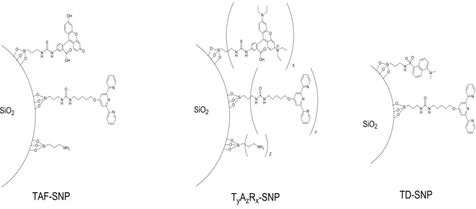
Figure 3- 1 Schematic representations of DDSNP surfaces for TD-SNP, TAF-SNP, and TyAzRx-SNP ... 47
Figure 3- 2. TEM Photos of FITC-Terpy-NP1 from UC-Berkeley Electron Microscope Lab (scale unit: 50 nm) .... 49
Figure 3- 3. TEM Photos of FITC-Terpy-NP1 from UC-Berkeley Electron Microscope Lab (scale unit: 0.2 µm) ... 50
Figure 3- 4. Scheme of Fluorescence Quenching upon Introduction of Metal Ions ... 51
Figure 3- 5 Fluorescein propyl triethoxysilane; λex: 490 nm; λem: 525 nm ... 53
Figure 3- 6 Overlay plot of FITC-terpyridine-NP1 (TAF-NP) fluorescence quenching in the presence of increasing amounts of Fe3+ (1:1 ethanol:ACN; 80µg/ml nanoparticle) ... 54
Figure 3- 7 Overlay plot of FITC-terpyridine-NP1 (TAF-NP) fluorescence quenching in the presence of Cu2+ (1:1 eth-anol:ACN; 80µg/ml nanoparticle) ... 55
Figure 3- 8 Overlay plot of FITC-terpyridine-NP1 (TAF-NP) fluorescence quenching in the presence of Fe2+ (1:1 etha-nol:ACN; 80µg/ml nanoparticle) ... 55
Figure 3- 9 Quenching of nanoparticle, FITC-terpyridine-NP1, fluorescence under the presence of Zn2+, spectral over-lap (solvent: 1:1 ethanol:ACN; 80µg/ml nanoparticle concentration) ... 56
Figure 3- 10 FITC-Terpy-NP1 quenching sensitivity versus metal ions at a range of concentrations. The nitrate salts were used for all of the metal ions except Fe3+ where the chloride salt was used. (20 µg/ml DDSNP solution; 1:1 Etha-nol:ACN) ... 57
Figure 3- 11 Stern-Volmer plot of FITC-Terpy-NP1 versus metal concentration for [Zn2+], [Ni2+],[ Fe3+], and [Pb2+]; (20 µg/mL DDSN solution in 1:1 Ethanol:ACN) ... 58
Figure 3- 12 Stern-Volmer plot of FITC-Terpy-NP1 versus metal concentration for [Zn2+], [Co2+], and [Cu2+]; (20 µg/ mL DDSN solution in 1:1 Ethanol:ACN) ... 58
Figure 3- 13 Polynomial Fit for Stern-Volmer plot of FITC-Terpy-NP1 versus metal concentration for [Cu2+] and [Co2+] ... 59
Figure 3- 14 Fluorescence quenching of RITC-Terpy-NP upon the introduction of Ni2+ up to 3 µM. (20 µg/ml DDSNP; in EtOH) ... 62
Figure 3- 15 Fluorescence quenching of RITC-Terpy-NP upon the introduction of Cu2+ up to 3 µM. (20 µg/ml DDSNP; in EtOH) ... 63
Figure 3- 17 Fluorescence quenching with RITC-Terpy-NP1. Metal ion solutions were made from metal nitrates. (20 µg/ml DDSNP; in EtOH; λex = 525 nm, λem = 575 nm) ... 63
Figure 3- 18 Stern-Volmer plot of RITC-Terpy-NP1 versus metal concentration for [Zn2+], [Fe3+], and [Pb2+]; (20 µg/ mL DDSNP in Ethanol) ... 64
Figure 3- 19 Stern-Volmer plot of RITC-Terpy-NP1 versus metal concentration for [Zn2+], and [Ni2+] (20 µg/mL DDSNP in Ethanol) ... 64
Figure 3- 20 Stern-Volmer plot of RITC-Terpy-NP1 versus metal concentration for [Zn2+], [Cu2+], and [Co2+]; (20 µg/ mL DDSNP in Ethanol) ... 65
Figure 4- 1 Diagram for our quencher displacement assay surface ... 70
Figure 4- 2. Effect of addition of five equivalents of substrate to a metal ion quenched terpy-DDSN. ... 72
Figure 4- 3 Lewis Structure of the 5 tested substrates.. ... 72
Figure 4- 4 Effect of substrate on emission recovery for FITC-NP1-Cu2+ (20 µg/ml NP1; 25 µM Cu2+) ... 73
Abstract
Chapter 1
Introduction
1.1 The Properties and Applications of Silica Nanoparticles
Research into nanoparticles is increasing due in part to the curious properties nanoparticles possess
due to their size. 1-3 Nanoparticles, defined as being in the range between 1-99 nm, are unique in that
they, as opposed to single molecules or bulk material, tend to undergo rapid Brownian motion when
dispersed in a solution and thus stay suspended at low concentrations. 2-4 As the particle size is smaller
than the wavelength of visible light, the suspension remains transparent despite bulk materials of the same
type reflecting or refracting light at larger particle sizes. 1-6 Nanoparticles are of particular interest to
analytical chemists and bioanalytical chemists because of their potential for use as benign sensors or drug
delivery systems. Due to their small size, nanoparticles can pass through the renal barrier unobstructed
leading to safe disposal from the body. The wide range of surface modification possibility then provides
sufficient customization to target potential antigens.2, 7-14
Silica nanoparticles (NPs) are receiving particular interest due to the rich chemistry available to
modify the surfaces and the relative cheapness and ease of synthesis compared to gold or iron NPs
respectively.12, 15-22 Synthesis of silica particles can follow either the top down or the bottom up approach,
but the former is typically reserved for micron sized particles and involves high energy mechanical
milling.23, 24 However, mechanical milling can only be effective and efficient up to a point, as the grain
required becomes smaller, more energy and time is required to bring down the median radius of the glass
beads.23 In 1968, Dr. Werner Stöber, building off of work by Dr. Kolbe, published a study on the
synthesis of colloidal silica particles of single micron sized silica nanoparticles through the use of
tetraorthosilane otherwise known as tetraethoxy orthosilane (TEOS) monomers reacting in a solution of
ethanol with an excess of water and ammonia. Their research demonstrated the ability to control size
2
time.25 Although the study had initially only focused on generating micron sized silica colloids, further
research on the topic has yielded the ability for the method to synthesize nanometer sized silica
nanoparticles.20, 22, 26-29
The exact mechanism for nanoparticle formation is still under debate but experiments in microgravity
and small-angle x-ray scattering experiments suggest that nanoparticle formation and growth is through
fast nucleation concurrent with controlled monomer aggregation increasing both nanoparticle
concentration and size over time.24, 30, 31 Additionally, the synthesis of the particles can be carried out in a
variety of pH environments utilizing different acids and bases as reaction catalysts.22, 24, 32 The pH of the
solution and the nature of the catalyst influences the rate of particle formation and particle structure
leading to a robust systems with a large amount of synthetic control.25, 29, 33
Commercial silica nanoparticles can be synthesized using monomers or through the industrial
process of seeded water glass.24, 25, 29 Water glass, or sodium silicate, is formed by mixing silica with
sodium carbonate under high heat and pressure.24 The resulting mixture is typically synthesized and
dissolved into aqueous environments resulting in a high viscosity clear liquid.24 Water glass is typically
used in cements, fire protection, and textile industries, but the silica can be nucleated under the right
conditions to form nanoparticles without additional acid or base introduction. 29 Industrial synthesis of
silica nanoparticles is typically through this route as it is cheaper than monomer condensation, despite
requiring high temperatures and pressures.24
Silica nanoparticles form via monomer condensation processes through silane chemistry and the
hydration of one of the ethoxy arms of TEOS.24, 25 Acid catalyzed reactions and base catalyzed reactions
are considered different during nucleation phase as they are believed to progress through slightly different
mechanisms based on pH, the acid catalyzed reactions are faster and favor chain formation, while
catalysis under basic conditions is believed favor the total hydrolysis of the silane monomer prior to
nucleation.24 A study done on the optimal pH for silica gel formation at high silane monomer
3
9.0 and would gel if straying in either direction.25 Despite this distinction, at low monomer
concentrations, there is no measureable difference between acid and base catalyzed react products.34-36
Additionally, at high concentrations with a suitable salt, the acid pathway is able to create gels, thus
emphasizing the importance of solvent composition during nanoparticle synthesis.24
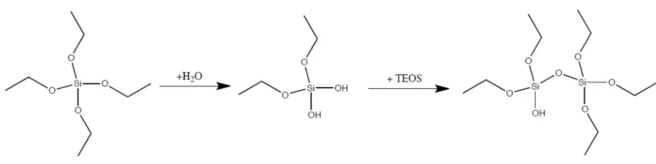
In the work to be reported here, we chose to proceed through the base catalyzed pathway for silica
nanoparticle formation (see Fig. 1-1) due the improved stability over the acid pathway even in the
presence of unanticipated salts.
Figure 1- 1. Synthetic Scheme of TEOS hydration and monomer formation used in our work
Surface modification is an essential part to making silica particles useful. Although silica could
theoretically bind oxygen or nitrogen groups in organic molecules or bind to metal ions through its
oxygen groups, the overall structure of the silica crystal will be overall positive or negative and the strain
on the crystal structure would therefore make it unfavorable.24 Compounds that bind directly to the silica
crystal are compounds that will typically increase the dissolution rate of silica such as HF or strong
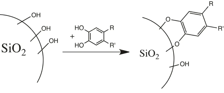
mineral acids.24 Of particular note are catechol compounds or catechol derivatives. Research done in the
1960s showed that catechol compounds will weakly bind to silica particles and to surface silyl atoms via
the same mechanism as nanoparticle formation, shown in Fig 1-2.24 This reaction will not directly affect
the effectiveness of modified silica nanoparticles, as it attacks silyl groups still baring two or more
hydroxyls, but is a concern when unmodified controls are used.4, 24 Additionally, the equilibrium is not
4
Figure 1- 2. Catechol attack of surface silyl groups24
Although direct binding of molecules to the silica crystal is unfavorable, silane chemistry, employing
similar chemistry to silica particle formation, is well researched and very robust.20, 27, 28, 32, 37-40 Bottom up
synthesis of silica particles via silane monomers leaves most if not all of the surface hydroxyl groups
available for further synthesis, thus removing the need to activate the silica surface.24, 41 Typical
modification techniques involve addition via silane chemistry or direct modification of surface
hydroxyls.4, 21, 22, 32, 42
Research into the surface of silica particles began in 1936 when Kiselev first proposed that the active
surface of silica was characterized by hydroxyl groups coating the surface of the SiO2 bulk and in the next
two odd decades research into the activity of silyl groups was probed by a number of groups each
contributing a different piece of the puzzle.24 Then in 1959, Belyakova et al. published a review showing
that the silica active layer is the only layer that participates in silane chemistry or organic molecule
chemisorption.24, 29 By summarizing the work of the preceding decade, Belyakova et al. were able to
surmise that unless micropores were involved, such as those in controlled pore glass, the SiO2 bulk in a
silica particle would not participate in the surrounding chemistry.29 Many groups since then have utilized
this principle to embed and protect dyes or other active species inside a silica core.32, 43-46 This finding also
means that most of the nanoparticle’s characteristics would be defined by its surface identity, enhancing
5
It is worth noting here that silica nanoparticle formation is a reversible reaction and the silica can
indeed dissolve into the surrounding solvent.24 Thus, any surface modification stability is limited by the
natural leeching of silica. Fortunately, trace impurities decrease the solubility of silica in water, from 16
to 60 ppm depending on particle size, and ethanol, up to 164 ppm in pure ethanol, and thus surface
modifications, acting as trace impurities, increase the stability of the silica nanoparticle.24, 48, 49 Not only is
the total equilibrium amount of dissolved silica in solution decreased upon the introduction of impurities
and surface modification, but the dissolving rate also decreases with impurities.24 This decrease in
dissolution rate is enhanced by a basic environment while being counteracted by the small size and high
specific area of nano-sized silica.21, 29 The rate of silica dissolution in a pure solution can be given as:24
= − (1)
where dc/dt is the rate of dissolution, k1 is rate constant of dissolution, k2 is rate constant of deposition, c is
the concentration of silica, and S is the specific area of the solid silica.24 Numerous studies using silica
nanoparticles as sensors or catalysts have demonstrated stable nanoparticle composition for upwards of 3
months.32, 38, 40, 50, 51 However, the stability of the nanoparticle over time is limited by the silica core
stability and by the dye or surface modification group stability.24, 49
Surface modification of silica particles has a long history dating back to around 1968 with the
modification of hydrophilic silica surfaces to create hydrophobic surfaces for dispersion of silica in
organic solvents.24 Initial efforts into altering silica surfaces mostly involved improving silica dispersion
into various media. 52 As shown in Figure 1-3, many of the techniques, such as silane modification22, 32, 52,
53, modification of the silane functional group13, 22, 47, and direct surface modification35, 53-57, have greatly
6
Figure 1- 3. Taken from Peng and coworkers, the figure presents the common modifications possible for silica surfaces52
Surface modified nanoparticles continue to be a promising field of research due to the ability to tailor
the surface chemistry of the nanoparticles for biochemical sensing and imaging purposes.28, 38, 58-61
Recently, groups have successfully tailored the surface of fluorescent particles to target cancer cells or
interesting marker proteins for relevant diseases.47, 62-67 This type of surface chemistry is of particular
interest in the work presented as it involves the covalent incorporation of a dye onto the surface o either
through thiol or silane chemistry.13, 27, 32, 65, 66 Many of these syntheses feature a silanized dye either on the
surface or incorporated into the matrix core of the particle.32, 40, 59, 65 This research thesis focuses on the
former in an attempt to synthesize silica nanoparticles with surface bound chromophores and functional
groups for further modifications with receptor groups that may affect the photochemistry of the dye..
1.2 Fluorescence Basics and Quenching Theories
When a molecule becomes excited via light absorption, there are a number of possible processes that can
7
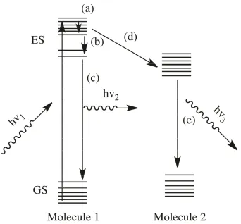
Fluorescence is the emission of photon from an excited molecule as it returns to the ground state. The
emitted photon is typically of a lower energy due to a large amount of alternative intermediate pathways
such as vibrational relaxation, contact energy transfer, or loss as heat shown in Fig 1-4.69,70, 71 This
phenomenon depends upon the difference between the initially populated molecules orbital’s energy
levels in a particular fluorescent molecule as a function of environment.69, 70 Research to alter the effect of
the environment of the dye molecule and their corresponding spectral changes has been reviewed
recently.11, 52, 61, 72-76
Figure 1- 4. Diagram of possible collisional energy loss pathways. (a) Thermal relaxation (b) Energy loss as heat (c) Fluorescence (d) Energy transfer (e) Phosphorescence70, 71
When light of the correct wavelength is absorbed, the electron is excited to a higher energy level, an
excited state (GS to ES in Figure 1-4).60, 69, 70 From that excited state, the electron can return to the ground
state by losing energy through a variety of pathways based on the electron states of the molecule.69, 70
8
emission.70, 77 The statistical likelihood of photon emission is dependent upon the identity of the molecule
based on the possible electron states, i.e. molecular orbitals, and the relative populations of electron
states.69 This statistical likelihood is known as quantum yield of a fluorescent molecule and quantitates
the efficiency of the fluorescence process.38, 69, 77, 78
ɸ = #
# (2)
Alternative relaxation states are possible photon emission.69 During electron relaxation, the
fluorescence pathway for energy loss may not be directly available, the electron could then drop down to
the nearby more available lower energy state before returning to ground state.69, 70 Both fluorescence
transitions and non-fluorescent transitions may not happen instantaneously as the electron needs time to
relax back to a lower energy state. The time between excitation and emission is known as the fluorescence
lifetime and typically can range from a manner of nanoseconds to half a microsecond.69, 70 With the
advent of new instrumentation, it is now possible to probe the fluorescence lifetime and use it as a
measure to determine whether the substrate is interacting with the fluorescent source.70 While we do not
utilize this method for probing fluorescence interactions here, it is necessary to describe, since as much
work is done in this area and many conclusions we draw are based on information obtained via this
technique.
Fluorescence dyes have garnered significant interest as a labeling technique and potential
analytical probe because of their high theoretical quantum yield, defined as the ratio of photons emitted
over photons absorbed.69, 77 Under the right conditions with the right fluorescent chromophore, a single
excitation photon is expected to be enough to incite the release of an emission photon thus making it a
more sensitive chromophore compared to UV-VIS active chromophores.69, 70 Additionally, the dyes can
be tailored to interact with certain molecules and turn-off or turn-on under set conditions.74, 79-82 Multiple
systems,13, 32, 47, 51, 52, 83-89 including the work presented here, exploit this characteristic of fluorescent
9
Besides quantum yield and fluorescence lifetime, the actual emission spectra may also shift under the
influence of interacting substrates or local matrix effects.70, 91-93 Both influences can cause a bathochromic
or hypochromic shift in the emission peak, as described below, suggesting interaction between the
molecule and surrounding environment.69-71
A bathochromic shift, or red shift, is the emission peak shifts to a longer wavelength, or lower energy,
whereas a hypochromic shift, or blue shift, is when the emission peak shifts to a shorter wavelength, or
higher energy.60, 70, 71 These shifts signify a change in energy for the emitted photon in fluorescence and
can be indicative of enhanced solvent molecules interactions or a change in the local matrix,60, 69, 70 such
as the surface modified nanoparticles. The possible explanations for these shifts are complex and
information is often presented on a per system basis.70, 94-97 A lack of a unifying theory to predict
fluorescence energy shifts has led to continued research in the field of fluorescent dye functionalized
nanoparticles. One such energy interaction whose mechanism is not fully understood is fluorescent
quenching.
Upon the addition of molecule 2 (figure 1-4), excited state electrons in molecules 1 can return to
ground state through intermolecular pathways.70, 71 When this pathway becomes competitive with the
native fluorescent emission pathway, the molecule will no longer emitt.69-71 In this case, the molecule is
said to be quenched.70
Fluorescent molecules can be quenched in a variety of ways, sometimes even the inclusion of a
charged salt molecule can decrease the quantum yield of a fluorescent dye.15, 46, 98, 99 Fluorescent
molecules sometimes exhibit quenching behavior when in proximity to metal ions,32, 90, 96 conjugated
organic molecules6, 100-102, and other dyes13, 75, 103-106. The specific quenching mechanism and its efficiency
is unique for each substrate, but can be categorized into two categories: static quenching or Forster
non-10
emissive complex, while energy transfer pathways tend to decrease the efficiency of the fluorescence
through a distance dependent effect.32 Rurack and coworkers recently40 reported the formation of a Cu2+
terpyridine single molecule complex that was able to favor distance based energy transfer over the more
common non-emissive complex formation typically seen for Cu2+ complexes or other single molecule
metal bound chromophores such as those synthesized by Lu and coworkers 107 and Wang and coworkers
90.
Forster resonance energy transfer (FRET) was first proposed by Theodore Forster in 1948 and refers
to the energy transfer between two chromophores.52 When the absorption band of Dye 1 overlaps with the
emission band of Dye 2 the emission can be quenched by Dye 2 absorbing the energy released in
emission from Dye 1.52, 61, 108, 109 During energy transfer, Dye 2, if fluorescent, could emit at a longer
wavelength than the initially excited chromophore. The result is that the system emits from Dye 2 after
being excited at Dye1’s absorption wavelength.52, 70 If the energy transfer pathway is then broken, Dye 1’s
fluorescence could be recovered.52, 75, 110 This property of the FRET system allows for in vivo probing of
biological systems without cell death or analysis of the interactions between systems or domains too small
for optical or electric microscopy.52, 61, 62, 111 FRET has been utilized in probing microcellular
interactions111, protein and enzyme structure analysis91, 112-116, ratiometric sensing32, 96, 109, 117-119, and solar
11
Figure 1- 5. Diagram for Forster resonance energy transfer13, 32, 52
FRET is affected by energy transfer efficiency, E, between the chromophores and the distance
between them52, 61. Their relationship is given below:
=
∑ (3)
An alternative equation looks strictly at the distance between the two chromophores given a distance
at which quantum efficiency is 50%:
=
( ) (4)
The quantum efficiency is found to be related to the distance by 1/(r6)52, due to the dipole-dipole
coupling nature of FRET75, 106, meaning that the efficiency of the interaction drops off by a factor of
distance to the power of six. This means typical FRET distances are less than 10 nm with exaggerated
signal drop off, thus making FRET especially useful for domain distance probing.61, 128, 129 In equation (4),
12
emission spectra of the transmitting chromophore; the larger the overlap between the two the higher the
expected efficiency.52, 75
= 9000 (ln 10) 128
Where Q0 is the fluorescence quantum yield of the donor, k2 is the dipole orientation, J represents the
spectral overlap, n is the refractive index of the solution and N is Avogadro’s number. Since the quantum
yield of the FRET system is defined as the number of photons emitted by the receiving chromophore, the
quantum yield fluorescence in the receiving chromophore must be accounted for as well. Most of the
variables in the last two equations are unique to each FRET pair.52, 61
Recent research into FRET quenching has seen a number of nanosensor based FRET probes
synthesized as metal ion detection probes with sensitivity in the micromolar range.13, 52, 84, 109 Research
presented by Mancin features a ratiometric dye system with FRET potential as metal ion sensors that
respond as both a turn-on and turn-off sensor depending on the wavelength of light chosen for
fluorescence sensing.13, 32 Their nanoparticles were synthesized with a silica core containing one part of a
FRET pair while the outer surface is decorated with the second dye in the FRET pair and a metal ion
binding site.32 Other research into the field of FRET based nanosensors used modified quantum dots.13, 52,
130 The quantum dot luminescence was altered upon the introduction of a target substrate and deactivated
13
Figure 1- 6. Figure reproduced from Peng and coworkers, diagram of nanorod functionalized gold nanoparticle being FRET quenched by quantum dots. With signal return upon the introduction of TNT52
Although the research presented here does not feature the use of FRET with two dyes. FRET
quenching for detection of alternative substrates allows for greater customization of the DDSN system to
probe into the intricacies of fluorescent molecules and their interactions with the surrounding species and
could be applicable to the system probed in this study.
1.3 Terpyridine as a Method Ion Binding Receptor
Research with terpyridine has been primarily focused on its incorporation into polymer chains132-138 and
the tuning of terpyridine properties via the metal ions it is bound to136, 139-148. Interest has increased in
recent years for terpyridine as a possible photon receiver and electron donator in solar cells due to their
customizability in polymer matrices.82, 149-153
Terpyridine is a tridentate chelator capable of binding to most all metal ions typically in a dimeric
14
Figure 1- 7. Diagram of metal ion bound terpyridine, presented with interchangable solvent molecules
Although not having a distinctive visible absorption spectra on its own, metal bound terpyridine can
take on the intense color due to the metal-ligand charge transfer band (MLCT).127, 153-157 The absorption
spectra of the metal-terpyridine complex can be further tuned by adding active groups onto the terpyridine
moiety during synthesis or coupling the terpyridine moiety onto conjugated polymer chains.133, 148, 158-162
This versatility led to research into its potential use as a co-polymer active site for polymer solar cells.141,
153
Figure 1- 8. Common modified terpyridine moieties
Research into the customizability of the terpyridine spectra typically utilizes terpyridine as a charge
transfer complex that can pass electrons down an attached conjugated polymer backbone chain or solid
15
the conditions during metal-terpyridine complex formation.153, 165, 166 Despite being highly customizable,
the synthetic cost of such a polymer may make commercialization difficult.146, 153, 162, 164 Thus research has
shifted to the self-assembly of terpyridine containing polymers, thus decreasing the cost for synthetic
design while ideally retaining terpyridine’s charge transfer ability.146, 153, 164, 165, 167
Other researcher explored terpyridine and its derivatives’ ability to act as metallo-enzymes through its
strong binding to most first row transitional metal ions.139, 153, 168, 169 Specifically, strong binding of Ni2+,
Cu2+,Fe2+/3+, and Pt2+ are of particular interest due to the bioavailablity of enzymes containing Ni2+, Cu2+,
and Fe2+/3+ and the strong catalytic activity of Pt2+ for a variety of reactions from hydrogenation to ring
formation.153, 170 When coupled with aromatic conjugated terpyridine moieties, the terpyridine is believed
to be able to transfer electrons to the metal ion center or absorb unwanted electrons into the ground during
catalytic activity.82, 153, 171
In addition to binding most first row transition metals153, terpyridine was shown to bind to other
larger metal ions.138, 171-173 Binding to these larger metal ions requires heat and purification via
crystallization due to the high energy barrier required to wrap the terpyridine molecule around the larger
metal ion.156, 174, 175 Once the complex is formed, it is resistant to both heat and light while retaining most
of its charge transfer properties.156, 175, 176 Additionally, the complexes tend to be luminescent allowing for
the creation of fluorescence terpyrdine complexes with tunable fluorescence profiles.176 This property
makes those metal-terpyrdine complexes attractive for single complex sensing since the terpyridine
moiety can be modified to contain an additional binding site or tuned for specific absorption-emission
wavelengths. Metal ions such as Ru2+ and Ir2+ can even undergo redox reactions while still attached to
terpyridine.160, 177
The potential redox reactivity and chromophore properties of the Ru2+ and Ir2+ terpyridine complexes
has led to their use as charge transfer complexes as well as catalysts,153 due to the transfer of 2 electrons
16
increases the possibility for artificial photosynthesis to occur because only two redox reaction pairs are
required to happen simultaneously in contrast to the four for a single electron transfer pathway.179
2H + e = H (6)
2H O → 4H + O + 4e (7)
When a current is applied, Ru2+ and Ir2+ terpyridine polymers have the potential to catalyze both
hydrogen reduction as well as oxygen oxidation in artificial photosynthesis. Metal-terpyridine containing
polymer with photosensitizing dyes or electron sinks were reported to increase the electron density in the
terpyridine polymer chain increasing the rate of artificial photosynthesis.146, 153, 160
Extending the photosensitive properties of terpyridine-Ru containing polymers, researchers have
cross-linked the terpyridine containing polymers with other polymers to increase electrical conductivity or
light absorption in metal organic frameworks, MOFs.180 MOFs are multiple polymers cross linked from
individual monomers to form a larger web like complex with a large amount of customizability.153 Much
like the coupling of metal-terpyridine units to a single polymer chain, construction of MOFs typically
uses self-assembling polymer monomers to create a MOF with the desired properties. By varying the ratio
of any single polymer or synthesis conditions, a MOF’s properties can be fine-tuned.
Another field of research utilizing terpyridine complexes revolves around their use as luminescent
analytical probes.82, 153, 181, 182 By modifying the terpyridine moiety, researchers have created metal ion
sensors able to selectively detect a variety of first row transitional metal ions.40, 82, 146
1.4 Metal Ion Sensing
Metal ion sensing in an aqueous environment is important due to widespread metal ion contamination
in water sources from industrial contamination or spillage.9 As more and more countries embrace modern
17
and waste treatment must be monitored and properly disposed.32, 183 Part of ensuring the correct and safe
disposal of waste is through the ability to monitor waste spillage and metal ion concentrations in water
sources.
Human consumption of contaminated water causes heavy metal build up in the body leading to renal
failure or heavy metal poisoning.184 Additionally, excess metal ions in the environment may affect the
local fauna and increase the rate at which they build up in the human body through the consumption of
said fauna.70, 185 With the potential impact in mind, the ability to sensitively and selectively monitor metal
ion concentrations in aqueous systems with a variety of matrix effects with high precision and
reproducibility remains an active research field.
Most analytical techniques for sensing metal ion concentrations at low levels typically employ atomic
spectrometry which requires the use of high purity gases to generate the plasma or flame required for the
spectrometer’s sensitivity.70, 186 This technique is not field work friendly and much research has been
devoted into developing cheaper alternative sensing methods with comparable selectivity and sensitivity
that can be employed at the site of sample gathering.70
One such field of research is molecular metal ion sensors, much like the well-known reaction between
metal ions and ethylenediaminetetraacetic acid (EDTA), the molecular sensors developed could change
color after binding with a particular metal ion. However, the sensitivity of colorimetric sensors does not
compare with that from atomic spectroscopy.70, 71 Research done by Wang and coworkers describes the
synthesis of a fluorescent molecular sensor utilizing a dansyl dye bound bipyridine ligand. The molecular
sensor targets Fe3+ ions, which bind to bipyridine and the dansyl signal is quenched. The sensitivity of
such a sensor is reported to be in the micromolar range but is restricted to organic solvent systems due to
the hydrophobic nature of both the dye and the chelate. 90 Another sensor was designed by Zhang and
coworkers as a ratiometric sensor was capable of Fe3+ detection in blood serum. The sensor consisted of
18
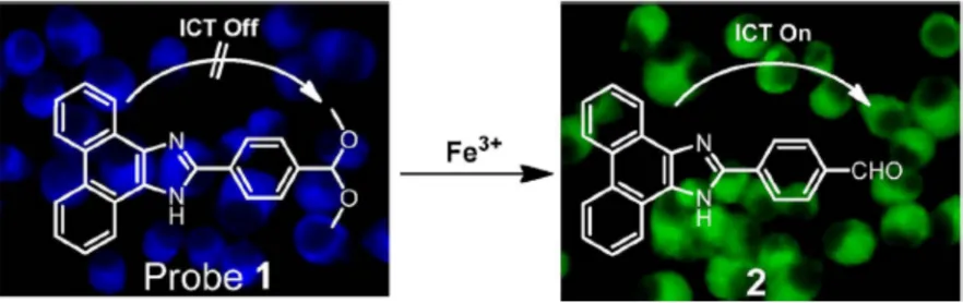
the metal catalyzed the deprotection of the ketone to form an aldehyde tail. The molecule undergoes
internal charge transfer, red shifting its fluorescence as a function of [Fe3+]. 96
Figure 1- 9. Graphic reproduced from Zhang and coworkers. The figure presented a molecular probe capable of ratiometric Fe3+ detection using fluorescence shifts96
Utilizing similar ideas, quantum dots (QD) have been presented as sensors for detection of metal ions.
Sun and coworkers, employed surface modified CdTe nanoparticles for the detection of Cd2+. The QD is
covered by surface bound phenanthroline attached the quantum dot’s thioglycolic acid functionalized
surface. The phenanthroline binds to the surface of the quantum dots and quenches the green fluorescence
of the CdTe quantum dot. Addition of Cd2+ returns the fluorescence. This research demonstrates the
ability of quantum dots to act as sensors that directly utilize their own excitation and emission spectra of
without the need for an additional chromophore. 187 The utilization of QD’s for metal ion detection has
often been hampered by the difficulty and cost of QD synthesis, modification, and stability.28 Other
quantum dots have been employed in a variety of schemes to detect cyanide, food toxins, and other
quantum dots.13, 28, 130, 188
In order to engineer robust systems capable of visual and stable analysis of metal ion contamination
in water, solid support based sensing has been proposed as a reusable sensing scheme. One such system
employs silane chemistry to modify glass slides with a chelate and dye that is able to change colors
visually in the presence metal ions.153, 189 The solid support system shows potential as a robust reusable
19
surface area of the modified glass slide.153, 190, 191 Further research is being conducted to convert these
solid support systems into disease related sensors as their robustness and relatively low cost is
appealing.89, 192, 193
Falling between solid support and quantum dots in terms of size and surface area are nanoparticles.
Nanoparticles can be dispersed in solvents while being easier to modify, synthesize, and isolate.24, 52
Modified nanoparticles were used as disease targeting systems66, 194, 195, biological target labeling
schemes196, 197, and sensors13, 22, 32, 198 due to their customizability, high functional group loading, and ease
of dispersion compared to micron sized particles.199, 200 As they can be chemically modified to suit a
variety of purposes, gold, silver, iron oxide, and silica nanoparticles have received particular interest in
recent years.10, 20, 32, 40, 199, 201-204
Gold nanoparticles were used in sensors due to their easy modification via disulfide bonding to the
surface and gold’s inherent inertness.10, 39, 202, 205 Initial efforts featured gold nanoparticles modified with
biological target binding groups such as antibodies that bind to cell targets staining the region with
different colors based on the size of the gold nanoparticle.15, 202 Drug molecules were attached to the
surface of 15 nm gold nanoparticles and were successfully delivered to bacterial and diseased cells in
vitro.128 The advantage of this type of delivery system is the ability to target cells or bacteria via surface
modification of the nanoparticle increasing drug potency. When enhanced by the large surface area to
volume ratio afforded by nanoparticles this can lead to a large increase in the local concentration of the
drug without increasing the dosage.15, 19, 128
Silver nanoparticles were similarly explored for their potential disease curing abilities. Of particular
note is a dendrimer modified silver nanoparticle that excreted renally from the human body when under
20 nm in diameter.206 Although the ability to target specific cancer cells was not demonstrated, this result
20
Iron oxide nanoparticles, primarily Fe2O3, have been developed primarily for sensor and catalytic
applications.207-209 Iron oxide nanoparticles, 10-50 nm in diameter, synthesized via oil emulsion followed
by surface modification gave particles for MRI imaging agents or sensors.210, 211 Iron oxide differs from
silver and gold nanoparticles in that the iron center is high spin resulting in a strong magnetic moment.39,
128, 194, 212 It is this property that allows the iron oxide core to function as targeted imaging agents after
surface modification.39, 210, 213 Recent research also looked into Fe
2O3 nanoparticles as magnetic storage devices much like current hard drives.214
In additional to these three types of nanoparticles, silica nanoparticles have also been ised for similar
sensing and catalytic applications.12, 15, 215 Silica nanoparticles can be cheaply prepared compared to gold
and silver nanoparticles.24, 25, 33, 54 Unlike the other metal based nanoparticles, silica is magnetically and
optically inert thus providing a blank canvas for which the behavior of the nanoparticle is almost solely
determined by the surface modifications applied.24, 185 Research was conducted for sensing and catalysis
similar to chemistry done on glass slides.22, 36 For the research described in this thesis, we focus on the use
of silica nanoparticles as metal ion sensors.
Silica nanoparticle based metal ion sensors have been successful in detectingPb2+, Cu2+, Fe3+, Ni2+,
and Co2+ with targeted surface modification.32, 40, 90 Lead ions were successfully detected using thiol
modified dansyl dye doped nanoparticles that were able to detect Pb2+ concentrations in the micromolar
range in acetonitrile.32 Copper(II) and iron(III) were successfully detected using a terpyridine
functionalized rhodamine dye doped silica nanoparticle similarly in acetonitle.40 Nickel(II) was similarly
detected through the use of a nitrilotriacetic acid functionalized tetramethyl rhodamine doped nanoparticle
system. 216 The surface of the nanoparticle was closely packed with these molecules resulting in a single
Cu2+ or Pb2+ being able to quench upwards of 13 surface-bound dye molecules.40, 41
Each of the systems presented above were able to detect specific metal ions with a great deal of
specificity, however those systems required the nanoparticle to be dispersed in an organic solvent40 or
21
polar modifiers in an optimized ratio, we predict that the surface-optimized nanoparticles will be able to
sensitively and selectively detect the desired metal ions while keeping the nanoparticles dispersible in
water or alcohol-water mixtures with controlled pH. In this work we will
The dye-doped silica nanoparticles (DDSN) synthesized here utilize a surface tethered 2’ 2:6’
2-terpyridine (terpy) as the metal binding site due to its high binding constant for most first row transitional
metals as shown in table 1.
Table 1. Stability Constants for Metal Bound Terpyrinde153
Mn2+ Fe2+ Co2+ Ni2+ Zn2+ Cd2+
log K1 4.4 7.1 8.4 10.7 6.7 5.1
log K1 13.8 9.9 11.1 5.2
Terpyridine was extensively studied for its use in metal organic frameworks (MOFs) as well as its
potential use in solar cells.132, 153, 154, 162 Additionally, terpyridine exhibits varied fluorescence and
absorbance behavior based on metal ion binding.153 Here, we aim to utilize terpy as a means to tailor the
response of the nanosensor to different metal ions and to study the quenching effects as a function of the
composition of the DDSN surface chemistry. It may even help us further probe Foster Resonance Energy
Transfer (FRET) dye quenching pathways.
1.5 Turn-on Sensors
In a conventional indicator displacement assay (IDA), a metal binding chelate with open coordination
sites binds to a specific metal ion to form a metal-chelate complex that can then bind a dye altering the
dye’s emission characteristics. A substrate is then added to compete with the dye for the chelate-metal
binding site, thereby releasing the dye into the surrounding substrate and coloring the solution.217, 218 A
similar scheme was devised for fluorescent dyes, as the fluorescent dye is quenched upon binding to the
22
dye affinity for the metal ion versus the substrate affinity for the metal ion determine selectivity of the
system. Some dyes bind directly to a variety of metal ions but do not offer the ability to tailor the binding
or spectroscopic behavior of the dye. By placing the metal ions on a chelate next to the dye rather than
have the dye directly interacting with the metal ion, this system potentially opens up more customizability
at the surface of the molecule. Coupled with the use of DDSNPs, a fluorescent dye that will be quenched
in the proximity of the metal ion-chelate complex allows for testing to be done directly on the suspended
nanoparticle without the need for separation.84, 219, 220
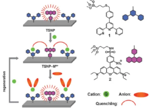
By modifying an older IDA concept, Rurack and coworkers proposed a scheme for a turn-on sensor
by introducing an anion to compete with the metal chelate whose nanoparticle fluorescence is quenched
under the presence of metal ions as shown in Figure 1-10, calling these quencher displacement assays
(QDA).40 By inducing a signal change through the removal of the metal ion, the sensor can now be used
to detect anions and other metal binding molecules.128, 218, 221, 222
23
Figure 1- 11. Figure reproduced from Rurack and coworkers Shows the results of added anions in the quencher displacement assay scheme at 5 equivalents versus metal ion concentration (1 µM)40
.
The rhodamine B functionalized SNP’s fluorescence was quenched by Cu2+, Fe2+, Ni2+, Mn2+, listed
in order of quenching efficiency, with Cu2+ chosen as the target metal-terpyridine system for quencher
displacement assays.40 The fluorescent lifetimes for first row transitional metal bound nanoparticles
increased with metal ion concentration suggesting a distance based effect between the bound metal ion
complex and the dye direct interaction between the dye and the metal ion, known as static quenching or
contact quenching. The team of researchers tested a variety of hard to detect anions, including fluoride,
chloride, bromide, iodide, nitrate, sulfate, and phosphate, and discovered that phosphate presented the
largest return in signal at 1 uM as shown in Figure 1-11. The team suggested that, due to the return in the
fluorescent lifetimes of the system upon the introduction of the anion, that the metal ion must have been
removed from the terpyridine in order to recover the fluorescence signal.40 This research lays the
foundation for a system that could potentially offer sensitive and selective sensing of anions in a variety
of solvent conditions.80, 218, 221 By altering the surface chemistry, the metal bound nanoparticles could be
used to detect these molecules by modifying the terpyridine or silica nanoparticle surface.
In this work we will present a new terpyridine surface functionalized DDSN, with alterations to
surface composition and dye identity which enable it to respond very sensitively to copper ions (see Fig
1-12). We will present data on this system activity as a potential QDA system much like the the system
24
species by substrate addition to recover the signal as the primary sensing technique (turn on sensor). We
propose that substrate binding to the terpy-Cu chelate may affect energy transfer quenching without
displacement of the metal ion.
Figure 1-12 the sensing scheme used in our work for both metal ion binding that turn-off dye
emission and the turn-on emission via binding of biologically relevant targets.4, 13, 76, 221, 223 This
hypothesis is tested by changing the terpyridine/dye/modifier ratio to study metal ion quenching and
subsequent addition of metal binding substrates to restore dye emission to offer some insight into FRET
or other type of energy transfer.40
25
Work Cited
1. Chen, L. Y.; Wang, C. W.; Yuan, Z.; Chang, H. T., Fluorescent gold nanoclusters: recent advances in sensing and imaging. Anal Chem 2015,87, 216-29.
2. Wang, W.; Tao, N., Detection, counting, and imaging of single nanoparticles. Anal Chem 2014, 86, 2-14.
3. Taniguchi, H.; Nishiya, M.; Tanosaki, S.; Inaba, H., Lasing behavior in a liquid spherical dye laser containing highly scattering nanoparticles. Opt Lett 1996,21, 263-5.
4. Qhobosheane, M.; Santra, S.; Zhang, P.; Tan, W., Biochemically functionalized silica nanoparticles. Analyst 2001,126, 1274-8.
5. Crooks, R. M.; Zhao, M.; Sun, L.; Chechik, V.; Yeung, L. K., Dendrimer-Encapsulated Metal Nanoparticles: Synthesis, Characterization, and Applications to Catalysis. Acc. Chem. Res. 2001,34, 181-190.
6. Lian, W.; Litherland, S. A.; Badrane, H.; Tan, W.; Wu, D.; Baker, H. V.; Gulig, P. A.; Lim, D. V.; Jin, S., Ultrasensitive detection of biomolecules with fluorescent dye-doped nanoparticles. Anal Biochem 2004,334, 135-44.
7. Gatselou, V. A.; Giokas, D. L.; Vlessidis, A. G., Determination of dissolved organic matter based on UV-light induced reduction of ionic silver to metallic nanoparticles by humic and fulvic acids. Anal Chim Acta 2014,812, 121-8.
8. Yu, X.; He, Y.; Jiang, J.; Cui, H., A competitive immunoassay for sensitive detection of small molecules chloramphenicol based on luminol functionalized silver nanoprobe. Anal Chim Acta 2014,812, 236-42.
9. Panichev, N.; Kalumba, M. M.; Mandiwana, K. L., Solid phase extraction of trace amount of mercury from natural waters on silver and gold nanoparticles. Anal Chim Acta 2014,813, 56-62. 10. Canbaz, M. C.; Simsek, C. S.; Sezginturk, M. K., Electrochemical biosensor based on self-assembled monolayers modified with gold nanoparticles for detection of HER-3. Anal Chim Acta 2014, 814, 31-8.
11. Descalzo, A. B.; Somoza, C.; Moreno-Bondi, M. C.; Orellana, G., Luminescent core-shell imprinted nanoparticles engineered for targeted Forster resonance energy transfer-based sensing. Anal Chem 2013,85, 5316-20.
12. Montalti, M.; Rampazzo, E.; Zaccheroni, N.; Prodi, L., Luminescent chemosensors based on silicananoparticles for the detection of ionic species. New J. Chem. 2013,37, 28-34.
13. Bau, L.; Tecilla, P.; Mancin, F., Sensing with fluorescent nanoparticles. Nanoscale 2011,3, 121-33.
14. Moragues, M. E.; Martinez-Manez, R.; Sancenon, F., Chromogenic and fluorogenic
chemosensors and reagents for anions. A comprehensive review of the year 2009. Chem Soc Rev 2011, 40, 2593-643.
15. Liang, H.; Zhang, X. B.; Lv, Y.; Gong, L.; Wang, R.; Zhu, X.; Yang, R.; Tan, W., Functional DNA-containing nanomaterials: cellular applications in biosensing, imaging, and targeted therapy. Acc Chem Res 2014,47, 1891-901.
16. Villalonga, R.; Diez, P.; Sanchez, A.; Aznar, E.; Martinez-Manez, R.; Pingarron, J. M., Enzyme-controlled sensing-actuating nanomachine based on Janus Au-mesoporous silica nanoparticles. Chemistry 2013,19, 7889-94.
17. Shrivastava, S.; McCallum, S. A.; Nuffer, J. H.; Qian, X.; Siegel, R. W.; Dordick, J. S.,
Identifying specific protein residues that guide surface interactions and orientation on silica nanoparticles.
Langmuir 2013,29, 10841-9.
18. Candel, I.; Calero, P.; Martínez-Máñez, R.; Sancenón, F.; Dolores Marcos, M.; Pardo, T.; Soto, J., Sensing properties of silica nanoparticles functionalized with anion binding sites and sulforhodamine B as fluorogenic signalling unit. Inorganica Chimica Acta 2012,381, 188-194.
26
20. Bonacchi, S.; Genovese, D.; Juris, R.; Montalti, M.; Prodi, L.; Rampazzo, E.; Sgarzi, M.; Zaccheroni, N., Luminescent chemosensors based on silica nanoparticles. Top Curr Chem 2011,300, 93-138.
21. Bagwe, R. P.; Hilliard, L. R.; Tan, W., Surface modification of silica nanoparticles to reduce aggregation and nonspecific binding. Langmuir 2006,22, 4357-62.
22. Rampazzo, E.; Brasola, E.; Marcuz, S.; Mancin, F.; Tecilla, P.; Tonellato, U., Surface modification of silica nanoparticles: a new strategy for the realization of self-organized fluorescence chemosensors. Journal of Materials Chemistry 2005,15, 2687.
23. Xia, D.; Li, D.; Ku, Z.; Luo, Y.; Brueck, S. R., Top-down approaches to the formation of silica nanoparticle patterns. Langmuir 2007,23, 5377-85.
24. Iler, R. K., The Chemistry of Silica. Wiley-Interscience: 1979.
25. Stober, W.; Fink, A.; Bohn, E., Controlled Growth of Monodisperse Silica Spheres in the Micron Size Range. Journal of Colloid and Interface Science 1968,26, 62-69.
26. Bau, L.; Bartova, B.; Arduini, M.; Mancin, F., Surfactant-free synthesis of mesoporous and hollow silica nanoparticles with an inorganic template. Chem Commun (Camb) 2009, 7584-6.
27. Arap, W.; Pasqualini, R.; Montalti, M.; Petrizza, L.; Prodi, L.; Rampazzo, E.; Zaccheroni, N.; Marchio, S., Luminescent silica nanoparticles for cancer diagnosis. Curr Med Chem 2013,20, 2195-211. 28. Knopp, D.; Tang, D.; Niessner, R., Review: bioanalytical applications of
biomolecule-functionalized nanometer-sized doped silica particles. Anal Chim Acta 2009,647, 14-30.
29. Bergna, H. E.; Roberts, W. O., Colloidal Silica Fundamentals and Applications. Taylor & Francis: 2006.
30. Pontoni, D.; Narayanan, T.; Rennie, A. R., Time-Resolved SAXS Study of Nucleation and Growth of Silica Colloids. Langmuir 2002,18, 56-59.
31. Nordstrom, J.; Sundblom, A.; Jensen, G. V.; Pedersen, J. S.; Palmqvist, A.; Matic, A.,
Silica/alkali ratio dependence of the microscopic structure of sodium silicate solutions. J Colloid Interface Sci 2013,397, 9-17.
32. Arduini, M.; Mancin, F.; Tecilla, P.; Tonellato, U., Self-organized fluorescent nanosensors for ratiometric Pb2+ detection. Langmuir 2007,23, 8632-6.
33. Vogel, R.; Surawski, P. P.; Littleton, B. N.; Miller, C. R.; Lawrie, G. A.; Battersby, B. J.; Trau, M., Fluorescent organosilica micro- and nanoparticles with controllable size. J Colloid Interface Sci 2007, 310, 144-50.
34. Nakamura, M.; Ishimura, K., One-pot synthesis and characterization of three kinds of thiol-organosilica nanoparticles. Langmuir 2008,24, 5099-108.
35. Bagwe, R. P.; Yang, C.; Hilliard, L. R.; Tan, W., Optimization of dye-doped silica nanoparticles prepared using a reverse microemulsion method. Langmuir 2004,20, 8336-42.
36. Santra, S.; Wang, K.; Tapec, R.; Tan, W., Development of novel dye-doped silica nanoparticles for biomarker application. J Biomed Opt 2001,6, 160-6.
37. <dual mode fluorophore NTA silica nanoparticle.pdf>.
38. Sharma, P.; Brown, S.; Walter, G.; Santra, S.; Moudgil, B., Nanoparticles for bioimaging. Adv Colloid Interface Sci 2006,123-126, 471-85.
39. Mahmoudi, M.; Serpooshan, V.; Laurent, S., Engineered nanoparticles for biomolecular imaging.
Nanoscale 2011,3, 3007-26.
40. Calero, P.; Hecht, M.; Martinez-Manez, R.; Sancenon, F.; Soto, J.; Vivancos, J. L.; Rurack, K., Silica nanoparticles functionalised with cation coordination sites and fluorophores for the differential sensing of anions in a quencher displacement assay (QDA). Chem Commun (Camb) 2011,47, 10599-601. 41. Moon, J. H.; Kim, J. H.; Kim, K. j.; Kang, T. H.; Kim, B.; Kim, C. H.; Hahn, J. H.; Park, J. W., Absolute Surface Density of the Amine Group of the Aminosilylated Thin Layers: Ultraviolet-Visible Spectroscopy, Second Harmonic Generation, and Synchrotron-Radiation Photoelectron Spectroscopy Study. Langmuir 1997,13, 4305-4310.
27
43. Gao, F.; Tang, L.; Dai, L.; Wang, L., A fluorescence ratiometric nano-pH sensor based on dual-fluorophore-doped silica nanoparticles. Spectrochim Acta A Mol Biomol Spectrosc 2007,67, 517-21. 44. Doussineau, T.; Trupp, S.; Mohr, G. J., Ratiometric pH-nanosensors based on rhodamine-doped silica nanoparticles functionalized with a naphthalimide derivative. J Colloid Interface Sci 2009,339, 266-70.
45. Lapresta-Fernandez, A.; Doussineau, T.; Moro, A. J.; Dutz, S.; Steiniger, F.; Mohr, G. J., Magnetic core-shell fluorescent pH ratiometric nanosensor using a Stober coating method. Anal Chim Acta 2011,707, 164-70.
46. Lapresta-Fernandez, A.; Doussineau, T.; Dutz, S.; Steiniger, F.; Moro, A. J.; Mohr, G. J., Magnetic and fluorescent core-shell nanoparticles for ratiometric pH sensing. Nanotechnology 2011,22, 415501.
47. Tapec, R.; Zhao, X. J.; Tan, W., Development of organic dye-doped silica nanoparticles for bioanalysis and biosensors. J Nanosci Nanotechnol 2002,2, 405-9.
48. Etienne, M., Analytical investigation of the chemical reactivity and stability of aminopropyl-grafted silica in aqueous medium. Talanta 2003,59, 1173-1188.
49. Pasternack, R. M.; Amy, S. R.; Chabal, Y. J., Attachment of 3-(Aminopropyl)triethoxysilane on Silicon Oxide Surfaces: Dependence on Solution Temperature. Langmuir 2008,24, 12963-12971. 50. Sharma, P.; Bengtsson, N. E.; Walter, G. A.; Sohn, H. B.; Zhou, G.; Iwakuma, N.; Zeng, H.; Grobmyer, S. R.; Scott, E. W.; Moudgil, B. M., Gadolinium-doped silica nanoparticles encapsulating indocyanine green for near infrared and magnetic resonance imaging. Small 2012,8, 2856-68.
51. Nooney, R. I.; McCormack, E.; McDonagh, C., Optimization of size, morphology and colloidal stability of fluorescein dye-doped silica NPs for application in immunoassays. Anal Bioanal Chem 2012, 404, 2807-18.
52. Chen, G.; Song, F.; Xiong, X.; Peng, X., Fluorescent Nanosensors Based on Fluorescence Resonance Energy Transfer (FRET). Industrial & Engineering Chemistry Research 2013,52, 11228-11245.
53. Haensch, C.; Hoeppener, S.; Schubert, U. S., Chemical modification of self-assembled silane based monolayers by surface reactions. Chem Soc Rev 2010,39, 2323-34.
54. Godoy-Navajas, J.; Aguilar-Caballos, M. P.; Gomez-Hens, A., Synthesis and characterization of oxazine-doped silica nanoparticles for their potential use as stable fluorescent reagents. J Fluoresc 2010, 20, 171-80.
55. Shan, L., Protoporphyrin IX and IR-820 fluorophore-encapsulated organically modified silica nanoparticles. In Molecular Imaging and Contrast Agent Database (MICAD), Bethesda (MD), 2004. 56. Senarath-Yapa, M. D.; Phimphivong, S.; Coym, J. W.; Wirth, M. J.; Aspinwall, C. A.; Saavedra, S. S., Preparation and characterization of poly(lipid)-coated, fluorophore-doped silica nanoparticles for biolabeling and cellular imaging. Langmuir 2007,23, 12624-33.
57. Soto-Cantu, E.; Turksen-Selcuk, S.; Qiu, J.; Zhou, Z.; Russo, P. S.; Henk, M. C., Silica-polypeptide composite particles: controlling shell growth. Langmuir 2010,26, 15604-13.
58. Bok, S.; Korampally, V.; Polo-Parada, L.; Mamidi, V.; Baker, G. A.; Gangopadhyay, K.; Folk, W. R.; Dasgupta, P. K.; Gangopadhyay, S., Confeito-like assembly of organosilicate-caged fluorophores: ultrabright suprananoparticles for fluorescence imaging. Nanotechnology 2012,23, 175601.
59. Choi, J. H.; Kang, S. R.; Kim, H.; Um, S. H.; Shin, K.; Choi, J. W.; Oh, B. K., Dye-doped silica nanoparticle with HIV-1 TAT peptide for bioimaging. J Biomed Nanotechnol 2013,9, 291-4.
60. Parkesh, R.; B., V. E.; T., G., Chemosensors: Principles, Strategies, and Applications. In 2011. 61. Gong, Y. J.; Zhang, X. B.; Zhang, C. C.; Luo, A. L.; Fu, T.; Tan, W.; Shen, G. L.; Yu, R. Q., Through bond energy transfer: a convenient and universal strategy toward efficient ratiometric fluorescent probe for bioimaging applications. Anal Chem 2012,84, 10777-84.
62. Babu, E.; Mareeswaran, P. M.; Rajagopal, S., Highly sensitive optical biosensor for thrombin based on structure switching aptamer-luminescent silica nanoparticles. J Fluoresc 2013,23, 137-46. 63. Bok, S.; Korampally, V.; Darr, C. M.; Folk, W. R.; Polo-Parada, L.; Gangopadhyay, K.;
28
immunoassay using nanoporous substrate and ultra-bright fluorescent suprananoparticles. Biosens Bioelectron 2013,41, 409-16.
64. Aswathy, J.; Jahnavi, S.; Krishna, R.; Manzoor, K.; Nair, S.; Menon, D., Targeted labeling of cancer cells using biotin tagged avidin functionalized biocompatible fluorescent nanocrystals. J Nanosci Nanotechnol 2011,11, 7611-20.
65. Bae, S. W.; Tan, W.; Hong, J. I., Fluorescent dye-doped silica nanoparticles: new tools for bioapplications. Chem Commun (Camb) 2012,48, 2270-82.
66. Chen, Q. Y.; Tao, G. P.; Liu, Y. Q.; Yang, X., Synthesis, characterization, cell imaging and anti-tumor activity of multifunctional nanoparticles. Spectrochim Acta A Mol Biomol Spectrosc 2012,96, 284-8.
67. Qian, J.; Li, X.; Wei, M.; Gao, X.; Xu, Z.; He, S., Bio-molecule-conjugated fluorescent
organically modified silica nanoparticles as optical probes for cancer cell imaging. Opt Express 2008,16, 19568-78.
68. Arduini, M.; Felluga, F.; Mancin, F.; Rossi, P.; Tecilla, P.; Tonellato, U.; Valentinuzzi, N., Aluminium fluorescence detection with a FRET amplified chemosensorElectronic supplementary information (ESI) available: experimental details and spectra. See
http://www.rsc.org/suppdata/cc/b3/b303195k. Chemical Communications 2003, 1606.
69. McQuarrie, D. A.; Simon, J. D., Physical Chemistry: A Molecular Approach. University Science Books: Sausalito, CA 94965, 1997.
70. Skoog, D. A.; Holler, F. J.; Crouch, S. R., Principles of Instrumental Analysis. Sixth ed.; David Harris: Belmont, CA, 2007.
71. Pavia, D. L.; Lampman, G. M.; Kriz, G. S.; Vyvyan, J. R., Introduction to Spectroscopy. Fourth ed.; Brooks/Cole, Cengage Learning: Belmont, CA, 2009.
72. Montalti, M.; Prodi, L.; Zaccheroni, N.; Battistini, G.; Marcuz, S.; Mancin, F.; Rampazzo, E.; Tonellato, U., Size effect on the fluorescence properties of dansyl-doped silica nanoparticles. Langmuir 2006,22, 5877-81.
73. Costero, A. M.; Parra, M.; Gil, S.; Gotor, R.; Mancini, P. M.; Martinez-Manez, R.; Sancenon, F.; Royo, S., Chromo-fluorogenic detection of nerve-agent mimics using triggered cyclization reactions in push-pull dyes. Chem Asian J 2010,5, 1573-85.
74. Huang, D.; Niu, C.; Wang, X.; Lv, X.; Zeng, G., "Turn-on" fluorescent sensor for Hg2+ based on single-stranded DNA functionalized Mn:CdS/ZnS quantum dots and gold nanoparticles by time-gated mode. Anal Chem 2013,85, 1164-70.
75. Genovese, D.; Bonacchi, S.; Juris, R.; Montalti, M.; Prodi, L.; Rampazzo, E.; Zaccheroni, N., Prevention of self-quenching in fluorescent silica nanoparticles by efficient energy transfer. Angew Chem Int Ed Engl 2013,52, 5965-8.
76. Sarkar, S.; Shunmugam, R., Unusual red shift of the sensor while detecting the presence of Cd2+ in aqueous environment. ACS Appl Mater Interfaces 2013,5, 7379-83.
77. Miessler, G. L.; Tarr, D. A., Inorganic Chemistry. third ed.; Pearson Prentice Hall: New Jersey, 2004.
78. Martinez-Manez, R.; Sancenon, F.; Hecht, M.; Biyikal, M.; Rurack, K., Nanoscopic optical sensors based on functional supramolecular hybrid materials. Anal Bioanal Chem 2011,399, 55-74. 79. Yu, C.; Luo, M.; Zeng, F.; Wu, S., A fast-responding fluorescent turn-on sensor for sensitive and selective detection of sulfite anions. Analytical Methods 2012,4, 2638.
80. Du, J.; Liu, M.; Lou, X.; Zhao, T.; Wang, Z.; Xue, Y.; Zhao, J.; Xu, Y., Highly sensitive and selective chip-based fluorescent sensor for mercuric ion: development and comparison of turn-on and turn-off systems. Anal Chem 2012,84, 8060-6.
81. Bhalla, V.; Sharma, N.; Kumar, N.; Kumar, M., Rhodamine based fluorescence turn-on
29
82. Fermi, A.; Bergamini, G.; Roy, M.; Gingras, M.; Ceroni, P., Turn-on phosphorescence by metal coordination to a multivalent terpyridine ligand: a new paradigm for luminescent sensors. J Am Chem Soc
2014,136, 6395-400.
83. Arduini, M.; Marcuz, S.; Montolli, M.; Rampazzo, E.; Mancin, F.; Gross, S.; Armelao, L.; Tecilla, P.; Tonellato, U., Turning fluorescent dyes into Cu(II) nanosensors. Langmuir 2005,21, 9314-21. 84. Meallet-Renault, R.; Herault, A.; Vachon, J. J.; Pansu, R. B.; Amigoni-Gerbier, S.; Larpent, C., Fluorescent nanoparticles as selective Cu(II) sensors. Photochem Photobiol Sci 2006,5, 300-10.
85. Shang, L.; Dong, S., Silver nanocluster-based fluorescent sensors for sensitive detection of Cu(ii).
Journal of Materials Chemistry 2008,18, 4636.
86. Veale, E. B.; Gunnlaugsson, T., Fluorescent sensors for ions based on organic structures. Annual Reports Section "B" (Organic Chemistry) 2010,106, 376.
87. Alvarez-Diaz, A.; Salinas-Castillo, A.; Camprubi-Robles, M.; Costa-Fernandez, J. M.; Pereiro, R.; Mallavia, R.; Sanz-Medel, A., Conjugated polymer microspheres for "turn-off"/"turn-on" fluorescence optosensing of inorganic ions in aqueous media. Anal Chem 2011,83, 2712-8.
88. Deng, Q.; Li, Y.; Wu, J.; Liu, Y.; Fang, G.; Wang, S.; Zhang, Y., Highly sensitive fluorescent sensing for water based on poly(m-aminobenzoic acid). Chem Commun (Camb) 2012,48, 3009-11. 89. Hu, R.; Liu, T.; Zhang, X. B.; Huan, S. Y.; Wu, C.; Fu, T.; Tan, W., Multicolor fluorescent biosensor for multiplexed detection of DNA. Anal Chem 2014,86, 5009-16.
90. Yang, M.; Sun, M.; Zhang, Z.; Wang, S., A novel dansyl-based fluorescent probe for highly selective detection of ferric ions. Talanta 2013,105, 34-9.
91. Sahl, S. J.; Moerner, W. E., Super-resolution fluorescence imaging with single molecules. Curr Opin Struct Biol 2013,23, 778-87.
92. Powe, A. M.; Das, S.; Lowry, M.; El-Zahab, B.; Fakayode, S. O.; Geng, M. L.; Baker, G. A.; Wang, L.; McCarroll, M. E.; Patonay, G.; Li, M.; Aljarrah, M.; Neal, S.; Warner, I. M., Molecular Fluorescence, Phosphorescence and Chemiluminescence Spectroscopy. Anal. Chem. 2010,82, 4865-4894.
93. Atkins, P. W.; Overton, T. L.; Rourke, J. P.; Weller, M. T.; Armstrong, F. A.; Shriver, D., Shriver and Atkins' Inorganic Chemistry. Fifth ed.; Oxford University Press: Great Britain, 2010.
94. Wu, C.; Zheng, Y.; Szymanski, C.; McNeill, J., Energy Transfer in a Nanoscale
Multichromophoric System: Fluorescent Dye-Doped Conjugated Polymer Nanoparticles. J Phys Chem C Nanomater Interfaces 2008,112, 1772-1781.
95. He, X.; Wang, Y.; Wang, K.; Chen, M.; Chen, S., Fluorescence resonance energy transfer mediated large Stokes shifting near-infrared fluorescent silica nanoparticles for in vivo small-animal imaging. Anal Chem 2012,84, 9056-64.
96. Long, L.; Zhou, L.; Wang, L.; Meng, S.; Gong, A.; Zhang, C., A ratiometric fluorescent probe for iron(III) and its application for detection of iron(III) in human blood serum. Anal Chim Acta 2014,812, 145-51.
97. Ros-Lis, J. V.; Martinez-Manez, R.; Sancenon, F.; Soto, J.; Spieles, M.; Rurack, K., Squaraines as reporter units: insights into their photophysics, protonation, and metal-ion coordination behaviour.
Chemistry 2008,14, 10101-14.
98. Heider, E. C.; Barhoum, M.; Peterson, E. M.; Schaefer, J.; Harris, J. M., Identification of single fluorescent labels using spectroscopic microscopy. Appl Spectrosc 2010,64, 37-45.
99. Peterson, E. M.; Harris, J. M., Imaging fluorescent nanoparticles to probe photoinduced charging of a semiconductor-solution interface. Langmuir 2013,29, 11941-9.
100. Descalzo, A. B.; Rurack, K.; Weisshoff, H.; Martinez-Manez, R.; Marcos, M. D.; Amoros, P.; Hoffmann, K.; Soto, J., Rational design of a chromo- and fluorogenic hybrid chemosensor material for the detection of long-chain carboxylates. J Am Chem Soc 2005,127, 184-200.