R E S E A R C H
Open Access
Correlation between p38 mitogen-activated
protein kinase and human telomerase reverse
transcriptase in sarcomas
Toshihiro Matsuo
1,2*†, Shoji Shimose
2†, Tadahiko Kubo
2†, Jun Fujimori
2†, Yuji Yasunaga
3†, Takashi Sugita
1†and
Mitsuo Ochi
2†Abstract
Background:One of the major components of telomerase is the human telomerase reverse transcriptase (hTERT) as the catalytic protein. hTERT mRNA expression are reported to be associated with prognosis and tumor
progression in several sarcomas. However, there is no clear understanding of the mechanisms of hTERT in human sarcomas. Recent studies have suggested that signals transmitted through p38 mitogen-activated protein kinase (MAPK) can increase or decrease hTERT transcription in human cells. The purpose of this study was to analyse the correlation between p38 MAPK and hTERT in sarcoma samples.
Methods:We investigated 36 soft tissue malignant fibrous histiocytomas (MFH), 24 liposarcomas (LS) and 9 bone MFH samples for hTERT and p38 MAPK expression. Quantitative detection of hTERT and p38 MAPK was performed by RT-PCR.
Results:There was a significant positive correlation between the values of hTERT and p38 MAPK in all samples (r = 0.445, p = 0.0001), soft tissue MFH (r = 0.352, p = 0.0352), LS (r = 0.704, p = 0.0001) and bone MFH samples (r = 0.802, p = 0.0093). Patients who had a higher than average expression of p38 MAPK had a significantly worse prognosis than other patients (p = 0.0036).
Conclusions:p38 MAPK may play a role in up-regulation of hTERT, and therefore, p38 MAPK may be a useful marker in the assessment of hTERT and patients’ prognosis in sarcomas.
Keywords:p38 mitogen-activated protein kinase, human telomerase reverse transcriptase, malignant fibrous histio-cytoma, liposarcoma
Background
Telomerase, an enzyme related to cellular immortality, stabilizes telomere length by adding DNA repeats onto telomere ends [1,2]. Many studies have revealed that tel-omerase activity is expressed in many different types of carcinomas, detected in more than 85% of the human carcinoma samples, and it has been found to be useful as a prognostic indicator [3-5]. Telomerase activity is mainly regulated by human telomerase reverse tran-scriptase (hTERT), which is the catalytic subunit of
telomerase [6,7]. Also, hTERT has been significantly detected in many types of sarcoma samples, and pre-vious reports have indicated that hTERT expression is associated with tumor aggressiveness, feature and clini-cal outcome in sarcomas [8-14]. Therefore, hTERT may play an important role in telomere maintenance mechanisms in human sarcomas. However, it is notable that thus far, there has been no clear understanding of the mechanisms of hTERT expression especially in sar-comas. p38 is a mitogen-activated protein kinase (MAPK) activated by phosphorylation on serine/threo-nine residue when cells are exposed to cellular stress, and has a wide variety of biological functions [15-17]. Recent studies have suggested that signals transmitted through MAP kinase can increase or decrease hTERT * Correspondence: [email protected]
†Contributed equally
1Department of Orthopaedic Surgery, National Hospital Organization Kure
Medical Center and Chugoku Cancer Center: 3-1, Aoyamacho, Kure, Hiroshima, 7370023 Japan
Full list of author information is available at the end of the article
transcription in response to various stimuli, depending on the downstream mediators [18-22]. This study was undertaken to analyze the clinical significance of p38 MAPK and hTERT expression in primary tumor sam-ples from soft tissue malignant fibrous histiocytomas (MFH), liposarcomas (LS) and bone MFH patients. In addition, with the broader aim of discovering regulation factors of hTERT in sarcomas, we investigated whether there is a correlation between hTERT and p38 MAPK.
Methods
Patients and tumor samples
A total of 69 (36 soft tissue MFHs, 24 LSs and 9 bone MFHs) sarcoma samples were obtained at the time of surgery, were immediately frozen and stored at -80°C until commencement of our study. Summarized clinical data at the time of last observation are shown in Tables 1, 2 and 3. All patients with these sarcomas were treated with tumor resection and/or chemotherapy between 1988 and 2005. We performed brachytherapy or exter-nal radiation therapy following conservative surgery for all soft tissue sarcoma patients who received marginal resection. Chemotherapy comprised of multiagent sys-temic chemotherapy in metastatic patients. High dose ifosfamide, doxorubicin and/or cisplatin were used. We collected all primary tumor samples by tumor resection or biopsy, and no patients had undergone chemotherapy before surgical specimens were collected. The study was approved by our institutional review board (Dai eki 133, and 263).
Quantification of hTERTand p38 MAPK mRNA expression Total cellular RNA was extracted using a Rneasy Mini Kit (Qiagen, Valencia, CA), and cDNA was synthesized
using 1 μg of total RNA using a Transcriptor First
Strand cDNA Synthesis Kit (Roche Applied Science, Mannheim, Germany). Quantitative detection of hTERT mRNA and p38 MAPK was performed with the Light-Cycler TaqMan Master using the LightLight-Cycler instru-ment (Roche Molecular System, Alameda, CA). The
primer pairs 5’-CGGAAGAGTGTCTGGAGCAA-3’and
5’-GGATGAAGCGGAGTCTGGA-3’ for hTERT, and
5’-ATGCCGAAGATGAACTTTGC-3’ and 5’
-TCTTATCTGAGTCCAATACAAGCATC-3’ for p38
MAPK were used for amplification. PCR used 10 sec-onds at 95°C, 30 secsec-onds at 60°C and 1 second at 72°C with 45 cycles. Expression of the gene glyceraldehyde-3-phosphate dehydrogenase (GAPDH) was also analyzed in each tumor sample as an indicator of RNA quality. A
3 × 10⁶ of HeLa cell was used as a positive control.
Quantification of mRNA expression was indicated by measuring mRNA expression levels of hTERT or p38 MAPK/mRNA levels of the Hela cell ratio.
Statistical analysis
The cumulative prospective of overall survival was calcu-lated using the method of Kaplan-Meier. Statistical signifi-cance of the differences between the survival curves was evaluated using the log-rank test. Pearson’s product-moment correlation coefficient (r and p values) was used to study the relationship between p38 MAPK and hTERT. Data are presented as the mean ± SD. In all analyses, a p value of < 0.05 was considered to indicate significance.
Results
Overall results of 69 samples
p38 MAPK and hTERT mRNA expression
p38 MAPK expression was demonstrated in 84.1% (58 of 69) and hTERT mRNA expression was demonstrated in 91.3% (63 of 69) of all 69 samples. The levels of p38 MAPK were 13.4 ± 27.7 (range: 0-191.1) and those of hTERT were 336.5 ± 554.8 (range: 0-2656.0) in all sam-ples. We previously reported the data of hTERT in bone and soft tissue MFHs [23,24].
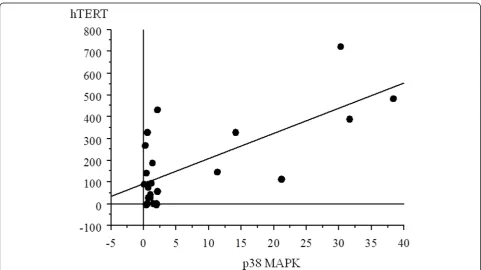
Correlation between levels of p38 MAPK and hTERT mRNA expression
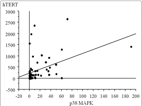
There was a significant correlation between the values of p38 MAPK expression and hTERT, with increased p38 MAPK expression with higher hTERT in all samples (r = 0.445, p = 0.0001) (Figure 1).
Prognostic factors
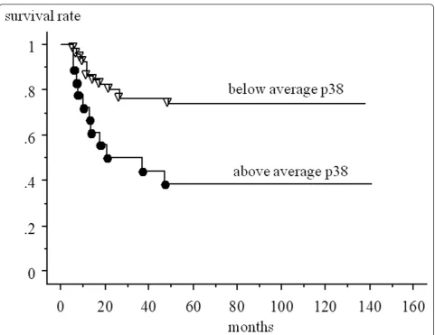
Patients who had a higher than average expression of p38 MAPK had a significantly worse prognosis (5-year survival rate; 38.1%) than other patients overall (73.8%) (p = 0.0036) (Figure 2). There were no significant differ-ences in prognosis between patients who had a higher than average expression of hTERT (5-year survival rate: 38.6%) and those who did not (71.1%) (p = 0.0585).
Soft tissue MFH samples
p38 MAPK and hTERT mRNA expression
p38 MAPK expression was demonstrated in 77.8% (28 of 36) and hTERT mRNA expression was demonstrated in 88.9% (32 of 36) of soft tissue MFH samples. The levels of p38 MAPK were 9.60 ± 17.5 (range: 0-71.1) and those of hTERT were 371.6 ± 695.9 (range: 0-2656.0).
Correlation between levels of p38 MAPK and hTERT mRNA expression
There was a significant correlation between the values of p38 MAPK expression and hTERT, with increased p38 MAPK expression with higher hTERT in soft tissue MFH samples (r = 0.352, p = 0.0352) (Figure 3).
Prognostic factors
those who did not (65.0%) (p = 0.213). There were no significant differences in prognosis between patients who had a higher than average expression of hTERT (41.7%) and those who did not (62.7%) (p = 0.610).
Liposarcoma samples
p38 MAPK and hTERT mRNA expression
p38 MAPK expression was demonstrated in 95.8% (23 of 24) and hTERT mRNA expression was demonstrated in 91.7% (22 of 24) of LS samples. The levels of p38 MAPK were 6.81 ± 11.5 (range: 0-38.2) and those of
hTERT were 171.3 ± 189.9 (range: 0-726.6) in LS samples.
Correlation between levels of p38 MAPK and hTERT mRNA expression
There was a significant correlation between the values of p38 MAPK expression and hTERT, with increased p38 MAPK expression with higher hTERT in LS samples (r = 0.704, p = 0.0001) (Figure 4).
Prognostic factors
Patients who had a higher than average expression of p38 MAPK (5-year survival rate: 50.0%) had a
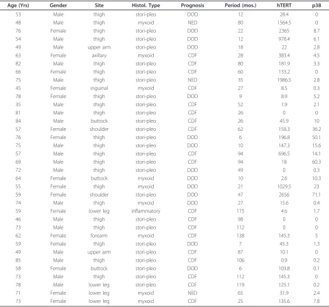
Table 1 Data in 36 patients with soft tissue MFH
Age (Yrs) Gender Site Histol. Type Prognosis Period (mos.) hTERT p38
53 Male thigh stori-pleo DOD 12 28.4 0
48 Male thigh myxoid NED 80 1564.5 0
76 Female thigh stori-pleo DOD 22 2365 8.7
54 Male thigh stori-pleo DOD 12 978.4 6.1
49 Male upper arm stori-pleo DOD 18 22 2.8
63 Female axillary myxoid CDF 28 383.4 4.5
82 Male thigh stori-pleo CDF 80 181.9 3.3
66 Female thigh stori-pleo CDF 60 133.2 0
75 Male thigh stori-pleo NED 35 1986.5 2.8
45 Female inguinal myxoid CDF 27 8.5 0.3
78 Female thigh stori-pleo DOD 9 8.9 5.2
35 Male thigh stori-pleo CDF 52 1.9 2.1
81 Male thigh stori-pleo CDF 26 0 0
84 Male buttock stori-pleo CDF 26 45.9 10
57 Female shoulder stori-pleo CDF 62 158.3 36.2
76 Female thigh stori-pleo DOD 6 196.8 50.1
75 Male thigh stori-pleo DOD 10 147.3 15.6
57 Male thigh stori-pleo CDF 94 696.5 14.1
69 Male thigh stori-pleo CDF 94 18 60.3
72 Male thigh stori-pleo DOD 49 0 0.3
64 Female buttock myxoid DOD 10 2.6 10.3
55 Female thigh myxoid DOD 21 1029.5 23
59 Female shoulder stori-pleo DOD 47 2656 71.1
74 Male thigh myxoid DOD 27 15.6 0.4
59 Female lower leg inflammatory CDF 115 4.6 1.7
46 Male thigh stori-pleo CDF 98 0 0
73 Male thigh stori-pleo CDF 112 0 0
62 Female forearm myxoid CDF 138 145.3 5
59 Female thigh stori-pleo DOD 7 45.3 1.3
49 Male upper arm stori-pleo CDF 87 10.1 0
85 Male thigh stori-pleo CDF 106 0.9 0.2
58 Female buttock stori-pleo DOD 6 103.8 0.1
73 Male thigh stori-pleo CDF 112 145.3 0
78 Male lower leg stori-pleo CDF 119 125.1 0.2
71 Female lower leg myxoid NED 65 31.9 2.4
73 Female lower leg myxoid CDF 25 135.6 7.8
significantly worse prognosis than other patients (88.9%) (p = 0.0448) in LS patients. There were no significant differences in prognosis between patients who had a higher than average expression of hTERT (62.5%) and those who did not (87.5%) (p = 0.110).
Bone MFH samples
p38 MAPK and hTERT mRNA expression
p38 MAPK expression was demonstrated in 77.8% (7 of 9) and hTERT expression was demonstrated in all (9 of
9) of bone MFH samples. The levels of p38 MAPK were 46.4 ± 58.2 (range: 0-191) and the levels of hTERT were 636.5 ± 453.3 (range: 241.7-1405.4) in bone MFH samples.
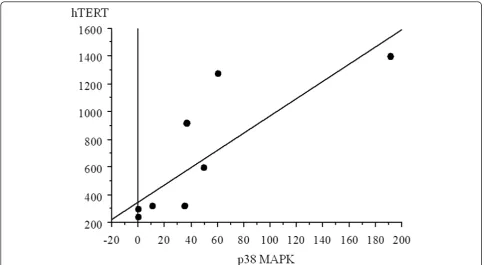
Correlation between levels of p38 MAPK and hTERT mRNA expression
There was a significant correlation between the values of p38 MAPK expression and hTERT, with increased p38 MAPK expression with higher hTERT (r = 0.802, p = 0.0093) (Figure 5).
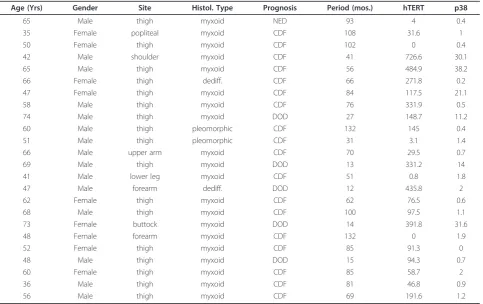
Table 2 Data in 24 patients with liposarcoma
Age (Yrs) Gender Site Histol. Type Prognosis Period (mos.) hTERT p38
65 Male thigh myxoid NED 93 4 0.4
35 Female popliteal myxoid CDF 108 31.6 1
50 Female thigh myxoid CDF 102 0 0.4
42 Male shoulder myxoid CDF 41 726.6 30.1
65 Male thigh myxoid CDF 56 484.9 38.2
66 Female thigh dediff. CDF 66 271.8 0.2
47 Female thigh myxoid CDF 84 117.5 21.1
58 Male thigh myxoid CDF 76 331.9 0.5
74 Male thigh myxoid DOD 27 148.7 11.2
60 Male thigh pleomorphic CDF 132 145 0.4
51 Male thigh pleomorphic CDF 31 3.1 1.4
66 Male upper arm myxoid CDF 70 29.5 0.7
69 Male thigh myxoid DOD 13 331.2 14
41 Male lower leg myxoid CDF 51 0.8 1.8
47 Male forearm dediff. DOD 12 435.8 2
62 Female thigh myxoid CDF 62 76.5 0.6
68 Male thigh myxoid CDF 100 97.5 1.1
73 Female buttock myxoid DOD 14 391.8 31.6
48 Female forearm myxoid CDF 132 0 1.9
52 Female thigh myxoid CDF 85 91.3 0
48 Male thigh myxoid DOD 15 94.3 0.7
60 Female thigh myxoid CDF 85 58.7 2
36 Male thigh myxoid CDF 81 46.8 0.9
56 Male thigh myxoid CDF 69 191.6 1.2
defiff. = dedifferentiated CDF = continuously disease-free DOD = died of disease
Table 3 Data in 9 patients with bone MFH
Age (Yrs) Gender Site Histol. Type Prognosis Period (mos.) hTERT p38
23 Female femur stori-pleo CDF 130 304 0
65 Female femur stori-pleo DOD 37 1405.4 191.1
46 Male femur stori-pleo CDF 141 921.8 36.2
27 Female clavicle stori-pleo CDF 92 323.1 10.3
57 Male femur stori-pleo CDF 93 241.7 0
69 Male femur stori-pleo DOD 8 1278.2 60.3
67 Male sacrum stori-pleo DOD 7 324.5 35.2
38 Male humerus stori-pleo DOD 18 603.6 49.3
57 Female ilium stori-pleo DOD 6 326.5 35
Prognostic factors
Patients who had a higher than average expression of p38 MAPK (5-year survival rate: 0%) had a worse prog-nosis than other patients (66.7%), but did not reach sig-nificant differences (p = 0.202). There were no significant differences in prognosis between patients who had a higher than average expression of hTERT (33.3%) and those who did not (50.0%) (p = 0.904).
Discussion
hTERT is the catalytic telomerase subunit component that copies a template region of its functional RNA sub-unit to the end of the telomere. In terms of carcinomas, hTERT mRNA expression and telomerase activity are closely associated, and quantification of hTERT mRNA has been reported as an alternative to the measure of telomerase activity [7,25,26]. Also, in sarcomas, the cor-relation between telomerase activity and hTERT has been reported [9,10,27]. However, in contrast, previous reports maintained that hTERT expression does not
correlate to telomerase activity [12,23], and hTERT mRNA expression was only studied in the absence of detectable telomerase activity on sarcomas [8,12,27,28]. There is no clear understanding of the discordance between hTERT and telomerase activity in sarcomas [23,29]. Recently, the presence of telomerase activity and alternative lengthening of telomeres (ALT) in several sarcomas was examined extensively, and these studies indicate a positive correlation between the telomere maintenance mechanism and tumor aggressiveness in several sarcoma types [29]. Furthermore, a positive cor-relation between hTERT and tumor aggressiveness in several sarcomas has been reported [8-14]. Therefore, it could be necessary to analyze hTERT, in order to eluci-date the telomere maintenance mechanisms and the tumorigenesis of sarcomas.
The predominence of large numbers of protein kinases involved in signal cascades following genotoxic stress is the p38 MAPK [30]. p38 MAPK is shown to induce a wide variety of intracellular responses, with
roles in tumorigenesis, cell-cycle regulation, develop-ment, inflammation and apoptosis [15-17]. Recent stu-dies have suggested that signals transmitted through MAP kinase can regulate hTERT transcription. Epider-mal growth factor (EGF) affects the up-regulation of hTERT transcription through the MAP kinase cascades [20]. E26 transformation-specific (Ets) transcription fac-tors, downstream of the mitogen signaling pathways of MAP kinase, regulates hTERT [31]. p38 MAPK may play an important role in the activation of the hTERT promoter by the upstream stimulatory factor (USF) in tumor cells [32]. In the present study, there was a signif-icant positive correlation between the values of p38 MAPK expression and hTERT, with increased p38 MAPK expression with higher hTERT in sarcoma sam-ples. This is the first report to show a correlation between the levels of hTERT mRNA expression and the levels of p38 MAPK in human sarcomas, and these results may suggest that p38 MAPK plays a role in
up-regulation of hTERT in soft tissue MFH, liposarcomas, and bone MFH, while we do not have a clear under-standing if some factor regulates both p38 MAPK and hTERT expression.
Recent studies have demonstrated that p38 MAPK has diverse roles in the pathogenesis of several cancers and have shown that they are also involved in regulating other functions including the differentiation and prolif-eration of various cell types [33]. The p38 MAPK path-way is most frequently associated with a tumor suppressor function, based on its negative regulation of proliferation and survival of cells [33,34]. However, con-tradictory effects have been observed, a fact that points to the pathway playing a positive role in cell-cycle pro-gression in some carcinoma cells [35-37]. In terms of sarcoma cells, inhibition of p38 MAPK activity rescues the antitumor agent fenretinide-mediated cell death in Ewing’s sarcoma family of tumors [38], and inhibition of p38 signals results showing a significant reduction in
chondrosarcoma cell proliferation mediated by complex effects of p38 signaling on cell-cycle gene expression
Figure 3Correlation between p38 and hTERT in soft tissue MFH samples. There was a significant correlation between the values of p38 expression and those of hTERT (r = 0.352, p = 0.0352).
[39], which suggests that p38 MAPK may play an important role in tumorigenesis in these sarcomas. In the clinical setting, p38 MAPK expression correlates to poor prognosis (p = 0.0036) in overall patients; of high expression of p38 MAPK, indicating the likelihood of a poor outcome and may indicate a positive role of p38 MAPK in tumor proliferation and aggressiveness, in patients with sarcomas. In terms of bone and soft tissue MFH, there were no significant differences in prognosis between patients who had a higher than average expres-sion of p38 MAPK and those who did not. However, patients who had above average p38 (5-year survival rate: soft tissue MFH; 41.7%, bone MFH; 0%) had a worse prognosis than other patients (5-year survival rate: soft tissue MFH; 65.0%, bone MFH; 66.7%), but did not reach significant differences. These results may be due to small numbers of patients. Patients who had a higher than average expression of p38 MAPK (5-year survival rate: 50.0%) had a significantly worse prognosis than other patients (88.9%) (p = 0.0448) in LS patients. Therefore, high expression of p38 MAPK may correlate with a worse prognosis especially for LS patients.
Conclusions
p38 MAPK may be a useful marker in the assessment of hTERT and prognosis. Given that more than 80% of sar-comas express p38 MAPK and hTERT, elucidation of the pathways and target genes of p38 MAPK in
sarcomas will yield additional understandings into the pathogenesis of several sarcomas and may lead to novel therapeutic strategies for their treatment.
Author details
1Department of Orthopaedic Surgery, National Hospital Organization Kure
Medical Center and Chugoku Cancer Center: 3-1, Aoyamacho, Kure, Hiroshima, 7370023 Japan.2Department of Orthopaedic Surgery, Graduate
School of Biomedical Sciences, Hiroshima University: 1-2-3, Kasumi, Minami-ku, Hiroshima, 7348551 Japan.3Department of Artificial Joints and Biomaterials, Graduate School of Biomedical Sciences, Hiroshima University, Hiroshima, Japan.
Authors’contributions
TM, SS, TK, JF, TS carried out literature research, experimental studies and data acquisition, participated in the study design, and drafted the manuscript. YY and OM proposed the study, participated in the design and coordination and helped to draft, and assisted writing the manuscript. All authors read and approved the final manuscript.
Competing interests
The authors declare that they have no competing interests.
Received: 6 December 2011 Accepted: 16 January 2012 Published: 16 January 2012
References
1. Kim NW, Piatyszek MA, Prowse KR, Harley CB, West MD, Ho PL, Coviello GM, Wright WE, Weinrich SL, Shay JW:Specific association of human telomerase activity with immortal cells and cancer.Science1994,
266:2011-2015.
2. de Lange T:Activation of telomerase in a human tumor.Proc Natl Acad Sci USA1994,91:2882-2885.
3. Hiyama E, Yokoyama T, Tatsumoto N, Hiyama K, Imamura Y, Murakami Y, Kodama T, Piatyszek MA, Shay JW, Matsuura Y:Telomerase activity in gastric cancer.Cancer Res1995,55:3258-3262.
4. Tatsumoto N, Hiyama E, Murakami Y, Imamura Y, Shay JW, Matsuura Y, Yokoyama T:High telomerase activity is an independent prognostic indicator of poor outcome in colorectal cancer.Clin Cancer Res2000,
6:2696-2701.
5. Hiyama E, Hiyama K:Clinical utility of telomerase in cancer.Oncogene 2002,21:643-649.
6. Weinrich SL, Pruzan R, Ma L, Ouellette M, Tesmer VM, Holt SE, Bodnar AG, Lichtsteiner S, Kim NW, Trager JB, Taylor RD, Carlos R, Andrews WH, Wright WE, Shay JW, Harley CB, Morin GB:Reconstitution of human telomerase with the template RNA component hTR and the catalytic protein subunit hTRT.Nat Genet1997,17:498-502.
7. Nakayama J, Tahara H, Tahara E, Saito M, Ito K, Nakamura H, Nakanishi T, Tahara E, Ide T, Ishikawa F:Telomerase activation by hTRT in human normal fibroblasts and hepatocellular carcinomas.Nat Genet1998,
18:65-68.
8. Schneider-Stock R, Jaeger V, Rys J, Epplen JT, Roessner A:High telomerase activity and high HTRT mRNA expression differentiate pure myxoid and myxoid/round-cell liposarcomas.Int J Cancer2000,89:63-68.
9. Tomoda R, Seto M, Tsumuki H, Iida K, Yamazaki T, Sonoda J, Matsumine A, Uchida A:Telomerase activity and human telomerase reverse transcriptase mRNA expression are correlated with clinical aggressiveness in soft tissue tumors.Cancer2002,95:1127-1133. 10. Schneider-Stock R, Boltze C, Jäger V, Epplen J, Landt O, Peters B, Rys J,
Roessner A:Elevated telomerase activity, c-MYC-, and hTERT mRNA expression: association with tumour progression in malignant lipomatous tumours.J Pathol2003,199:517-525.
11. Ohali A, Avigad S, Cohen IJ, Meller I, Kollender Y, Issakov J, Gelernter I, Goshen Y, Yaniv I, Zaizov R:Association between telomerase activity and outcome in patients with nonmetastatic Ewing family of tumors.J Clin Oncol2003,21:3836-3843.
12. Sanders RP, Drissi R, Billups CA, Daw NC, Valentine MB, Dome JS:
Telomerase expression predicts unfavorable outcome in osteosarcoma.J Clin Oncol2004,22:3790-3797.
13. Fuchs B, Inwards C, Scully SP, Janknecht R:hTERT Is highly expressed in Ewing’s sarcoma and activated by EWS-ETS oncoproteins.Clin Orthop Relat Res2004,426:64-68.
14. Sabah M, Cummins R, Leader M, Kay E:Immunohistochemical detection of hTERT protein in soft tissue sarcomas: correlation with tumor grade.
Appl Immunohistochem Mol Morphol2006,14:198-202.
15. Ambrosino C, Nebreda AR:Cell cycle regulation by p38 MAP kinases.Biol Cell2001,93:47-51.
16. Bradham C, McClay DR:p38 MAPK in development and cancer.Cell Cycle 2006,5:824-828.
17. Coulthard LR, White DE, Jones DL, McDermott MF, Burchill SA:p38(MAPK): stress responses from molecular mechanisms to therapeutics.Trends Mol Med2009,15:369-379.
18. Wang Z, Kyo S, Takakura M, Tanaka M, Yatabe N, Maida Y, Fujiwara M, Hayakawa J, Ohmichi M, Koike K, Inoue M:Progesterone regulates human telomerase reverse transcriptase gene expression via activation of mitogen-activated protein kinase signaling pathway.Cancer Res2000,
60:5376-5381.
19. Alfonso-De Matte MY, Yang H, Evans MS, Cheng JQ, Kruk PA:Telomerase is regulated by c-Jun NH2-terminal kinase in ovarian surface epithelial cells.Cancer Res2002,62:4575-4578.
20. Maida Y, Kyo S, Kanaya T, Wang Z, Yatabe N, Tanaka M, Nakamura M, Ohmichi M, Gotoh N, Murakami S, Inoue M:Direct activation of telomerase by EGF through Ets-mediated transactivation of TERT via MAP kinase signaling pathway.Oncogene2002,21:4071-4079.
21. Goueli BS, Janknecht R:Upregulation of the Catalytic Telomerase Subunit by the Transcription Factor ER81 and Oncogenic HER2/Neu, Ras, or Raf.
Mol Cell Biol2004,24:25-35.
22. Takakura M, Kyo S, Inoue M, Wright WE, Shay JW:Function of AP-1 in transcription of the telomerase reverse transcriptase gene (TERT) in human and mouse cells.Mol Cell Biol2005,25:8037-8043.
23. Matsuo T, Shay JW, Wright WE, Hiyama E, Shimose S, Kubo T, Sugita T, Yasunaga Y, Ochi M:Telomere-maintenance mechanisms in soft-tissue malignant fibrous histiocytomas.J Bone Joint Surg Am2009,91:928-937.
24. Matsuo T, Shimose S, Kubo T, Fujimori J, Yasunaga Y, Ochi M:Alternative lengthening of telomeres as a prognostic factor in malignant fibrous histiocytomas of bone.Anticancer Res2010,30:4959-4962.
25. Nakamura TM, Morin GB, Chapman KB, Weinrich SL, Andrews WH, Lingner J, Harley CB, Cech TR:Telomerase catalytic subunit homologs from fission yeast and human.Science1997,277:955-959.
26. Meyerson M, Counter CM, Eaton EN, Ellisen LW, Steiner P, Caddle SD, Ziaugra L, Beijersbergen RL, Davidoff MJ, Liu Q, Bacchetti S, Haber DA, Weinberg RA:hEST2, the putative human telomerase catalytic subunit gene, is up-regulated in tumor cells and during immortalization.Cell 1997,90:785-795.
27. Yan P, Benhattar J, Coindre JM, Guillou L:Telomerase activity and hTERT mRNA expression can be heterogeneous and does not correlate with telomere length in soft tissue sarcomas.Int J Cancer2002,98:851-856. 28. Yan P, Coindre JM, Benhattar J, Bosman FT, Guillou L:Telomerase activity
and human telomerase reverse transcriptase mRNA expression in soft tissue tumors: correlation with grade, histology, and proliferative activity.Cancer Res1999,59:3166-3170.
29. Matsuo T, Shimose S, Kubo T, Fujimori J, Yasunaga Y, Ochi M:Telomeres and telomerase in sarcomas.Anticancer Res2009,29:3833-3836. 30. Yang J, Yu Y, Duerksen-Hughes PJ:Protein kinases and their involvement
in the cellular responses to genotoxic stress.Mutat Res2003,543:31-58. 31. Dwyer J, Li H, Xu D, Liu JP:Transcriptional regulation of telomerase
activity: roles of the the Ets transcription factor family.Ann N Y Acad Sci 2007,1114:36-47.
32. Goueli BS, Janknecht R:Regulation of telomerase reverse transcriptase gene activity by upstream stimulatory factor.Oncogene2003,
22:8042-8047.
33. Nebreda AR, Porras A:p38 MAP kinases: beyond the stress response.
Trends Biochem Sci2000,25:257-260.
34. Ono K, Han J:The p38 signal transduction pathway: activation and function.Cell Signal2000,12:1-13.
35. Neve RM, Holbro T, Hynes NE:Distinct roles for phosphoinositide 3-kinase, mitogen-activated protein kinase and p38 MAPK in mediating cell cycle progression of breast cancer cells.Oncogene2002,
21:4567-4576.
36. Recio JA, Merlino G:Hepatocyte growth factor/scatter factor activates proliferation in melanoma cells through p38 MAPK, ATF-2 and cyclin D1.
Oncogene2002,21:1000-1008.
37. Pomérance M, Quillard J, Chantoux F, Young J, Blondeau JP:High-level expression, activation, and subcellular localization of p38-MAP kinase in thyroid neoplasms.J Pathol2006,209:298-306.
38. Myatt SS, Redfern CP, Burchill SA:p38MAPK-Dependent sensitivity of Ewing’s sarcoma family of tumors to fenretinide-induced cell death.Clin Cancer Res2005,11:3136-3148.
39. Halawani D, Mondeh R, Stanton LA, Beier F:p38 MAP kinase signaling is necessary for rat chondrosarcoma cell proliferation.Oncogene2004,
23:3726-3731.
doi:10.1186/1756-9966-31-5
Cite this article as:Matsuoet al.:Correlation between p38
mitogen-activated protein kinase and human telomerase reverse transcriptase in sarcomas.Journal of Experimental & Clinical Cancer Research201231:5.
Submit your next manuscript to BioMed Central and take full advantage of:
• Convenient online submission
• Thorough peer review
• No space constraints or color figure charges
• Immediate publication on acceptance
• Inclusion in PubMed, CAS, Scopus and Google Scholar
• Research which is freely available for redistribution