DETERMINATION OF AMOXYCYLLIN AND CLAVULANIC ACID IN
PHARMACEUTICAL DOSAGE FORMS BY HPLC WITH
AMPEROMETRIC DETECTION
A. Aghazadeh
*and G. Kazemifard
Department of Pharmaceutics, College of Pharmacy, Tehran University of Medical Sciences, Islamic Republic of Iran
ABSTRACT
A simple and sensitive high performance liquid chromatographic method with
electrochemical detection is described for the quantitative determination of the
amoxycillin and clavulanic acid in pharmaceutical dosage forms. Separation of
sample components occurs on a reversed phase C18 column with a mobile phase
consisting of methanol-water-phosphate buffer. For the amperometric detection,
the potentials of +1.180 and +1.250 V were set on the working electrode for
amoxycillin and clavulanic acid, respectively. High linearity over a concentration
range of (25 ng to 250 ng, r = 0.998) and (31.25 ng to 1 µg, r = 0.999) was
observed. This method is convenient and reproducible for analysis of these two
components in different dosage forms.
*
E-mail: [email protected]
Introduction
Amoxycillin, an aminopenicillin, is commonly prescribed with clavulanic acid as potassium salt, the naturally occurring β-lactamase inhibitor produced by fermentation of Streptomyces clavuligerus, for treatment of infection caused by β-lactamase producing bacteria that are resistant to amoxycillin alone.
Determination of antimicrobial drugs is mainly carried out using microbiological technique [1]. A disadvantage of this technique is relatively long time required for analysis, especially when results are
Keywords: Amoxycillin; Clavulanic acid; Amperometric
detection; LCEC-assay
urgently needed. In addition, when there are more than one antibiotic in formulation, some interactions may
occur.
In addition to microbiological assay, a number of other analytical methods have been reported for the determination of these two components in pharma-ceutical preparation which include; enzymatic assay [2], iodometric titration [3], spectrofluorometry [4,5], UV spectrophotometry [6,7], polarography [8,9] and HPLC assay involving pretreatment of amoxycillin and clavulanic acid with imidazole [10]. Some precolumn [11] and postcolumn derivatization [12-15] and ion-pair HPLC methods [16] have been introduced. Methods for simultaneous assay of these two components in pharmaceutical products by RP-HPLC with UV detection [17] or β -cyclodextrin stationary Phase [18] have also been reported.
amperometric detection for quantitative analysis of both components as raw materials or in pharmaceutical preparations. Electrochemical methods such as normal and differential pulse polarography have been used for estimating clavulanic acid in pharmaceutical preparations. Unfortunately the polarographic waves for this compound overlapped, severely limiting the usefulness of these electrochemical techniques. Liquid chromatography with amperometric detection (LCEC) provides significant improvements by combining the resolution of a chromatographic system with the sensitivity of electrochemical measurements and is suitable for determining trace levels. The usefulness of this technique has been demonstrated in the analysis of various classes of electrochemically active compounds, most notably aromatic amines and phenols. In this regard LCEC has been used for analysis of those antibiotics or NSAIDs having phenolic or amine functionality such as, tetracycline, piroxicam and indomethacin [19, 20].
Experimental Instrumentation
The HPLC system consisted of pumping system (Model Perkin-Elmer series 4), loop injector (Model 7125-075 Cotati. California), and glassy carbon liquid chromatographic detector (Model TL-5A) which controlled by a potentiostat (Model LC-4b). It was
operated in the direct current mode at +1.180 V vs.
Ag/AgCl for amoxycillin and at +1.25 V vs. Ag/AgCl
for clavulanic acid at attenuation range of 100-500
nA/V. The column used was 15 cm×4.6 mm i.d.
stainless steel prepacked with Octadecyl Silane (Perkin-Elmer/HS-5 C18). Chromatographic recording was made on an integrator (Model Perkin-Elmer LCI-100).
Materials
All materials were of analytical reagent grade. The methanol was obtained from Merck Co. (Frankfurt, Germany). Amoxycillin trihydrate and Lithium clavulanate were purchased from U.S.P.C. INC Rockville MD. Augmentin tablets (Beecham, Jurong, Singapore), Co-amoxyclav (Kosar Co. Iran) and the suspension of Co-amoxyclav (Kosar Co. Iran) were purchased from a local drug store. Each tablet was labeled as containing 500 mg amoxycillin (as trihydrate) and 125 mg clavulanic acid (as potassium salt), while the suspension was labeled as containing 125 mg amoxycillin (as trihydrate) and 31 mg clavulanic acid (as potassium salt) per each 5 ml after reconstitution.
Chromatographic Conditions
The mobile phase used was 5%(v/v) methanol in
0.05 M phosphate buffer (pH 3.2-3.4). The isocratic
mobile phase at flow rate of 1 ml/min was used and 50
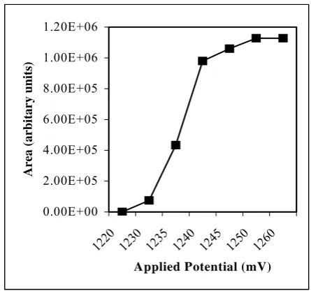
µl sample solutions were injected. The optimal detector cell potential for the oxidation of amoxycillin and clavulanic acid was explored by using reference standards dissolved in mobile phase. The resulting hydrodynamic voltammograms are shown in Figures 1 and 2. Based on these results +1.180 and +1.25 V was chosen for amoxycillin and clavulanic acid, respectively.
0.00E+00 2.00E+07 4.00E+07 6.00E+07 8.00E+07 1.00E+08 1.20E+08 1.40E+08 1.60E+08 1.80E+08 2.00E+08
960 1000 1040 1080 1120 1160 1200
Applied Potential (mV)
A
rea (A
rbitary units
)
Figure 1. Voltammogram of Amoxycillin.
0.00E+00 2.00E+05 4.00E+05 6.00E+05 8.00E+05 1.00E+06 1.20E+06
1220 1230 1235 1240 1245 1250 1260 Applied Potential (mV)
Area (arbitary units)
Figure 2. Voltammogram of Clavulanic acid.
Standard Solutions
accurately weighed and added to a volumetric flask for preparing the stock solution. Amoxycillin standard solutions were prepared by diluting stock solution with mobile phase. Five standard solutions containing 0.5, 1.0, 2.0, 4.0 and 5.0 µg/ml of amoxycillin were prepared. Clavulanic acid standard solutions were similarly prepared, concentration of clavulanic acid solutions were 0.625, 1.25, 2.5, 5, 10 and 20 µg/ml. Duplicate reading of 50 µl injections for each sample of these two components were recorded and calibration
curves (peak area or height vs. concentration) were
constructed.
Procedure for Samples
Tablets. Ten tablets of each (Augmentin 625 and
Co-amoxyclav 625) were weighed and grounded to powder. Six samples from the each powder were accurately weighed. As each sample of them contained about 50 mg amoxycillin and 12.5 mg clavulanic acid. Each sample transferred quantitatively into a 100 ml volumetric flask and mixed with 80 ml of mobile phase. The solution was shaken for 30-40 min, then the volume of each was adjusted to 100 ml and filtered. The filtrate was diluted with mobile phase to reach a suitable concentration for each drug. Duplicate injections (50 µl) were carried out and average peak responses were computed. The concentrations of the unknown sample were calculated using the standard calibration curve.
Suspensions. Each of six samples containing about 125
mg amoxycillin and 31 mg clavulanic acid were separately weighed and transferred quantitatively into six 100 ml volumetric flask and adjusted to desired volume with mobile phase. Further dilutions were made and assayed as above.
Results and Discussion
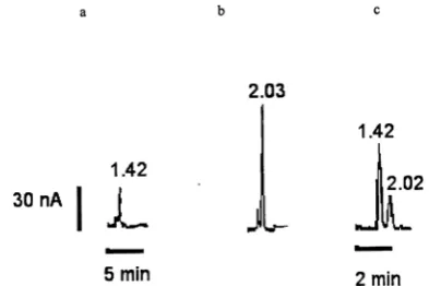
Combination of HPLC with the oxidative amperometric detection yields a highly sensitive and specific assay for amoxycillin and clavulanic acid. Typical chromatograms of amoxycillin and clavulanic acid standard solutions and formulation samples are shown in Figs. 3 and 4. There is not any interaction between the peak of these compounds and other additives presenting in pharmaceutical preparations. The linearity of the chromatographic system was verified by injection of five solutions containing amoxycillin from 0.5 to 5 µg/ml and six solutions containing clavulanic acid from 0.625 to 20 µg/ml. Straight lines for amoxycillin and clavulanic acid were obtained when the area or height of the peak plotted versus concentration (Table 1). Conducting replicate measurements using standard solutions gives a RSD value of less than 8.03%, which indicates a good
reproducibility of the proposed method.
Determination of intra-day variations of assay was carried out by injection of standard solution in three occasions in the same day. For inter-day study the standard solutions of amoxycillin were assayed in three different days. The results of this study are shown in Tables 2 and 3. Due to lack of stability, inter-day variation of clavulanic acid assay was not performed. This compound will be decomposed in less than one week at 8 °C and two days at room temperature [21].
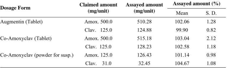
Three types of formulation were assayed as represented in Table 4. These results indicate that the variation between the amount detected in this study and the amount claimed by the manufacturer are in the range of USPXXIV standard.
Figure 3. Typical chromatograms of (a) blank mobile phase,
(b) amoxycillin standard solution (5.0 µg/ml) and (c) amoxycillin (about 2.5 µg/ml) in sample solution of Co-amoxyclav 625 tablets (Applied potential E=1180mV).
Figure 4. Typical chromatograms of (a) blank mobile phase,
Table 1. Calibration data for the standard curves of the peak response vs.
concentration of amoxycillin and clavulanic acid
Compound Concentration (µg/ml)
Correlation
coeff.(r) Slope Intercept
Amoxycillin 0.5 - 5 0.9987 597193 47931 Clavulanic acid 0.625 - 20 0.9994 13.493 -2.0121 Results are the mean of six replicate analysis.
Table 2. Intra - day variability of assay for amoxycillin and clavulanic acid Measured concentration (µg/ml) Compound
Initial concentration
(µg/ml) Mean S. D. R. S. D (%)
Amoxycillin 1.0 0.97 0.02 2.23
4.0 3.86 0.31 8.03
5.0 5.11 0.27 5.3
Clavulanic acid 2.5 2.54 0.08 3.1
5.0 4.55 0.23 5.14
10.0 9.88 0.545 5.52
Results are the mean of six replicate analysis.
Table 3. Inter - day variability of assay for amoxycillin
Measured concentration (µg/ml) Compound
Initial concentration
(µg/ml) Mean S. D. R. S. D (%)
Amoxycillin 1.0 0.93 0.06 6.45
4.0 3.91 0.28 7.16
5.0 5.09 0.27 5.3
Results are the mean of six replicate analysis.
Table 4. Determination of amoxycillin and clavulanic acid in pharmaceutical dosage forms
Assayed amount (%) Dosage Form Claimed amount
(mg/unit)
Assayed amount
(mg/unit) Mean S. D.
Augmentin (Tablet) Amox. 500.0 510.28 102.06 1.28 Clav. 125.0 124.88 99.90 0.82 Co-Amoxyclav (Tablet) Amox. 500.0 515.18 103.04 2.12 Clav. 125.0 128.23 102.58 1.18 Co-Amoxyclav (powder for susp.) Amox. 125.0 126.43 101.14 0.98
Conclusion
In summary, the LC procedure reported here has shown to be simple, concise and reproducible. The method described can be the method of choice for analysis of electroactive antibiotics amoxycillin and clavulanic acid as raw materials and in dosage forms. It is superior to any previous method in terms of sensitivity and specificity.
References
1. A. P. Ball, P. G. Davey, A. M. Geddes, I. D. Farrei and G. Brookes, Lancet., i, 620-23, (1980).
2. W. Cullmann and W. Dick, Immun. Infect., 14(5):
188-90, (1986).
3. Code Federal Regulation., Title, 21: 436, 204, (1976).
4. D. F. Davidson, Clin. Chem. Acta., 69: 67-71, (1976).
5. R. H. Barhaiya, P. Turner and E. Shaw, Ibid., 77: 373-77,
(1977).
6. P. Izquierdo, A. Gomez-Hens and D. Perez-Bendito, J. Pharm. Biomed. Anal., 11(10): 927-31, (1993).
7. A. E. Bird, J. M. Bellis and B. C. Gasson, Analyst., 107:
1241-45, (1982).
8. C. G. Perez, I. G. Martin and B. R. V. De Aldana, J. Pharm. Biomed. Anal., 9(5): 383-86, (1991).
9. L. J. Nunez-Vergara, J. A. Sequella and M. M. Silva,
Farmaco, Ed. Prat. 35: 409-15, (1980).
10. M. Foulstone and C. Reading , Antimicrob. Agents Chemother., 22(5): 753-62, (1982).
11. J. Martin and R. Mendez, J. Liq. Chromatogr., 11(8): 1697-1705, (1988).
12. J. Haginaka, H. Yasuda, T. Uno and T. Nakagawa, Chem. Pharm. Bull., 31(12): 4436-47, (1983).
13. J. Haginaka, J. Wakai. H. Yasuda, T. Uno and T. Nakagawa, J. Liq. Chromatogr., 8(13): 2521-34, (1985).
14. J. Haginaka, J. Wakai. and H. Yasuda, Chem. Pharm. Bull., 34(4): 1850-52, (1986).
15. J. Haginaka, J. Wakai. and H. Yasuda, Anal. Chem., 59(2): 324-33, (1987).
16. S. Chulavatnatol and B. G. Charles, J. Chromatogr., 615:
91-96 (1993).
17. M. A. Abounassif, E. M. Abdel-Moety, M. E. Mohamed and E. A. Gad-Karim, J. Pharm. Biomed. Anal., 9(9):
731-35, (1991).
18. T. Tsou, J. Wu, C. Young and T. Wang, Ibid., 15:
1197-1205, (1997).
19. G. Kazemifard and D. E. Moore, Ibid., 16: 689-96,
(1997).
20. G. Kazemifard and D. E. Moore, J. Chromatogr., 533:
125-31, (1990).
21. A. C. Mehta, S. Hart-Davies, J. Payne and R. W. Lacey,