PHYSIOLOGICAL CORRELATES OF WORKING MEMORY BEHAVIOR AND COGNITIVE EFFICIENCY:
IMPLICATIONS FOR CONCUSSION MANAGEMENT
Christina B. Vander Vegt
A dissertation submitted to the faculty at the University of North Carolina at Chapel Hill in partial fulfillment of the requirements for the degrees of Doctor of Philosophy in the Human
Movement Science Curriculum in the School of Medicine.
Chapel Hill 2020
Approved by:
©2020
ABSTRACT
Christina B. Vander Vegt: Physiological Correlates of Working Memory Behavior for Cognitive Efficiency: implications for concussion management
(Under the direction of Johna K. Register-Mihalik)
Cognitive efficiency—characterized via robust behavioral and physiological response
dynamics, may provide clinically meaningful information with respect to dynamic concussion
injury response and return to play considerations. Moreover, a feasible and ecologically valid
method to assess dynamic behavioral and physiological metrics is also needed to best
characterize cognitive efficiency in athletes and soldiers at a greater risk for concussion. This
project was designed to examine the clinical utility and feasibility of a cognitive efficiency
assessment designed to be completed within a virtual reality environment, which may hold
significant implications for improved concussion clinical management. As such, we aimed to
first further understand the relationships between heart rate variability and pupillary response as
physiological correlates of digit-span task behavior in the context of cognitive efficiency, then to
examine the effects of prior concussion on these responses. Additionally, we applied innovative,
reliably sound, and validated psychophysiological methods using a feasible instrumentation
option (virtual reality headset with pupillometry and heart rate monitor watch and strap). We feel
these methodological considerations allowed us to best address our study aims and inform future
directions of this line of inquiry to have direct ecologically valid applications. Collegiate club
sports athletes, (N=59; 40% with a concussion history; age = 20.48 ± 1.86 years; 58% male),
reality environment while recording pupil size. Linear mixed effects models showed a
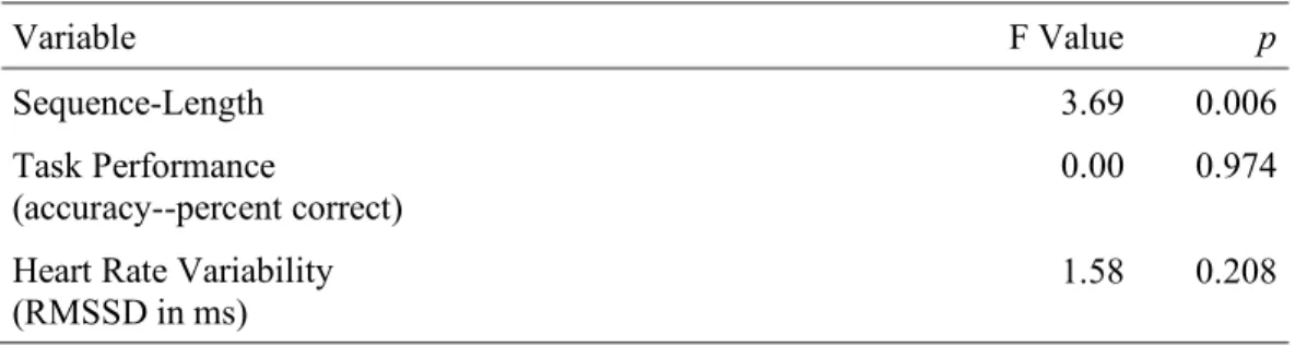
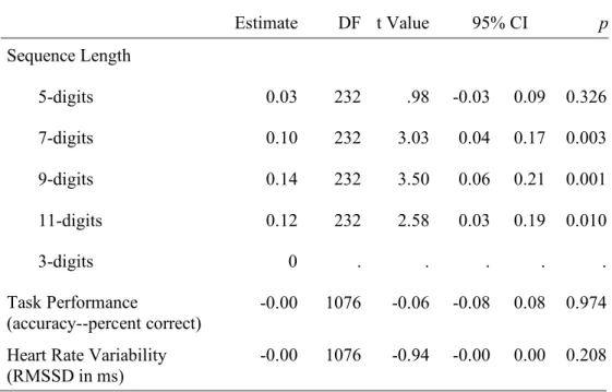
significant effect of cognitive load (digit sequence-length) on pupillary response (F4,232=3.67,
p=0.006). Negligible effects were seen in task performance, heart rate variability or concussion
history p<0.05 Pupillary response shows potential in informing cognitive load and efficiency in
applied settings, using VR and eye tracking technology. Future investigations should consider
ACKNOWLEDGEMENTS
I would like to express my deepest appreciation and gratitude for the many people who
helped to make this work possible. To my mentor and friend Johna Register-Mihalik: thank you
for your unyielding support and guidance throughout this journey, both personally and
professionally. Your devotion to your faith, family, and career is unparalleled, and balanced with
such grace. I would also like to thank my committee members, Greg Appelbaum, Adam Kiefer,
Jason Mihalik, and Kevin Guskiewicz, for sharing your time, wisdom, and expertise with me.
Each of you contributed such unique and vital perspectives to support the success of this project,
and I am grateful for your guidance.
I would like to thank my research assistants Kou Yang and Emily Barron for their time
and dedication to the success of this project. I’d also like to thank Ryan MacPherson for his
assistance modifying the digit-span task to the hardware and software platforms used in this
TABLE OF CONTENTS
LIST OF TABLES ... x
LIST OF FIGURES ... xi
ABBREVIATIONS ... xiii
CHAPTER 1: INTRODUCTION ... 1
1.1 Overview of Concussion Injury and Associated Deficits ... 1
1.2 Cognitive Efficiency Following Concussion ... 3
1.3 Physiological Correlates of Cognitive Efficiency ... 4
1.4 Problem Statement ... 7
1.5 Specific Aims ... 8
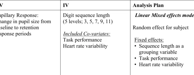
1.6 Independent Variables ... 9
1.7 Dependent Variable: ... 10
1.8 Potential Co-variates: ... 10
1.9 Definition of Terms ... 10
1.10 Delimitations ... 12
1.11 Limitations ... 12
1.12 Assumptions ... 12
1.13 Summary of Study Significance ... 12
CHAPTER 2: LITERATURE REVIEW ... 14
2.1 Overview of Concussive Injury ... 14
2.1.2 Injury Mechanics and Response ... 16
2.1.3 Physiological Response ... 17
2.1.3.1 Neurometabolic Cascade ... 17
2.1.3.2 Role of the Autonomic Nervous System ... 18
2.1.4 Clinical Response ... 18
2.2 Clinical Versus Physiological Considerations ... 21
2.2.1 Advanced Physiologic Measures ... 22
2.2.1.1 Advanced Neuroimaging Techniques ... 22
2.2.1.2 Heart Rate Variability ... 25
2.2.1.3 Current Visual Metrics ... 26
2.2.2 Considerations for Advanced Clinical and Physiological Assessments ... 27
2.2.2.1 Behavior Specific Considerations ... 28
2.2.2.2 Physiologic Specific Measures ... 29
2.3. Pupillary Response as a Potential Solution ... 30
2.3.1 Neurophysiological Underpinnings ... 30
2.3.2 Pupillary Response Correlates with Behavioral Outcomes ... 31
2.3.3 Pupillary Response in Clinical Populations ... 31
2.4 Methodological Considerations ... 33
2.4.1 Study Design and Participants ... 33
2.4.2 Digit-Span Task Design and Administration Considerations ... 34
2.4.3 Task Performance and Physiological Response Considerations ... 34
2.4.3.1 Task performance ... 34
2.5 Summary—Study Rationale ... 36
CHAPTER 3: METHODS ... 38
3.1 Experimental Design and Participants ... 38
3.1.1 Experimental Design and Study Setting ... 38
3.1.2 Participants ... 38
3.1.2.1 Inclusion and Exclusion Criteria ... 38
3.1.2.2 Recruitment Strategy ... 39
3.2 Digit-span Working Memory Task ... 40
3.2.1 Task Design ... 40
3.2.1.1 Traditional Design and Administration Parameters ... 40
3.2.1.2 Present Study ... 41
3.2.1.3 Control Elements of Task Design ... 43
3.3 Task Performance ... 44
3.4 Heart Rate Variability ... 45
3.5 Pupillary Response Measures ... 45
3.6 General Testing Session Procedures ... 46
3.7 Statistical Approach ... 48
3.7.1 Power Analysis ... 49
CHAPTER 4: RESULTS ... 51
4.1 Descriptive results ... 51
4.2 Aim 1 Results ... 52
CHAPTER 5: DISCUSSION ... 82
5.1 General Findings Informing Cognitive Efficiency ... 82
5.1.1 Overall descriptives ... 82
5.1.2 Heart Rate Variability ... 83
5.2 Discussion for Specific Aim 1 ... 85
5.3 Discussion for Specific Aim 2 ... 87
5.4 Summary of Findings Related to Original Hypotheses ... 88
5.5 Limitations ... 89
5.6 Future Research ... 90
5.6 Conclusions ... 90
APPENDIX A. TESTING SESSION MATERIALS ... 92
APPENDIX B. REFINEMENT PROJECT RESULTS ... 104
Preliminary Analyses: ... 104
Pupillary response evoked by a digit-span working memory task ... 104
Task performance—Digit-span Working Memory Task ... 105
Sex Differences in Pupillary Response ... 107
APPENDIX C. DISSERTATION MANUSCRIPT – MEDICINE & SCIENCE IN SPORT & EXERCISE ... 108
APPENDIX D. EXECUTIVE SUMMARY ... 140
Background: ... 140
Methods Overview: ... 140
Summary Results and Discussion Points: ... 142
LIST OF TABLES
Table 3.1. Statistical analysis plan by aim ... 50
Table 4.1. Participant demographic information ... 55
Table 4.2. Self-reported performance ... 56
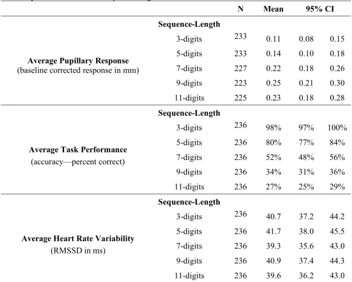
Table 4.3. Average pupillary response, task performance, and heart rate
variability summarized across sequence-length and 95% confidence intervals ... 57
Table 4.4. Bivariate correlation matrix among numeric variables of interest ... 58
Table 4.5. Aim 1 mixed effects model for the effect of cognitive load,
task performance, and heart rate variability on pupillary response (type III results) ... 59
Table 4.6. Aim 1 mixed effects model for the effect of cognitive load,
task performance, and heart rate variability on pupillary response (simple effects) ... 60
Table 4.7. Concussion history group average study measures
across sequence lengths and 95% confidence intervals ... 61
Table 4.8. Pupillary response means by sequence-length for concussion
history subgroups ... 62
Table 4.9. Aim 2 mixed effects model for the effect of concussion history, cognitive
load, task performance, and heart rate variability on pupillary response (type III results) ... 63
Table 4.10. Aim 2 mixed effects model for the effect of concussion history, cognitive
LIST OF FIGURES
Figure 1.1. Conceptual model ... 13
Figure 3.1. Digit-span randomized blocked design ... 41
Figure 3.2. Sample five-digit-sequence ... 44
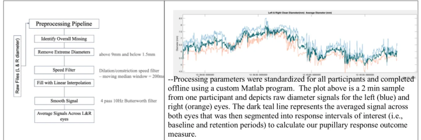
Figure 3.3. Pupil data preprocessing ... 46
Figure 3.4. Participant set up ... 47
Figure 4.1. Prototypical time trace for pupillary response dynamics by sequence-length ... 65
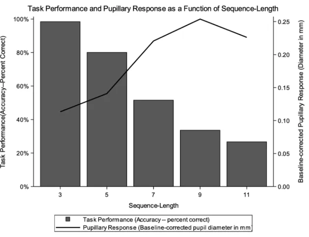
Figure 4.2. Grand means: pupillary response and task performance by sequence-length ... 66
Figure 4.3. Grand means: heart rate variability and task performance by sequence-length ... 67
Figure 4.4. Participants’ pupillary response by sequence-length ... 68
Figure 4.5. Participants’ task performance by sequence-length ... 69
Figure 4.6. Participants’ heart rate variability by sequence-length ... 70
Figure 4.7. Aim 1 model predicted pupillary response means by sequence-length ... 71
Figure 4.8. Group differences in pupillary response and task performance by sequence-length ... 72
Figure 4.9. Group differences in heart rate variability and task performance by sequence-length ... 73
Figure 4.10. Average pupillary response by sequence-length–split by total lifetime concussion subgroups ... 74
Figure 4.11. Average pupillary response by sequence-length–split by concussion chronicity subgroups ... 75
Figure 4.13. Pupillary response by sequence-length for each participant–
split by concussion history sub-groups for lifetime concussions ... 77
Figure 4.14. Pupillary response by sequence-length for each participant–
split by concussion history sub-groups for concussion chronicity ... 78
Figure 4.15. Task performance by sequence-length for each participant–
split by concussion history groups ... 79
Figure 4.16. Heart rate variability by sequence-length for each participant–
split by concussion history groups ... 80
Figure 4.17. Aim 2 model predicted pupillary response means by
ABBREVIATIONS
ANS Autonomic Nervous System
CBF Cerebral Blood Flow
DLPFC Dorsolateral Prefrontal cortex
fMRI Functional Magnetic Imaging
HR Heart Rate
HRV Hear Rate Variability
ImPACT Immediate Post-concussion Assessment and Cognitive Test
PFC Prefrontal cortex
RTP Return to Play
SOT Sensory Organization Test
SRC Sport-Related Concussion
VR Virtual Reality
CHAPTER 1: INTRODUCTION 1.1 Overview of Concussion Injury and Associated Deficits
Repetitive head impact exposures and concussion are major athletic health concerns1 for
which the physiological response and recovery dynamics are poorly understood. Concussion
among college aged athletes participating in National Collegiate Athletic Association (NCAA)
sports is estimated around 10,560 nationally with the overall concussion rate of 4.47 per 10,000
athlete-exposures.2 Recent literature regarding the effects associated with repetitive head impact
exposures remains unclear. However, studies suggest greater functional impairments and
potentially long-term structural changes in those who have experienced multiple prior
concussions.3–5 Contact and collision sport athletes (e.g., American football, rugby, men’s
lacrosse, etc.) are likely at the greatest risk of such deficits, as they experience relatively high
numbers of head impact exposures and greater concussion rates over the course of a single
season and their athletic careers.65 5,6Understanding the relationships between clinical and
physiological response dynamics following repetitive head impact exposures and concussion is
therefore important to ensure proper care and management.
Neuropathophysiological changes following concussion and repetitive head impacts—
and the associated clinical impairment manifestations7,8 do not adhere to fixed response and
recovery timelines. As such, these changes are difficult to fully characterize with currently
available clinical measures. The recommended concussion assessment paradigm6,10,119 uses a
etc.) known to demonstrate ceiling effects10 and rapidly lose signal detection11,12 in the
days following concussion injury.13–15 The current battery functions to guide clinical decisions
such as return to play (RTP), often made following clinical measure normalization. Clinical
normalization for collegiate aged athletes typically occurs within 7 to 10 days following
injury.16,17 Recent neuroimaging studies however, report prolonged physiological impairment
exhibited by increased spatial and temporal activity disproportional to task demands.8,18,19
Moreover, the sole monitoring of performance deviations relative to baseline or normative
values, further limits dynamic clinical and/or physiological response characterization.8,9,18
Prolonged post-concussion physiological impairment has been described as
compensatory neural resource utilization to meet cognitive demands, which previous studies
have described as neurophysiological cognitive inefficiency. 7,8,19 These compensatory
mechanisms are posited to provide some explanation for early clinical normalization in
performance-based outcomes.8 Studies using advanced neuroimaging measures have contributed
valuable evidence regarding compensatory physiological mechanisms, and suggest that they may
result in prolonged neural vulnerability and associated negative consequences (e.g. increased risk
for neurodegenerative disease, neuropsychiatric deficits, etc.).8,18,19 However, the poor ecological
validity (e.g., cost, time, availability) of these studies,18 limit our ability to further elucidate
neurophysiological responses and recovery associated with various head trauma exposure types
(e.g., documented concussion injury7,8, repeat concussions,3,4,18 repetitive head impacts,6,20,20 and
participation in a single season of collision sport play6,21). Overall, these issues highlight the need
for more ecologically valid assessment options to better capture concussion injury response
Recent studies concerning the clinical concussion assessment paradigm have suggested
the need for critical modifications to include more robust, reliable, and validated measures that
capture the dynamic nature of post-injury clinical and physiological responses. Adaptations to
the current assessment battery that fulfill these needs may support clinical decision-making
regarding athlete/soldier readiness for return to activity and/or duty, potentially mitigating
negative and injurious consequences associated with premature return. 22,23
1.2 Cognitive Efficiency Following Concussion
Current concussion assessments lack neurophysiologic and performance-based
measurement options that are feasible and demonstrate adequate utility, to fully inform cognitive
efficiency. Combining these two measurements and examining them in the context of task
demands allows extends their meaning to that of efficiency—beyond effectiveness. Whereby,
efficiency refers to the assessment of the dynamic interplay between elements of cognitive task
demands (task difficulty), physiological characteristics associated with cognitive effort and
capacities, and performance-based outcomes24,25 is not represented in the current clinical battery.
Current neurocognitive assessments within the concussion assessment battery statically capture
two of these three elements—i.e., task demands (design and difficulty) and performance
outcomes. However, important adaptations within these two elements may be necessary to
improve our understanding of task performance dynamics as they relate to cognitive efficiency
following concussion. For example, task performance on the SAC’s digit-span task is one of the
most sensitive to acute concussion.11,12 Previous findings in healthy adult samples suggest that
relatively minor (though necessary) task adaptations (i.e., adapted task demands, scoring, and
administration parameters) elicit better dynamic working memory processes and should therefore
efficiency—physiological characteristics of cognitive effort and capacities—have yet to be
established.
An objective physiological marker for neural resource utilization and/or cognitive effort
would need to demonstrate sensitivity, dynamic responsivity, and robustness, with
discriminatory abilities across varying levels of cognitive efficiency associated with the
concussion response acutely and in the longer-term. Specific consideration should also be given
to the clinical feasibility of potential metric solutions to best examine their overall utility.
1.3 Physiological Correlates of Cognitive Efficiency
Pupillary responses have demonstrated significant associations with various cognitive
and emotional constructs, to examine information processing. Research efforts in cognitive
pupillometry specifically, have shown that pupillary responses reflect changes in cognitive
processing load.28,29 As such, pupillary response dynamics serve as an indication of the
allocation of neural resources, relative to variations in cognitive load and information processing.
Cognitive pupillometry metrics have further demonstrated associations with advanced spatial and
temporal measures of neural activation using electroencephalography (EEG) and functional
magnetic resonance imaging (fMRI).29–33 Concurrent validation with advanced imaging
concluded that cognitive pupillary response metrics are modulated by the noradrenergic Locus
Coeruleus neuromodulatory system (LC-NE) with widespread projections that extend to nearly
all cortical and subcortical regions.30,33
Autonomic Nervous System (ANS) contributes dual ciliary innervation of both
sympathetic and parasympathetic branches, to regulate pupil dilation and constriction
task demands and/or difficulty until the point of capacity, at which point pupil size plateaus or
constricts.34–36 As such, researchers often examine pupillary responses to the varying cognitive
demands of digit-span tasks, with respect to working memory and working memory
capacity.28,35,70 28,32,35,37–39 Pupillary response dynamics to cognitive demands may therefore
prove meaningful with respect to physiological and task performance dynamics following
concussion, given its valid association with increasing cognitive demands across various
cognitive processes.31,32,37,40 including those often affected by concussion (e.g., attention,
processing speed, and working memory) and ANS regulatory involvement considering known
dysfunction in both sympathetic and parasympathetic activity following injury.
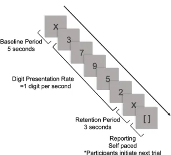
Pupillary response dynamics to the backwards digit-span task (Figure 1.1) include
incremental dilation as each digit is encoded, reaching maximum dilation following final digit
presentation (while manipulating and reordering) or at the point of encoding capacity, whichever
comes first.28,32,35 Upon recall, pupils recover to pre-trial size.28,32,37 Pupillary response
magnitudes are often summarized for each trial as the average pupil size change following final
digit presentation, while encoding and reordering numbers, compared to pre-trial size—where
longer digit sequence lengths elicit greater dilation responses until the point of capacity.28,34
Outcomes represent individual neural resource utilization for a given sequence-length—which
may inform individuals’ cognitive efficiency.30,35 Current literature supports resultant decreases
in performance with increased resource utilization via cognitive effort in response to higher
levels of task difficulty29, also seen in dual-task concussion literature.3931,41 Therefore,
incorporating simultaneous pupillary response recording during the digit-span task may be a
meaningful physiologic metric for concussion assessment, with respect to the individual
Pupillary response dynamics may be useful to inform cognitive efficiency in isolation—
though recent psychophysiological investigations suggests monitoring multiple metrics when
examining associations between physiological and behavioral outcomes associated with complex
cognitive constructs and processes.29,34,41,42 Juxtaposed with concussion literature, recent
systematic reviews regarding the physiological response to injury concludes that a single ‘perfect
metric’ that accounts for the complexities associated with concussion is highly unlikely—rather,
a combination of measures may be more appropriate.8,18 Moreover, physiological metrics
regulated by the ANS require careful consideration given the many latent variables that may
contribute to variability in response dynamics (e.g., stress, emotional response, etc.).41,43–45 Heart
rate variability (HRV) may be a meaningful supplement to pupillary response during digit-span
task completion to inform cognitive efficiency, as an index of neurocardiac function associated
with the ANS.44,46,47 Limited research is available to fully describe HRV and ANS dysfunction
following concussion, though most studies report HRV response dynamics to physical
activity/movement task demands.46–49 The majority of HRV investigations in the concussion
space focus on persistent cerebral metabolic deficiencies related to reduced cerebral blood flow
at rest, and threshold determinants for exercise tolerance testing.18,50,51 Applications of this
measure to estimate potential thresholds for cognitive load are unclear.
Cognitive neuroscience data clearly acknowledges environmental factors that influence
current pupil size monitoring. Specifically, environmental luminance and accommodation
responses, have previously hindered scientific inquiry progression in this space and other
physiologic outcomes, with respect to internal validity.32 Recent advancements in virtual reality
(VR) head mounted displays with embedded infrared eye tracking technology provide a
for pupillary response parameter assessment to inform cognitive efficiency. Examination of HRV
as it relates to ANS activity may serve as a useful supplement to describe the resolution of
post-concussion compensatory mechanisms and physiological impairment, combined with an
ecologically valid marker for neural resource utilization.46,52,53
1.4 Problem Statement
Clinicians and researchers need objective measures to better characterize behavioral and
physiological response dynamics associated with cognitive inefficiency following concussion.
While cognitive efficiency can be described relative to various cognitive processes, working
memory is most often examined with respect to relationships between task demands and
available cognitive resources. Working memory is also one of the most common cognitive
impairments following concussion—where associated post-injury clinical measures (digit-span
task) demonstrate high diagnostic sensitivity.11,12
Clinical task performance and physiological response dynamics associated with cognitive
efficiency are difficult to characterize following concussion due to rapidly deteriorating signal
detection and poor ecological validity of current clinical assessments and advanced
neuroimaging modalities.8,18 Dynamic cognitive efficiency characterization via clinical task
performance and physiological and metrics may better inform concussion recovery response
dynamics. Results may therefore have important implications for improved concussion clinical
assessment and management regarding readiness to return to athletic and or military activity.
The study aims were to first examine relationships between clinical task performance,
heart rate variability, and cognitive load associated with a digit-span working memory task, on
1.5 Specific Aims
Specific Aim 1: To examine associations between task performance accuracy, heart rate
variability, and pupillary response dynamics, across levels of task difficulty within a
digit-span working memory, task in healthy collegiate club sports athletes.
Hypothesis: We anticipate significant associations to exist across participants between
task performance accuracy, heart rate variability, and pupillary response dynamics with
respect to the levels of task difficulty within a digit-span working memory task.
Specifically, we anticipate individuals will demonstrate decreasing heart rate variability
and task performance accuracy across increasing digit sequence-lengths—while
pupillary responses will increase (dilation) to a point of working memory capacity, then
plateau or decrease (constriction).
Significance: The digit-span task within the current recommended concussion
assessment battery is known to be one of the most sensitive to acute injury. Rapid loss in
this signal detection is likely attributable to limitations associated with clinical task
performance factors (e.g., task design, administration and interpretation, etc.) and lack of
psychophysiological characterization of cognitive efficiency. If combined task
performance and physiological (heart rate variability and pupillary response) assessments
can objectively inform individual differences in cognitive efficiency, the potential exists
to also provide insight regarding neurocognitive deficits. Dual physiological assessment
monitoring during the digit-span task by both pupillary response and heart rate variability
may provide a more robust picture of cognitive efficiency with respect to physiological
Specific Aim 2: To examine the effect of prior concussion injury, and task performance
accuracy and heart rate variability response dynamics on pupillary response dynamics,
across levels of task difficulty within a digit-span working memory task in healthy
collegiate club sport athletes.
Hypothesis: We anticipate that those who report prior concussion, task performance
accuracy, and heart rate variability dynamics will exhibit worse physiological outcomes
of pupillary response (increased dilation responses) across digit-sequence lengths,
though performance-wise they may not differ from those without a concussion history.
Significance: Recent studies examining the individual ability of potential physiological
biomarkers to discriminate between those who have a concussion history —with respect
to neurocognitive deficits—have exhibited various threats to internal validity.
Examination of the effects of prior injury as it relates to cognitive efficiency may provide
insight regarding the potential utility of a more dynamic behavioral and physiological
assessment for post-concussion assessment and monitoring.
1.6 Independent Variables
1. Digit sequence length: the number of digits within a given sequence. Discussed in terms of
cognitive load as a representation of task difficulty level associated with longer sequences.
(Aims 1 & 2)
2. Task performance—average percent of correctly identified digits (by serial position) with
respect to sequence-length for each trial. (Aims 1 & 2)
3. Heart rate variability— Total HRV as the root mean square of successive differences
4. Concussion history—Self-reported by first providing athletes with the definition for
concussion from included in Section 1.8, then asking them to consider their concussion
history following provision of a definition. Participants included in the history group were
those who reported their most recent concussion occurring between the years in which they
attended secondary school (grades 9-12) until 6 months prior to their study participation date.
(Aim 2)
1.7 Dependent Variable:
1. Pupillary response: Pupillary Response represented as the baseline corrected pupil diameter
in mm, during the retention period–measured by trial, whereby greater dilation response
reflects greater neural resource utilization.28,32,56
1.8 Potential Co-variates:
1. Sex—male versus female
2. Prior contact/collision sport participation—examined via 2 variables using questions from the
(Head Impact Exposure Index—HIEI57)
i. Total number of years participating in contact/collision sport
ii. Total number of hours participating in contact/collision sport
1.9 Definition of Terms
1. Concussion: The definition provided in the Berlin Concussion Consensus statement will be
applied throughout as follows: A change in brain function following a force to the head,
which may be accompanied by temporary loss of consciousness and is identified in awake
individuals with measures of neurologic and cognitive dysfunction. Common concussion
dizziness, balance problems/loss of balance, fatigue/loss of energy, feeling in a fog,
irritability, drowsiness, nausea, memory loss, sensitivity to light/noise, and blurred vision.
2. Sequence-length: the numbers of digits in a digit-sequence.
3. Cognitive load: cognitive demands relative to the task—i.e., task difficulty associated with
longer sequences.
4. Neural Resource Utilization: brain activity used to accomplish a cognitive task.
5. Baseline Period: two seconds prior to each digit-span being presented to allow for pupils to
rest and stabilize.
6. Loading Phase: the portion of the digit-span task in which participants are presented with a
sequence of digits at the rate of 1 per second and asked to remember the number sequence.
7. Retention Period: three second period after each digit-span is presented when participants
process/encode the information and prepare to recall.
8. Task performance: percent correctly identified digits (by serial position) across trials for each
sequence-length.
9. Pupillary Response: average pupil size (diameter) across trials for each sequence length.
10.Cognitive Efficiency: the ability to maximize neural resource utilization while maximizing
task performance.
11.Heart Rate Variability: The root mean square of successive differences (RMSSD). A reliable
estimate of vagally mediated changes in heart rate variability (i.e., beat-to-beat variance in
heart rate), from ultra-short-term measurement durations—shown to capture acute mental
1.10 Delimitations
1. Individuals who were not a collegiate club sport athlete were not included in this study.
2. Individuals with permanent vision loss, strabismus, amblyopia, or eye surgery in the last 6
months were not included in this study.
3. Individuals participating in visual or vestibular therapy were excluded to prevent
confounding variables.
1.11 Limitations
1. Participants with a concussion history were responsible for reporting their own medical
history. Being so, there is potential for participants to be included in the concussion history
group who should not have been or vice versa.
2. Study sample may predominantly consist of males, and therefore, we may be unable to
examine sex differences within the proposed study aims.
1.12 Assumptions
1. Participants accurately reported past medical and sport participation history.
2. Participants remembered and accurately reported all concussion injuries.
3. Participants remained engaged and gave full effort during the task.
1.13 Summary of Study Significance
This study is the first to examine an assessment for cognitive efficiency that accounts for
both task performance and physiological response dynamics, which may provide meaningful
insight for concussion injury response and recovery. Consideration for necessary adaptations to
the digit-span task within the clinical battery to better elicit dynamic working memory processes,
digit-span task performance outcomes, will allow us to better describe efficiency.(Figure 1.1) We will
also examine the effect of concussion history on these measures as a preliminary step towards
improving our ability to capture the dynamic clinical and physiological aspects of cognitive
efficiency.
CHAPTER 2: LITERATURE REVIEW 2.1 Overview of Concussive Injury
Repetitive head impact exposures and concussion are major athletic health concerns1 for
which the physiological response and recovery dynamics are poorly understood. The 5th
International Conference on Concussion in Sport defines concussion as: “A change in brain
function following a force to the head, which may be accompanied by temporary loss of
consciousness and is identified in awake individuals with measures of neurologic and cognitive
dysfunction. Common concussion symptoms include: headache, feeling slowed down, difficulty
concentrating or focusing, dizziness, balance problems/loss of balance, fatigue/loss of energy,
feeling in a fog, irritability, drowsiness, nausea, memory loss, sensitivity to light/noise, and
blurred vision.”9 Prolonged neurophysiological abnormalities in concussed individuals assessed
using advanced imaging techniques suggest a prolonged over-excitatory period, when the brain
remains physiologically-compromised requiring greater neural resource allocation and metabolic
energy to balance task demands with available cognitive resources.8,19 Consequences of these
persistent deficits suggest decreased cognitive efficiency which may leave the brain at an
increased risk for repeat injury, new musculoskeletal injury, prolong recovery, neurocognitive
impairment, and persistent symptom presence.4,22,50
Despite growing evidence of persistent compensatory mechanisms and associated
neurophysiological cost—dynamic clinical and physiological response dynamics for cognitive
relevant literature supporting the need for a physiological measure associated with task
performance response dynamics of working memory, and sensitive to potential deleterious
repetitive head impact exposures and concussion to better inform cognitive efficiency. We then
propose pupillary response dynamics to cognitive task demands as a physiological index for
neural resource utilization to meet these assessment needs and inform cognitive efficiency. We
also suggest the simultaneous measurement and inclusion of heart rate variability to characterize
cognitive efficiency as a secondary physiological measure in order to best account for cognitive
effort input relative to task demands whist still accounting for task performance outcomes.
Finally, we review relevant literature to support the methodological considerations associated
with the proposed study in the context of design, instrumentation, and overall ecological validity
to allow for interpretation of future directions in this space.
2.1.1 Epidemiology
Estimated prevalence for recreational and sport-related concussion (SRC) in the United
States from the national injury databases report prevalence between 1.1 to 1.9 million, in
pediatric and adolescent populations.12 Concussion injury rates for this population are 2.5 per
10,000 athlete exposures and are higher in collision/high-contact sport athletes. Concussion
among college aged athletes participating in National Collegiate Athletic Association (NCAA)
sports is estimated around 10,560 nationally with the overall concussion rate of 4.47 per 10,000
athlete-exposures.2 Recent literature regarding the effects associated with repetitive head impact
exposures remains unclear—though studies suggesting greater functional impairments in those
who have experienced multiple prior concussions and potentially greater long-term structural
changes, relative to cumulative exposure provide sufficient evidence for further
football, rugby, men’s lacrosse, etc.) are at the greatest risk, as they experience relatively high
numbers of head impact exposures over the course of a single season and athletic career.5,6,21
Many research- and clinically-based challenges limit the understanding for the true SRC
epidemiology. Injury definitions lack consensus across disciplines and may contribute to
decreased concussion injury-based knowledge among patient populations, and therefore athlete
self-report/disclosure.2,59
Annual participation rates for collegiate club sports is currently unknown though
estimated to make up a large percentage of competitive athletes at risk for concussion given the
discrepancy between high school varsity and college varsity level athletes.60,61 The majority of
these athletes have prior experience participating in their respective sports and maintain a
relatively high level of competition, though lack the medical coverage and clinical resources
available to those rostered on varsity teams. These teams are also often larger than varsity
collegiate sports and therefore still represent a large at-risk population.
2.1.2 Injury Mechanics and Response
Concussion is theorized to result from linear and/or rotational biomechanical forces
(direct or indirect) to the head, neck, or body resulting in an impulsive force to the head and
brain, causing axonal shearing and increased pressure gradients, resulting in diffuse axonal injury
and subsequent altered neuronal function.7,62 The neuropathophysiological process that follows is
described as a neurometabolic cascade, posited to drive clinical deficits and dysfunction.
Moreover, there are growing concerns regarding the elevated risk of repeat injury following
concussion and associations with slower recovery, prolonged symptoms, etc.63–65
Additional concerns exist regarding potential effects of repetitive head impact exposure
Studies examining these effects suggest greater functional impairments in those who have
experienced multiple prior concussions and potentially greater long-term structural changes.3,58
Attempts to better understand the cumulative effects between repetitive head impacts and
long-term neurological consequences (e.g., increased risk for neurodegenerative disease,
neuropsychiatric deficits, etc.) demonstrate limited correlational associations and causal
relationships have yet to be established.4,20,58 Continued investigation regarding physiological
and clinical response dynamics following concussion with respect to symptoms, neuro-cognitive
functioning, and motor control/postural stability, is needed to further inform these concerns.
2.1.3 Physiological Response
2.1.3.1 Neurometabolic Cascade
The physiological response to concussion injury is multifactorial—primarily informed by
animal models, and a few recent human studies.4,27,29 The neurometabolic cascade of events
following the biomechanical insult mentioned above, has been described in detail by Giza et al.
relative to associated clinical impairments.27 Initial potassium efflux causes a dramatic release of
excitatory neurotransmitter glutamate causing neuronal depolarization and further ionic
disruption. Initial hyperglycolysis supports sodium potassium pumps as they respond to
homeostatic disruption, requiring additional adenosine triphosphate to restore ionic balance.
Calcium influx is appropriated into mitochondria for short-term relief but eventually leads to
mitochondrial dysfunction and decreased oxidative capacity. Glucose metabolism then shifts to
support this energy demand, resulting in a state of hyperglycolysis. Concurrent reduction in
cerebral blood flow during this time of high energy demands is posited to result in the ‘energy
metabolism further contribute to the crisis as neurons attempt to restore homeostatic intracellular
calcium levels.27
Post-concussion pathophysiological recovery is such that potassium and glutamate
stabilization occur within 24 hours, and calcium levels within the first 3-4 days, and glucose
levels and disruptions in cerebral blood flow within 7 to 10 days. Recent advanced imaging
studies, however, suggest these physiological deficits indicating increased neural activity may
persist beyond these timeframes. Implications of these prolonged physiological deficits indicate a
window of cerebral vulnerability that extends beyond clinical measure normalization when the
brain remains physiologically-compromised and at a greater risk of repeat injury.7,8
2.1.3.2 Role of the Autonomic Nervous System
Autonomic nervous system (ANS) function following concussion has been posited to
play a role in the above-mentioned cerebrovascular-related alterations, with respect to
sympathetic and parasympathetic activity balance.46,55 Balanced ANS function is essential from a
neurocardiac perspective with respect to its role in regulating cerebral blood flow to meet
neurometabolic demands. Dysfunction within this system following concussion may
significantly influence the prolonged energy crisis and subsequent secondary injury—though the
mechanism behind these alterations are unknown.7,8,50 Centers within the brain responsible for
ANS regulation of neurocardiac function are suggested to be uncoupled following concussion,
though more evidence is need to further elucidate its role in physiological injury response and
recovery.18,49,53,55,66
2.1.4 Clinical Response
Clinical impairments are widely variable and often reflected in a multitude of subjective
visual/oculomotor, and vestibular function—all associated with the pathophysiological response
to concussion injury.62,66 Given the complexities in clinical presentation, clinicians rely on a
comprehensive assessment battery for injury identification and recovery monitoring (i.e., clinical
measure normalization).9 The clinical recovery time course (e.g., symptoms, neurocognitive,
balance) has been well documented in large prospective studies over the last 2 decades with
current available measures. Specifically, balance deficits and neurocognitive function often
recover to baseline levels within 3-5 days.11,12 Symptom severity scores are known to be the
longest lasting clinical deficit, recovering around 7-10 days in most uncomplicated cases.7,65
The prolonged energy crisis (i.e., decreased glucose levels and cerebral blood flow),
reportedly lasting up to 10 days in the neurometabolic cascade is theorized to drive secondary
injury in the brain and associated symptom reports and neurocognitive deficits.7 Subjective
symptom reports post-concussion often include (e.g., headache, dizziness, fatigue, sensitivity to
light, and difficulty with memory or concentration) though are widely varied. Symptom
checklists including the most commonly reported post-concussion symptoms—and associated
Likert scales for severity, are heavily relied upon by clinicians for recovery monitoring (i.e.,
clinical normalization).63,65 In those experiencing prolonged symptoms (beyond 7 to 10 days
post-injury), recent studies suggest closer clinical examination to identify potential neurological
sub-system involvement (i.e., visual/vestibular, cervicogenic, and physiologic/metabolic) and
alternated management strategies.66 Additionally, concussion-like symptoms, in the absence of
diagnosed injury, are common.67 This complicates clinical decision making around the
recommended graded exertion protocol and eventual return to play (RTP), given protocol
objective physiological markers to aid in the further elucidation of the relationship between
clinical and physiological response dynamics post-concussion.
Visual and vestibular impairments are also common following concussion given axonal
injury and impaired neurotransmission, or direct damage to their respective special sensory
organs.68,69 Specifically, impairments associated with binocular visual skills such as
accommodation, convergence, smooth pursuits, and saccadic eye movement, are common
following concussion.69–71 Symptoms associated with these deficits specifically include blurred
or double vision, headache, eyestrain, dizziness, nausea, and difficulty concentrating; which
contribute to functional impairments such as difficulty reading and tracking, and trouble with
near tasks.72,73 Given the impact post-concussion visual and vestibular impairments can have on
daily activity levels and quality of life, and their association with prolonged recovery, clinicians
are encouraged to incorporate earlier screening and management for these specific deficits.68
Common assessments used to capture these impairments (e.g., King Devick, Vestibular Ocular
Motor Screen, etc.) have demonstrated clinical utility as physiological measures for dysfunction
within their respective sensory sub-systems—though provide little insight with respect to global
neurophysiological dysfunction following concussion.69,74,75
Neurocognitive deficits arise from initial axonal injury and impaired neurotransmission—
where common deficits involve information processing, attention and reaction time, often
captured using neuropsychological assessments.16,76,77 These pathophysiologic factors may also
affect the regulatory central and peripheral neural networks that contribute to motor control and
balance.7,63,79 Slowed information processing and reaction time, along with attention deficits play
major roles in the maintenance of balance and postural control whereby specific cognitive
responsible.9,11,78 Neurocognitive deficits are often identified using clinical measures such as the
Standard Assessment for Concussion (SAC) or many of the different computerized
neurocognitive testing platforms such as the Immediate Post-Concussion Assessment and
Cognitive Test.9
Motor control and balance deficits are most often assessed clinically using the balance
error scoring system.9,78 Of noteworthy concern, these neurocognitive and balance assessments
are known to lose signal detection as quickly as within the first 24 hours post-concussion and
demonstrate ceiling effects.11,12 More advanced assessments using the Sensory Organization Test
(SOT) or dual-task gait assessment protocols aim to examine more complex motor
control/balance impairments with respect to higher order integration of sensory-motor
information.79–82 Feasibility and ecological validity concerns associated with advanced balance
assessments such as the SOT are limiting for further clinical consideration. Clinical utility of
dual-task ‘cost’ outcomes as they relate to post-concussion physiological response dynamics
continue to be examined though may be useful in later stages of concussion recovery with
respect to functional testing.82,83 Overall, clinical response dynamics following concussion are
complex and may not follow a fixed recovery time frame or be appropriately captured using the
current assessment battery components. Combined dynamic clinical and physiological
assessments may provide a better characterization of the neurophysiological concussion injury
response and recovery to aid in improved concussion management paradigms.
2.2 Clinical Versus Physiological Considerations
While most individuals recover from concussion within 2-4 weeks, a substantial number
may experience a prolonged recovery and persistent symptoms.65,66,84 Physiological deficits
suggesting that the current battery of assessments may not be sensitive enough to identify injury
recovery.16,19 The clinical and physiological response to concussive injury, is best described in
the context of the integrated recovery model proposed by Dr. Mike McCrea first in 2009 and
revised in 2015.85 This model proposes a progression of recovery, characterized by an acute
period of clinical signs and concurrent physiological dysfunction, followed by persistent
physiological dysfunction in the sub-acute period, and finally, complete clinical and
physiological recovery. This model has been further supported in a recent systematic review
highlighting key physiological considerations in the context of concussion injury response and
recovery.8 Persistent physiological deficits cultivate a neural environment in which the brain is
susceptible to injury, during a time in which most athletes are actually beginning an RTP. Too
much or too little physical and/or cognitive activity during this time may further delay the
recovery process)51, therefore it is imperative that we understand the time course of the
physiological response to injury in order to continue the development of appropriate
management paradigms.
2.2.1 Advanced Physiologic Measures
2.2.1.1 Advanced Neuroimaging Techniques
Numerous advanced assessment techniques have been examined to improve our
understanding regarding the time course of clinical and physiological recovery following
concussion. Further characterization of the relationships between physiological disturbance and
clinical outcomes continue to be pursued by many researchers to identify more objective
diagnostic and recovery criteria for improved concussion management paradigms. Neuroimaging
techniques in particular have been used to examine widespread neural systems that cross
though the level of evidence to support their clinical utility is low and therefore not
recommended at this time.18 Results from a recent systematic review summarizes the
contributions from neuroimaging modalities (e.g., functional magnetic resonance imaging
(fMRI), diffusion tensor imaging (DTI), and electroencephalography (EEG) to our current
understanding regarding physiological and clinical responses following concussion. Specific
results from fMRI studies report varied results regarding blood oxygen level dependent (BOLD)
responses during resting state and task-based examinations following concussion.18 Task-based
fMRI studies often examine this measure to describe neural activity in task related networks of
working memory (e.g., dorsolateral prefrontal cortex)—as one of the most commonly affected
neurocognitive processes following concussion.11,16,76 Varied results have been reported showing
both lower and higher levels of activity have been exhibited in concussed individuals.18
Prefrontal cortex (PFC) related activity is modulated by working memory cognitive loading,31,77
where fMRI studies using a digit-span task show that higher digit sequence-lengths, representing
a higher cognitive load is consistently associated with greater cortical activation, including
critical PFC regions.8,18,19 Results from these fMRI studies provide support for compensatory
neural resource utilization associated with prolonged physiological impairment as brain activity
extends beyond regions of interest in the dorsolateral prefrontal cortex and inferior parietal areas
in these studies. Resting state fMRI is currently the most extensively studied network in SRC –
though similar varied results have been reported where both increased and decreased
connectivity between default mode network regions are observed following injury.8
Similarly, the use of fluid biomarkers has advanced our understanding of concussion
pathophysiology, though the validation of these markers is in the preliminary stages.18,86,87
by examining serum and blood biomarkers indicative of axonal injury.18,87 Additional
investigations using genetic testing has sought to inform prognostic factors associated with
concussion injury risk, prolonged recovery and long-term neurological heath with respect to
potential life-long consequences of injury and repetitive head impact exposure.39,538,18The
majority of these investigations have been completed in more traumatic brain injury cases,
though increasing in athletic populations. The major limitations affecting fluid biomarkers
pertain to the time needed for analysis and results and access to a basic science laboratory—thus
currently not a feasible option for sports medicine clinical settings.18 Future studies with
longitudinal designs to further elucidate the dynamic recovery of these compensatory
neural-mechanisms following concussion are not a viable option as fMRIs are very expensive.
Overall, authors of a recent and comprehensive systematic review including biomarkers
(e.g., blood serum and plasma markers, salivary cortisol, cerebrospinal fluid, etc.)8,18 highlight
the current limitations challenging this research initiative to generalizability of findings
including: small homogenous sample sizes across studies (primarily male participants), varied
study designs, limited number of studies overall, differences in outcome measures and analytic
methods, and lack of consistency post-injury data collection time points. Recommendations from
this review emphasize continued research efforts to further characterize the pathophysiological
response to concussion and repetitive head impact exposure in the absence of diagnosed
concussion that include larger samples sizes inclusive of both sexes, standardized protocols,
more stringent study designs that allow for baseline comparisons, appropriate controls, blinded
analyses that include clinically applicable outcome measures.8,18 Moreover, limited
generalizability in neuroimaging outcomes stems from the underlying issue of clinical feasibility
validity. Investigations using outcomes known to be indirect measures associated with these
advanced techniques such as heart rate variability and pupillary response may provide a more
clinically feasible option that allows for the dynamic characterization of post-concussion
physiological response and recovery.
2.2.1.2 Heart Rate Variability
Heart Rate Variability (HRV) measures function as indices of neurocardiac function used
to inform the dynamic responses associated with ANS dysfunction following concussion.55
These measures represent the fluctuation in the time intervals between adjacent heartbeats with
respect these subsystem dynamics as individuals adapt to environmental and psychological
challenges.54 Recent investigations have examined these measures at rest and in stressed states
(both physically and psychologically), where specific outcomes have been linked to performance
of executive functions like attention and emotional processing by the prefrontal cortex.41,45
Common outcomes for HRV in the concussion literature space include heart rate and time- and
frequency-domain indices of HRV. Heart rate (HR) is also a common metric used in concussion
literature with respect to persistent cerebral metabolic deficiencies related to reduced cerebral
blood flow at rest, and threshold determinants for exercise tolerance testing.47,48,53,66 Specifically,
HR is represented as the average difference between the highest and lowest HRs during each
respiratory cycle (HR Max − HR Min). Examination of HRV measures is less common in this
population. Time domain measures are used to quantify the amount of variability in
measurements of the inter-beat interval (IBI) (i.e., the time period between successive
heartbeats); and Frequency-domain measurements estimate the distribution of absolute or
relative power into four frequency bands.52 Overall, current understanding of the effects of
though generally suggest post-concussion increases in sympathetic activity and lower
parasympathetic compared to controls.18,46 Individuals with concussion have been found to have
higher rates of sympathetic nervous system output than controls, as exemplified by higher resting
heart rates and higher heart rates during cognitive activity. 8,18,55
Response dynamics associated with HRV measures may provide meaningful information
regarding the concussion physiological response characterization given associations with clinical
recovery and symptom resolution due to resolved metabolic impairment. Altered autonomic
nervous system regulation is evident as individuals recover following concussion though when
HRV measures are examined in isolation during task-based paradigms associated with working
memory results are limited given the potential influence of the stress response associated with
increasing cognitive demands.14 Therefore, examination of HRV as it relates to ANS activity
may serve as a useful supplement to describe the resolution of compensatory mechanisms and
physiological impairment, combined with an ecologically valid marker for neural resource
utilization.6
2.2.1.3 Current Visual Metrics
Visual impairments are among the most prevalent following concussion occurring in up
to 60% of children and adolescents.68,73 Visual disturbances following concussion are most often
reported in oculomotor, and visual processing contexts,4,72,75 given the widespread, neural
architecture of the visual system within the brain, with over half of neural pathways related to
vision.71,75 This network widely expands fronto-parietal circuits and subcortical nuclei, cranial
nerves, and interconnections between these areas, all of which are particularly vulnerable to head
Oculomotor deficits in accommodation, convergence, smooth pursuits, and saccadic eye
movement, are commonly affected following concussion. Symptoms of these deficits include
blurred or double vision, headache, eyestrain, dizziness, nausea, and difficulty
concentrating.68,88,89 Moreover, recent studies identifying predominant visual impairments
following concussion highlight their relationships with neurocognitive processes of memory and
attention—also often impaired following concussion.68,73
Additionally, deficits in the pupillary light reflex have been reported following
concussion due to diffuse axonal injury resulting in abnormal static and dynamic responsivity.90
Specifically, the pupillary light reflex following concussion has been showed to be symmetric,
though delayed, slowed, and reduced; additionally smaller initial baseline pupil diameters have
been reported following concussion, compared to uninjured controls.90,91 While there are limited
studies published in this space, findings suggest dysfunction within afferent pupillary pathways,
and the parasympathetic and sympathetic efferent pathways of the ANS. These findings
underscore the role of the ANS in post-concussion physiological impairments and need for
further response characterization.90 Overall, this review echoes results from two of the most
recent and comprehensive systematic reviews regarding the concussion pathophysiological
response in that clinical utility of a physiological marker is more likely to derive from
measurement combinations rather than by any one in isolation. 18,43
2.2.2 Considerations for Advanced Clinical and Physiological Assessments
Task performance and physiological responses following concussion do not adhere to a
fixed recovery time course, therefore assessments for impairment within these areas should
2.2.2.1 Behavior Specific Considerations
Neurocognitive impairment following concussion describes deficits in memory, attention
and processing speed as a result of diffuse axonal injury and the energy crisis that results in
secondary neuronal injury. Neurocognitive assessments are used to capture task performance
responses associated with each of these cognitive constructs following concussion. Longitudinal
examination of working memory (WM) task performance outcomes are theorized to provide
insight regarding cognitive efficiency given their representation of task performance across
varying levels of difficulty.25,77 Deficits in WM are common following concussion and are
typically assessed using recall tasks in the Standard Assessment for Concussion (SAC).9,76 The
digit-span task in the SAC in particular is sensitive to injury as a performance-based
representation of working memory capacity and/or the relationship between task demands and
available cognitive resources. 11,12
The digit-span is one of the most commonly used tests of working memory in clinical
research and practice and can include forward and backwards administration. Backwards
digit-span administration typically involves digit-sequence presentation (verbal or visual), a brief
retention period, followed by a recall period during which participants are asked to recite the
presented digits in exact reverse order (e.g., 3-5-8 correctly recalled is 8-5-3). Task difficulty is
characterized by digit-sequence length (i.e., number of ‘to be recalled’ digits) and increases,
every 1 to 2 trials depending on recall accuracy. Individuals who fail to demonstrate perfect
recall on the first trial attempt for a given sequence-length are typically permitted a second
attempt—with a new digit sequence. Traditional task administration implements a ‘discontinue’
rule following sequence length at which both trials are inaccurately recalled. This task requires
and theoretical utility.31,77 However, recent evidence suggests that current administration of the
backwards digit-span task, using all or nothing scoring and discontinuing when behavior is less
than 100% limits our ability to understand how working memory may be affected when demands
exceed capacity. All-or-nothing scoring assigns credit only for perfectly recalled sequences,
whereas partial-credit scoring counts each digit recalled in the correct serial position. These
adaptations to this task are suggested is suggested as a more robust examination of task
performance responses during the digit-span task with more scoring variability and increased
ability to detect individual differences.27,92 Overall, this new evidence highlights the fluidity of
working memory and cognitive efficiency, rather than having a fixed capacity)—worthy of
examination in concussion populations with respect to known compensatory neural resource
utilization post injury that demonstrate dynamic changes across recovery.27
2.2.2.2 Physiologic Specific Measures
Post-concussion physiological response and recovery remains poorly characterized by
current clinical and advanced imaging techniques. Recent systematic reviews emphasize the
dynamic nature of physiological changes following concussion injury that cannot be constrained
within a single window of ‘physiological recovery’.8 Neurocognitive assessment paradigms that
elicit a behavioral response are often used under advanced imaging to functionally characterize
regional brain activity and neural resource utilization—though not feasible with respect to cost
effectiveness for long term monitoring. Clinically based physiological measures such as heart
rate variability have also been described in concussion literature to describe dynamic aspects of
ANS function, though when used in isolation may not be adequate to give a full picture of the
2.3. Pupillary Response as a Potential Solution
Examination of pupillary response during a cognitive control task of working memory
may provide clinicians with a more sensitive assessment of functional neurophysiological
impairment and recovery following concussion, ultimately bridging the gap between clinical and
physiological recovery. While pupillary response to cognitive load has been described in healthy
and other cognitively impaired populations,28,32,36 little is known about this measure in
concussion populations. Clinical and advanced physiological evidence has described higher
order cognitive impairment in attention, processing speed, and working memory utilization
following concussion.9,12 Many of the common testing paradigms used to examine these
cognitive constructs in concussion populations (e.g., digit-span)— both clinically and with
fMRI— have also been examined using pupillometry to reflect pupillary response changes to
cognitive load in healthy and diseased states.28,31,38,39
2.3.1 Neurophysiological Underpinnings
The neurophysiological underpinnings of this response suggest that pupil dilation
response in particular, is modulated by the noradrenergic Locus Coeruleus neuromodulatory
system (LC-NE).30,33,93 This system has widespread projections that extend to nearly all cortical
and subcortical regions.94 The LC-NE is a small collection of nuclei in the brainstem’s
pontomedullary reticular formation and plays a central role in behavioral adaptation, task
performance, attention, functional reorganization of cortical activity when environmental
contingencies change that allow for cognitive and behavioral adaptation, and working
memory.33,95 Current evidence validates pupillary response to cognitive load across various
conclude that pupil dilation response to cognitive load is correlated with activity in brain regions
engaged by current task demands.29,30,33,93
2.3.2 Pupillary Response Correlates with Behavioral Outcomes
Previous literature has shown that pupil dilation is associated with a broad range of
cognitive processes (e.g. attention, memory, etc.) in healthy individuals. In this context, pupil
dilation refers to a stimulus-induced increase in pupil diameter relative to a pre-stimulus baseline
period, or a task-evoked pupillary response.96 Pupillary responses during cognitive control tasks
have been extensively examined in healthy populations ages 10-83 years, where pupil dilation
increases with increasing tasks demands.29,31,37,38 Previous studies have further supported pupil
dilation as a valid physiological marker of cognitive load via comparison to EEG and fMRI
measures of brain activity. 30,97
2.3.3 Pupillary Response in Clinical Populations
More recently, pupil dilation and cognitive processes have been examined across varying
diseases states such as Alzheimer’s and Parkinson’s disease.38,39 One study examined pupillary
response during a digit-span task in adults who are cognitively normal and those across varying
levels of mild cognitive impairment (MCI), both single domain (S-MCI) and multiple
domain(M-MCI). The results of this study suggested that pupillary responses during a digit-span
task reflect compensatory effort —exhibited by greater pupil dilation—to achieve equal task
performance in those with lower levels of MCI based on working memory capacity.38
Additionally, those with M-MCI exhibited no significant changes in cognitive effort across
varying levels of cognitive load suggesting that in later stages of this degenerative disease
individuals lack the ability to appropriately adapt cognitive effort.38 One study has examined