PEROXIDE DEPENDENT EFFECTS IN HUMAN AIRWAY EPITHELIAL CELLS EXPOSED TO OXIDANT AIR POLLUTANTS
Phillip A. Wages
A dissertation submitted to the faculty at the University of North Carolina at Chapel Hill in partial fulfillment of the requirements for the degree of Doctor of Philosophy in the Curriculum
in Toxicology in the School of Medicine.
Chapel Hill 2015
Approved by:
James M. Samet
Bernard E. Weissman
Avram Gold
Urmila Kodavanti
iii ABSTRACT
Phillip A. Wages: Peroxide dependent effects in human airway epithelial cells exposed to oxidant air pollutants
(Under the direction of James M. Samet)
Air pollution is a global public health problem. The induction of oxidative stress, or the
cellular response to avoid cytotoxicity due to an increase in reactive oxygen species or decrease
in antioxidants, is frequently cited as a mechanism of toxicity for air pollutants. In addition to the
role of oxidative stress in disease, there is growing evidence that oxidative processes including
the generation of reactive oxygen species is essential for normal cellular function. This
dissertation provides evidence that the reactive oxygen species, hydrogen peroxide, is a key
mediator in air pollutant-induced adverse cellular responses. First, it is demonstrated that the
divalent metal, zinc, induces hydrogen peroxide-dependent adaptive gene expression in human
airway epithelial cells. This builds upon previous work to establish that zinc-induced pro-oxidant
effects and electrophilic activity are both critical in its mechanism of toxicity. We next show that
the organic component, 1,2-naphthoquinone, increases protein sulfenylation of regulatory
proteins via hydrogen peroxide. This is the first report that protein sulfenylation is effected by an
environmentally relevant exposure, establishing a potential new mechanism of toxicity as well as
a new biomarker for future studies. Finally, two approaches to utilize readouts of oxidative stress
in a translational manner are discussed. Specifically, the biological basis of a genetic risk factor
of a susceptible population to air pollution is explored using a primary human airway epithelial
iv
glutathione in the air pollutant-induced outcomes, and that the genetic risk factor, GSTM1-null,
enhances the effect of 1,2-NQ to induce the novel readout protein sulfenylation. Furthermore, we
were able to use a live cell imaging analysis of oxidative stress to rank the toxicity of fibers of
importance to respiratory toxicity and show that asbestos fibers obtained from the Libby
Montana Superfund Site have similar toxicity to that of crocidolite asbestos fibers. Together the
data suggests a vital and important role of hydrogen peroxide in air pollutant-induced adverse
responses and provides the basis to use redox-based readouts as biomarkers to improve public
v
vi
ACKNOWLEDGEMENTS
I am truly thankful and indebted to my family for being with me through every hardship
and celebration. Above all, I am grateful for my parents, Eugene and Evelyn Wages, for being
the most supportive and encouraging parents a son could ask for. They have always challenged
me to be the best I could be, and for that I credit all my success to their inspirational nature. I am
also thankful for my siblings, Michael, Melissa, and Stephen as they have been a blessing in my
life providing me with goals to achieve and advice when asked.
I must express my sincere appreciation to my research advisor, Dr. James M. Samet, for
providing me the opportunity to train as a scientist under his guidance. He is an excellent mentor
and, simply, a good person. Likewise, I am also fortunate to have been mentored by Dr. Philip
Bromberg as well, whom I have learned much from and would certainly not be as critical of a
thinker without our interactions. I am similarly thankful to my thesis committee members for
their commitment to my doctoral training to ensure my preparedness for the future.
Although there are many others who have helped me succeed, and survive, as a graduate
student, I would like to especially thank Ilona Jaspers, Rob Silbajoris, Wan-Yun Cheng, Eugene
Gibbs-Flournoy, Jaime Mirowsky, Shaun McCullough, Katelyn Lavrich, Peyton Bohnsack,
Orrin Stone, Quyen Tang, Abi Agoglia, Kristen Fisher, Sarah Muse, Dana Walsh, Desinia
vii
TABLE OF CONTENTS
LIST OF TABLES ... xi
LIST OF FIGURES ... xii
LIST OF ABBREVIATIONS ... xiv
CHAPTER 1: Introduction ... 1
1.1: Air Pollution ... 1
1.1.1: Global Public Health Burden ... 1
1.1.1.1: Susceptible Populations ... 2
1.1.2: Traffic Related Air Pollutants ... 3
1.1.2.1: Zinc ... 4
1.1.2.2: 1,2-Naphthoquinone... 5
1.2: Redox Biology ... 7
1.2.1: Reactive Oxygen Species ... 7
1.2.1.1: H2O2 ... 8
1.2.2: Protein and Peptide Thiols ... 10
1.2.2.1: Glutathione ... 10
1.2.2.2: Oxidative Posttranslational Modifications ... 11
1.3: Redox Toxicology ... 14
1.3.1: Oxidative Stress ... 14
1.3.1.1: Antioxidant Response Element ... 15
1.3.2: Perturbation of Redox-Dependent Processes ... 17
1.3.2.1: Activation of Signaling Pathways ... 17
1.3.2.2: Bioenergetics ... 18
1.4: Approaches to Study Redox Toxicology ... 20
1.4.1: Small Molecule Redox Sensors ... 21
1.4.2: Genetically Encoded Redox Sensors ... 22
1.4.2.1: HyPer ... 22
1.4.2.2: rxYFP, Oba-Q Proteins and roGFP... 23
viii
1.4.3.1: DYn-2 and DAz-2 ... 27
1.4.4: In Vitro Models ... 27
1.5: Summary ... 28
CHAPTER 2: Role of H2O2 in the Oxidative Effects of Zinc Exposure in Human Airway Epithelial Cells ... 30
2.1: Introduction ... 30
2.2: Materials and Methods ... 32
2.2.1: Reagents ... 32
2.2.2: Cell culture ... 32
2.2.3: Viral transduction... 33
2.2.4: Live Cell Imaging ... 34
2.2.5: Gene Expression Analysis ... 35
2.2.6: Statistical Analysis ... 35
2.3: Results ... 36
2.3.1: Catalase Expression Ablates Zn2+-Induced H 2O2 Generation ... 36
2.3.2: H2O2 Contributes to Zn2+-Induced Oxidant Stress ... 43
2.3.3: EGSH Changes Do Not Mediate HO-1 Gene Expression ... 45
2.3.4: H2O2 Mediates Zn2+-Induced Adaptive Gene Expression ... 48
2.4: Discussion ... 48
2.5: Summary ... 52
CHAPTER 3: Protein Sulfenylation: A Novel Readout of Environmental Oxidant Stress ... 53
3.1: Introduction ... 53
3.2: Materials and Methods ... 55
3.2.1: Reagents ... 55
3.2.2: Cell Culture ... 55
3.2.3: Viral Transduction ... 56
3.2.4: Detection of Intracellular Sulfenylation ... 56
3.2.4.1: Staudinger Ligation ... 56
3.2.4.2: Immunoblotting... 57
3.2.4.3: Copper Catalyzed Azide Alkyne Cyclo-addition ... 58
3.2.5: Detection of Recombinant Protein Sulfenylation ... 58
3.2.6: Live Cell Imaging ... 59
ix
3.3: Results ... 60
3.3.1: Exposure to 1,2-Naphthoquinone Induces Protein Sulfenylation in BEAS-2B Cells ... 60
3.3.2: 1,2-Naphthoquinone-Induced Protein Sulfenylation is Dependent on H2O2 ... 61
3.3.3: 1,2-Naphthoquinone-Induces Sulfenylation of Regulatory Proteins ... 64
3.3.4: 1,2-Naphthoquinone Induces Sulfenylation of the Catalytic Cysteine (150C) in GAPDH ... 67
3.4: Discussion ... 69
3.5: Summary ... 72
CHAPTER 4: The Role of Glutathione in Redox Toxicology ... 73
4.1: Introduction ... 73
4.2: Materials and Methods ... 75
4.2.1: Reagents ... 75
4.2.2: Cell Culture ... 75
4.2.3: Live Cell Imaging ... 76
4.2.4: Immunoblotting of Intracellular Sulfenylation ... 76
4.3: Results ... 77
4.3.1: Glutathione depletion perturbs the dynamic nature of redox homeostasis ... 77
4.3.2: EGF-induced changes in protein sulfenylation are not impacted by GSTM1 status ... 78
4.3.3: GSTM1-null cells show more pronounced protein sulfenylation in response to exposure to 1,2-Naphthoquinone ... 79
4.4: Discussion ... 80
4.5: Summary ... 82
CHAPTER 5: Ranking Asbestos Fiber Toxicity Using Live Cell Imaging of EGSH ... 83
5.1: Introduction ... 83
5.2: Materials and Methods ... 84
5.2.1: Fiber Preparation ... 84
5.2.3: Cell Culture ... 85
5.2.4: Confocal Analysis ... 85
5.2.5: Statistical Analysis ... 86
5.3: Results ... 86
5.3.1: Exposure to Asbestos Does Not Compromise Cell Viability Acutely ... 86
x
5.3.3: Crocidolite Induces Cytosolic EGSH ... 91
5.4: Discussion ... 92
5.5: Summary ... 94
CHAPTER 6: Summary Discussion ... 95
6.1: Redefining Oxidative Stress in Environmental Exposures ... 95
6.1.1: Defining Redox Toxicology ... 95
6.1.1.1: H2O2 ... 97
6.1.1.2: Glutathione Redox Potential ... 97
6.1.1.3: Protein Sulfenylation ... 98
6.1.1.4: Energy Equivalents (NADPH and NADH) ... 99
6.1.2: Environmental Exposure-Induced Redox Toxicology ... 100
6.1.3: Oxidative Stress or Electrophilic Stress ... 100
6.2: The Thin Line Separating Redox Biology and Redox Toxicology... 102
6.2.1: Redox Second Messengers ... 103
6.2.2: Mitochondria ... 104
6.2.3: Extrapolating the Redox Basis of Disease to Redox Toxicology ... 106
6.2.3.1: Redox Theory of Asbestos-Induced Mesothelioma ... 106
6.3: Redox Toxicology as a Translational Science ... 109
6.3.1: Biomarkers of Redox Toxicology ... 110
6.3.2: Clinical implications of redox toxicology ... 112
6.3.3: Redox toxicology use in susceptible populations ... 114
6.3.3.1: Preexisting Disease ... 114
6.3.3.2: Genetic Polymorphisms ... 115
6.4: The Future of Redox Toxicology ... 116
6.4.1: Reconciling redox chemistry with redox biology ... 116
6.4.1.1: Flood-Gate Hypothesis of Redox Regulation ... 117
6.4.1.2: Chaperone Theory of Redox Regulation ... 118
6.4.2: Emerging Technologies and Needs... 120
6.4.2.1: Detecting H2O2 ... 121
6.4.2.2: Detecting Protein Sulfenylation ... 122
6.5: Conclusions ... 123
xi
LIST OF TABLES
xii
LIST OF FIGURES
Figure 1: Quinone Redox Cycling Reaction. ... 6
Figure 2: Cysteine Posttranslational Modifications. ... 13
Figure 3: Nrf2-Depdenent Activation of the Antioxidant Response Element. ... 15
Figure 4: Midpoint Potentials for EGSH Sensors. ... 25
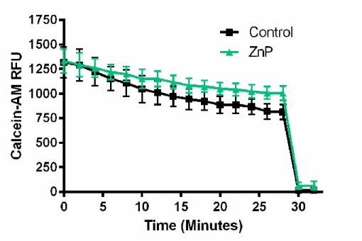
Figure 5: Zinc Pyrithione Does Not Induce Cytotoxicity. ... 36
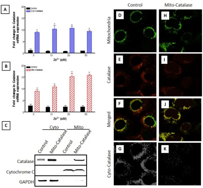
Figure 6: Targeted catalase expression abrogates Zn2+-induced cytosolic and mitochondrial increases in H2O2 levels in BEAS cells.. ... 38
Figure 7: Mito-HyPer is specifically localized in the mitochondria of BEAS cells.. ... 39
Figure 8: Exposure to pyrithione alone does not induce intracellular H2O2 or EGSH responses. ... 39
Figure 9: Zn2+-induced elevation in H2O2 reported by HyPer are not mediated by changes in pH. ... 40
Figure 10: Ectopic expression of catalase in the mitochondria of BEAS-2B cells.. ... 42
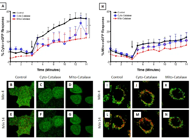
Figure 11: H2O2 mediates Zn2+-induced elevation in cytosolic but not mitochondrial EGSH. ... 44
Figure 12: Zn2+ -induced changes in E GSH are properly reported by roGFP2.. ... 45
Figure 13: Depletion of glutathione levels does not alter Zn2+-induced HO-1 mRNA levels... 46
Figure 14: Depletion of glutathione levels does not lead to HO-1 mRNA expression in BEAS cells. ... 47
Figure 15: Targeted catalase expression blunts Zn2+-induced HO-1 mRNA levels in BEAS cells... 48
Figure 16: H2O2 mediates Zn2+-induced increases in cytosolic EGSH and HO-1 mRNA expression. ... 51
Figure 17: Schematic of cysteinyl-thiol post-translational modifications.. ... 54
xiii
Figure 19: 1,2-naphthoquinone induces a rapid increase in cytosolic intracellular H2O2.. ... 62
Figure 20: Catalase overexpression blunts 1,2-napthoquinone-induced protein sulfenylation. ... 63
Figure 21: 1,2-naphthoquinone induced protein sulfenylation is H2O2 dependent.. ... 64
Figure 22: Detection of 1,2-naphthoquinone-induced protein sulfenylation through biotinylation in human airway epithelial cells. ... 65
Figure 23: 1,2-naphthquinone-induces sulfenylation of regulatory proteins with differential kinetics.. ... 66
Figure 24: 1,2-naphthoquinone-induces sulfenylation of specific regulatory proteins with differential kinetics. ... 67
Figure 25: 1,2-naphthoquinone induces sulfenylation of the GAPDH catalytic cysteine. ... 68
Figure 26: 1,2-naphthoquinone produces H2O2 in vitro to oxidize
recombinant GAPDH. ... 68
Figure 27: Depletion of the glutathione pool leads to perturbed response to H2O2.. ... 78
Figure 28: GSTM1 Does Not Directly Regulate Protein Sulfenylation.. ... 79
Figure 29: 1,2-Naphthoquinone-Induced Protein Sulfenylation is Enhanced in GSTM1-Null Cells. ... 80
Figure 30: Cells Remain Visibly Viable throughout Direct, Acute Exposure to Asbestiforms. ... 88
Figure 31: BEAS-2B Cells Respond Variably to Asbestiform Exposure. ... 90
Figure 32: Cells Respond to Acute Exposures to UICC Crocidolite in a Dose Dependent Manner... 91
xiv
LIST OF ABBREVIATIONS
1,2-NQ 1,2-naphthoquinone
1O
2 Singlet oxygen
2-AAPA 2-acetylamino-3-[4-2-acetylamino-2-carboxyethylsufanylthio-carbonylamino phenylthiocarbamoylsufanyl] propionic acid
4-HNE 4-hydroxynonenal
8-OHdG 8-hydroxyguanosine
A-2 Aldrithiol-2
ARE Antioxidant response element
BEBM Bronchial epithelial basal media
BEGM Bronchial epithelial growth media
Biotin azide PEG4 carboxamide-6-azidohexanyl biotin
BSO Buthionine sulfoximine
cpYFP Circularly permutated YFP
CP Peroxidatic Cysteine
CR Resolving Cysteine
Cu2SO4 Copper sulfate
CuAAC Copper-catalyzed alkynyl-azo cycloaddition
d6-dimedone Deuterated-dimedone
DTT Dithiothreitol
EGFR epidermal growth factor receptor
EGSH Glutathione redox potential
xv GFP Green fluorescent protein
Gpx Glutathione peroxidase
GR Glutathione reductase
Grx Glutaredoxin
GSH Reduced glutathione
GSSG Glutathione disulfide
GST Glutathione S-transferase
H2O2 Hydrogen peroxide
HAEC Human airway epithelial cells
HO· hydroxyl radicals
HO-1 Heme oxygenase-1
I-dimedone Iodo-dimedone
KBM Keratinocyte basal media
KEAP1 Kelch-like ECH-associated protein 1
KGM Keratinocyte growth media
LDH Lactate dehydrogenase
NAC N-acetylcysteine
N-HRP NeutrAvidin-HRP
NO· Nitric oxide
Nox NADPH oxidase
Nrf2 NF-E2-related factor 2
O2·- Superoxide
xvi PBS Phosphate buffered saline
PDI Protein disulfide isomerases
PM Particulate matter
Prx Peroxiredoxin
PTP Protein tyrosine phosphatase
PTP1B Protein tyrosine phosphatase 1b
PTM Posttranslational Modification
PYRI Pyrithione (2-mercaptopyridine N-oxide sodium salt)
Redox Reduction-oxidation
RNS Reactive nitrogen species
roGFP Redox sensitive green fluorescent protein
ROS Reactive oxygen species
rxYFP Redox sensitive yellow fluorescent protein
SA Sodium ascorbate
SOD Superoxide dismutase
TBTA Tert-butyl 2,2,2-trichloroacetimidate
Trx Thioredoxin
YFP Yellow fluorescent protein
1
CHAPTER 1: Introduction
1.1: Air Pollution
Outdoor air pollution is a global public health problem, responsible for approximately 3.3
million premature deaths per year (1). Air pollution originates from both anthropogenic sources
including the burning of carbon-based fuels as well as natural sources including volcanic
eruptions. The negative health impact of air pollution on public health was first noted during the
industrial revolution. By the mid-1900’s cities including London, UK (2) and Donora, Pa, USA
(3) were impacted by an increase in human morbidity and mortality that was closely linked to
ambient air pollution levels. Despite continuing improvements over the past decades to improve
air quality (4) there is still evidence to suggest air pollution contributes to the adverse health
outcomes in humans throughout the world (5).
1.1.1: Global Public Health Burden
The initial target of air pollution is the respiratory system. Even though there is
epidemiological evidence for air pollution-induced acute respiratory disease, the mechanistic
data to reinforce such findings is lacking (6). There is little argument that the nares and lung are
the initial targets of air pollution exposure, but there is sufficient epidemiological evidence that
suggests air pollution is linked to the prevalence of other adverse health outcomes including
cancer (7), diabetes (8), and cardiovascular disease (9). In addition, there is strong experimental
evidence in animal models (10,11) and controlled human exposure (12,13) and panel studies (14)
2
disease. These findings correlate well with mechanistic data afforded by in vitro studies that
suggest a potential link between air pollution exposure and adverse health outcomes (15-17). The
involvement of air pollution in many adverse health outcomes including both acute and chronic
human disease supports the need to understand the fundamental basis of toxicity associated with
air pollution. Of particular concern are those individuals living in developing countries as they
are at higher risk to air pollution, due to fewer regulatory and engineering safeguards (18).
1.1.1.1: Susceptible Populations
In addition to the impact of air pollution on the general population, there are specific
subpopulations that are susceptible to the negative health effects associated with exposure to air
pollution. Individuals with preexisting disorders such as asthma (19) and cardiovascular disease
(20) are at particular risk to air pollution. In fact, it has been documented that high air pollution
days correlate with increases in hospital visits (21-23). Persons with existing ailments are not the
only populations negatively affected by air pollution, as certain genetic polymorphisms can also
lend to susceptibility (24). One such polymorphism leads to perturbed translation of the
glutathione S-transferase mu 1 (GSTM1) gene. The prevalence of the GSTM1-sufficient allele is
actually less prevalent than the GSTM1-null allele, with approximately 40% of the global
population being GSTM1-null (25). Epidemiologically, GSTM1-null individuals are particularly
susceptible to the acute effects of inhaled pollutants (26-28). Although developing countries have
a significant health burden associated with air pollution derived from many different combustion
sources including those linked with industrial processes, developed countries still observe a
3
of current air pollution are mobile sources, which is prevalent in developed countries and tends
to be localized to large metropolitan areas.
1.1.2: Traffic Related Air Pollutants
Broadly speaking air pollution is a complex mixture of both gaseous and particulate
components, of which the U.S. EPA monitors and regulates six. Although five are well defined
(ozone, nitric oxides, sulfur oxides, carbon monoxide and lead) the sixth regulated air pollutant is
particulate matter (PM), which can be defined by size or composition. Currently PM is
monitored by size with regulations set at coarse (> 10 μm) and fine (10 uM-2.5 μm). However,
PM is characteristically different with regards to composition geographically and based on
source (29,30). Air pollution of large metropolitan areas, as mentioned previously, is largely
influenced by the surrounding traffic (31).
Air pollution originating from traffic primarily fall under two categories: the combustion
of fuels (gasoline and diesel) and the wear and tear of surfaces (tires, brake-pads, and the road
itself). Traffic-related pollution leads to an increase in both primary and secondary air pollutants.
Primary pollutants are those that are immediately produced, while secondary pollutants are
generated through the interaction of primary pollutants with environmental factors including
sunlight, as in case of ozone. The incomplete combustion of fuels is a major source of primary
pollutants including PM, as it provides the carbon core necessary for many particulates to form.
Metals, both essential and heavy, are distributed throughout the air by the wear and tear of solid
components. These metals as well as other organic materials can either anneal to particulates or
can originate from the particulates eroded from the solid components of vehicular parts.
4
components there are individual components that are of particular interest in regards to human
health.
1.1.2.1: Zinc
Zinc is a ubiquitous metallic component of air pollution primarily derived from the wear
and tear of galvanized vehicular parts as well as tires (32), where tires are approximately
0.4-4.3% zinc by weight (33). Epidemiological and observational studies have implicated zinc as the
causative metallic pollutant for the adverse health effects attributed to air pollution (34-37).
Furthermore, zinc-induced health effects can be modeled by the occupational health disorder
known as “metal fume fever.” Individuals that work with metal, such as welders, experience a
self-limiting flu-like state when they are exposed to zinc particulates in the air (38). This
demonstrates the capacity for inhaled zinc to cause a systemic inflammatory response in humans.
It should be noted that zinc is an essential nutrient and when ingested is utilized physiologically,
even at high chronic exposure (39,40). However, the inhalation of zinc and zinc particulate-laden
particulates represents a route of exposure that causes an adverse toxicological response (41).
At the cellular level, zinc can induce an adverse inflammatory response. The cellular and
molecular mechanisms of zinc-induced inflammatory responses have been described (42). First,
zinc is known to be thiol reactive allowing it to interact with important cysteine residues on
proteins to alter the protein’s function (43). One such example is zinc’s interaction with the
catalytic cysteine of protein tyrosine phosphatases (PTP) to inhibit their ability to
dephosphorylate proteins (44-46). This can lead to an increase in the activation of signaling as
kinases can freely activate pathways through phosphorylation without the balance of PTPs to
5
The second mechanism through which zinc elicits an inflammatory response is by
elevating intracellular reactive oxygen species (ROS). Unlike most transition metals, zinc cannot
redox cycle under physiological conditions to generate ROS, but it can interfere with
mitochondrial respiration which leads to an accumulation of ROS (41,47). These ROS can then
lead to the induction of adaptive and signaling responses (46,48). Even though both of these
mechanisms could occur simultaneously within cells exposed to zinc, the relative contribution of
the initiating effects of zinc-induced adverse responses is unknown.
1.1.2.2: 1,2-Naphthoquinone
1,2-naphthoquinone (1,2-NQ) is an organic component of air pollution that people are
exposed to through direct and indirect sources. 1,2-NQ is primarily generated through the
incomplete combustion of diesel exhaust (49), but its presence in the air can also be linked to
industrial operations and second hand smoke (50). Additionally, naphthalene, the most prevalent
organic chemical in the air, can be metabolized to 1,2-NQ through three secondary enzymes
(51,52). Although the toxicity of 1,2-NQ has not been well described in humans, it is considered
to have similar toxicity as 1,4-naphthoquinone, which has been demonstrated to be
pro-inflammatory and described as a potential carcinogen when inhaled (53).
Whereas the pathophysiological effects of 1,2-NQ are not as well characterized as other
organic chemicals and quinones, the effects of 1,2-NQ at the cellular and molecular level have
been studied. Similar to most quinones, 1,2-NQ has two distinct mechanisms of toxicity: direct
electrophilic attack by Michael addition (54,55) and generation of ROS through redox cycling
(56,57). 1,2-NQ contains an electrophilic carbon that can attack nucleophilic sites on biological
6
1,2-NQ redox cycles through the semiquinone and hydroquinone state (Figure 1) to provide
electrons to generate ROS which could cause cellular stress (58). Of particular interest, with
regards to 1,2-NQ’s ability to redox cycle is that when adducted to proteins it can continue to
redox cycle to generate ROS as harmful by-products, which other quinones including
1,4-naphthoquinone cannot (59). This would suggest that 1,2-NQ has the unique capability of being
redox active even when adducted to proteins.
Figure 1: Quinone Redox Cycling Reaction. 1,2-naphthoquinone (left) is converted to the semiquinone (center) and hydroquinone (right) by electron transfer from the action of flavor-containing enzymes. The chemical can push electrons to oxygen and superoxide to generate reactive oxygen species, where the reaction between the semiquinone and oxygen is more favored over the hydroquinone and superoxide reaction. The hydroquinone is able to interact with oxygen to generate superoxide; however, this is even less chemically favored than the interaction between the hydroquinone and superoxide.
Mechanistically, 1,2-NQ can induce a variety of signaling pathways within the cell either
through its electrophilic or pro-oxidant activity, leading to unwanted biological outcomes.
Regarding physiological outcomes, exposure to 1,2-NQ has been observed to induce a
pro-inflammatory response in mice (60). Additionally, 1,2-NQ can lead to the inhibition of important
cellular processes including dephosphorylation of kinases by targeting phosphatases, which
result in tracheal contraction (61) and suppression of vasorelaxation in blood vessels (62).
However, like zinc, due to the multiple mechanisms of toxicity of 1,2-NQ, the relative
7 1.2: Redox Biology
Similar to how physiological processes at the tissue level can be explained by cellular
adaptations and activities, many cellular functions can be explained through the biochemical
transfer of electrons. This transfer of electrons is described through the tuning of
reduction-oxidation (redox) state of biological macromolecules (nucleic acid, lipid, protein). As in most
cases with regards to balance in biology this is a homeostatic process, wherein the cell actively
avoids a loss of viability due to an excess of either oxidized or reduced molecules within the cell
(63). Homeostasis is maintained through both constitutive factors as well as adaptive processes.
Constitutive factors include the microenvironment of the macromolecule such as sterics and
subcellular characteristics including pH, which directly influences the protonation state of atoms
in molecules. For example the subcellular pH of mitochondria matrix is markedly more alkaline
(pH ~8) than the cytosol (pH~7.2), which allows for a location specific function of a protein or
molecule (64). Additionally, the cell can utilize adaptive processes such as the induction of the
antioxidant peptide glutathione in response to an elevation of ROS, where the ROS are largely
responsible for the dynamic nature of redox-dependent processes.
1.2.1: Reactive Oxygen Species
Reactive oxygen species (ROS) have a high capacity to oxidize other molecules (65-67).
Reactive nitrogen species (RNS) are distinguished from ROS by the content of a nitrogen group.
However, for this document RNS will be referred to as ROS, wherein ROS will define any small
chemical that is an oxidant. Within this work the ROS are separated by their function in the cells
as some are promiscuously active and able to react readily with any available site such as
hydroxyl radicals (HO·), superoxide (O
8
species are relatively less functional in redox-dependent processes that are dynamic or
regulatory; however, other ROS such as nitric oxide (NO·) and hydrogen peroxide (H
2O2) have
chemical characteristics that make them more capable to serve as effectors of cellular processes
(70). These relatively less reactive species can also serve as second messengers. For instance
NO· is a gaseous second messenger that is notably important in vasodilation (71). Despite this
physiological role, if the levels of these ROS exceed the homeostatic capacity of the cell they
will exert deleterious effects that may lead to cytotoxicity.
1.2.1.1: H2O2
H2O2 is arguably the most important ROS in regards to redox-dependent signaling. H2O2
is “intentionally” produced through the enzymes NADPH oxidase (Nox) and superoxide
dismutase (SOD) (70,72). The Nox enzymes facilitate the transfer of electrons from intracellular
NADPH or FAD to the extracellular compartment by reducing molecular oxygen to H2O2 (73).
The H2O2 generated from this reaction has been linked to many cellular processes, and mutations
within the Nox enzymes that perturb the generation of H2O2 is linked to pathophysiology (74).
Intracellular H2O2 also originates from the mitochondria through the metabolism of O2·
-generated from complex 1 and 3 of mitochondrial respiration (75). The mitochondria maintain a
high basal level of the antioxidant protein SOD, which rapidly and efficiently converts O2·- to
H2O2 (76,77). Under some circumstances, the levels of H2O2 exceed the peroxidatic activity
available in the mitochondria, and are able to diffuse to the surrounding cytosol and subcellular
compartments. As H2O2 is generated in multiple sites in the cell, there are localized elevations of
9
Due to its relative stability and reactivity compared to other ROS, H2O2 is considered a
better candidate to act as a second messenger than most other ROS. For these reasons H2O2,
evolutionarily, has likely become a tightly regulated ROS within the cell (79,80). H2O2, as
mentioned above, is generated through the function of the family of Nox enzymes as well as a
byproduct of mitochondrial respiration (78). In addition to non-specific antioxidant molecules
such as glutathione that serve as sinks for oxidant species, the cell expresses enzymes that
specifically target H2O2. These enzymes are more efficient mechanism to lower H2O2 levels
within the cell. Peroxiredoxins (Prx) have a high reactivity with H2O2; glutathione peroxidases
(Gpx) reduce H2O2 through the interaction of glutathione; and catalase catalyzes the
decomposition of H2O2 to molecular oxygen and water (81,82). The combination of these three
enzyme families (Prx, Gpx, and catalase) serve as the first line defense to lower H2O2 levels as
they are more energetically and kinetically efficient to metabolize H2O2 than the direct
interaction of H2O2 with antioxidant small molecules such as glutathione. Accordingly, levels of
these major enzymes, which are differentially expressed at the tissue and subcellular level,
largely dictate the intracellular levels of H2O2. Furthermore, the induction of these proteins or
inhibition of their function can dramatically impact redox-dependent signaling, since it would
ultimately affect H2O2’s second messenger functions.
H2O2 is electrophilic and as such acts on nucleophilic sites on biological molecules
including the thiol (SH) of glutathione (83) and nucleophilic amino acids including methionine
and cysteine (84). The interaction between H2O2 and cysteine is a highly studied topic in many
research groups (85). When the thiol (SH) group of the cysteine is deprotonated, usually as a
result of pH of the subcellular compartmental or the microenvironment of the molecule, it forms
10
H2O2 leading to the formation of a sulfenic acid (SOH) (86). The oxidation of cysteine thiols to
the sulfenic acid of glutathione or proteins is an important regulator of redox-dependent
processes (87).
1.2.2: Protein and Peptide Thiols
The sulfenylation of intracellular thiols by H2O2 is vital and necessary for physiology
(87). Even though the elevation of H2O2 is the event leading to these redox-dependent processes,
it is also important to discuss the importance of the target: cysteine thiols. The availability of
cysteine is negligible within the cell and consequently is the limiting substrate to synthesize
glutathione (88). Cysteine is also one of the rarest, and most conserved amino acids in biological
systems (84,85). Despite the nucleophilic capability of the thiol portion of the cysteine to serve
in oxidant reactions in the cell, it is restricted by availability. Thus, it is imperative to understand
that protein sulfenylation, while an essential physiological process, is rare because of the
availability of cysteine thiol targets as well is being tightly regulated through the generation and
decomposition of H2O2.
1.2.2.1: Glutathione
Glutathione is the most prevalent non-protein thiol that serves as an antioxidant in
mammalian cells (89). Frequently glutathione is present in concentrations in the millimolar range
(90). Structurally, glutathione is a three amino acid peptide consisting of cysteine, glutamate, and
glycine. It is synthesized in two steps, the first links cysteine to L-glutamate by the enzyme
glutamate cysteine ligase to form gamma-glutamylcysteine. The second step adds glycine to
11
an important cellular antioxidant, by itself it has a weak interaction with H2O2. Even though,
thermodynamically H2O2 should be able to oxidize the thiol of glutathione, the pKa of the thiol
makes the reaction chemically unfavorable and slow (91).
Despite glutathione’s slow reaction with H2O2 by itself, through the enzymatic interaction
of Gpx, glutathione serves as an important physiological antioxidant target for H2O2 (92). Gpx
enzymatically reacts with two molecules of reduced glutathione (GSH) with one molecule of
H2O2 to form glutathione disulfide (GSSG) and water. GSSG is then reduced to glutathione by
glutathione reductase (GR) at the cost of energy, specifically NADPH. In fact the cell expends a
significant amount of energy to maintain glutathione in its reduced form (90). A quantitative
measurement of GSH/GSSG is the glutathione redox EGSH (mV), as defined by the Nernst
Equation:
𝐸𝐺𝑆𝐻= 𝐸𝐺𝑆𝐻°′ −𝑅𝑇
2𝐹ln(
[𝐺𝑆𝐻]2
𝐺𝑆𝑆𝐺 )
The EGSH can be utilized as an important marker of redox metabolism that has been linked to a
many cellular states including differentiation and apoptosis; however, it should be noted that
EGSH in itself is a poor marker of redox-dependent signaling (91).
1.2.2.2: Oxidative Posttranslational Modifications
As opposed to the interaction between H2O2 and glutathione, it is currently thought that
the interaction between protein thiols and H2O2 is a spontaneous, non-enzymatic reaction (93).
However, the number of cysteines in the proteome is finite and this number is further restricted
as many of those cysteines are sterically unavailable for oxidation (85). In addition to the
minimal number of protein thiols available for H2O2 to form sulfenic acids, there is an additional
12
The thiol that becomes sulfenylated is referred to as the peroxidatic residue, and its oxidation is
dictated by mutliple factors including its pKa. The relative reactivity of the peroxidatic thiol is a
major factor dictating the ranking of which proteins are targeted by H2O2 (94).
The spontaneous oxidation of thiols to the sulfenic acid is also characteristically
short-lived. However, sulfenic acids serve as a precursor for the formation of other post-translational
modifications (PTM) (Figure 2). One of the most important PTMs is the disulfide bond between
two cysteines. The oxidation of the peroxidatic cysteine (CP) interacts with another cysteine, the
resolving cysteine (CR), to form a relatively more stable disulfide bond. This disulfide bond can
then be reduced back to the thiol through enzymes including thioredoxin (Trx) or glutaredoxin
(Grx). Sulfenic acids can also interact with other resolving residues, such as protein amines, to
form a sulfenamide residue (Figure 2). The final oxidative PTM to discuss is S-glutathionylation
leading to the formation of a mixed disulfide bond between glutathione and the cysteine thiol.
All of these oxidative PTMs either stabilize the effect of the oxidation of the thiol, which results
in a longer lasting effect on the protein, or can result in a completely different protein
13
Figure 2: Cysteine Posttranslational Modifications.Protein thiols (RS-H) can undergo oxidation to the sulfenic acid OH), which then can react with intracellular glutathione to form a mixed disulfide (RS-SG) or with another protein moiety such as another thiol to form a disulfide bond (RS-SR’) or an amine to form a sulfenamide (RS-NHR’). These modifications are reversible and can be reduced back to the thiol, enzymatically, at the expense of NADPH. The sulfenic acid can be further oxidized to the irreversible modifications sulfinic acid (RS-O2H) and sulfonic acid (RS-O3H). *It has been demonstrated that the
hyperoxidized sulfinic acid in the peroxiredoxin can be enzymatically reduced to the sulfenic acid at the expense of ATP, and as such it is possible that other unidentified proteins may utilize a reversible sulfinic acid in their function.(95)
Proteins known to be sulfenylated are frequently referred to as “redox switches” (96).
This term, “redox switches,” is used to describe these proteins because their activity is regulated
by their redox state. In other words, the protein’s constitutive function is completely reversed
once oxidized (97,98). The other important factor of “redox switches” is their reversibility, so the
protein’s function can be turned “on” or “off” without permanently effecting the protein.
However, this dynamic nature can be influenced by subsequent oxidation by ROS, including
H2O2, to the sulfinic acid and sulfonic acid (Figure 2). The hyperoxidation of proteins is
14 1.3: Redox Toxicology
Using ROS as the regulators of redox biology comes at a risk as they also have the
potential to be harmful. Under physiological conditions the cell is able to replete ROS levels
through multiple cellular antioxidant defense mechanisms. However, when ROS levels are
elevated beyond the antioxidant capacity of the cell, the resulting outcome can be pathologic or
toxicological in nature (67). Virtually all diseases (cancer, cardiovascular, neuro-degeneration,
metabolic syndrome, etc.) (99-101) as well as most environmental (102,103) and
pharmacological (104) exposures have been reported to have an associated oxidative stress
component. For instance, even a slight change in the redox status(es) of the cell can lead to
perturbed cellular function that if unchecked leads to cellular senescence by irreversibly
damaging the cellular machinery (105). In other words there appears to be two distinct thresholds
when one discusses redox toxicology, the first inappropriate activation of physiological cellular
processes, while the second is overt cytotoxicity, which results in irreversible damage (106,107).
1.3.1: Oxidative Stress
Of the two thresholds of redox toxicology, the irreversible damage has been studied more
extensively. This form of redox toxicology is often referred to as oxidative stress. The term
oxidative stress as a descriptor has been used nebulously referring to both electrophilic and
oxidative species and lacking no definitive meaning to characterize the effect of the stress. The
vagueness of the term oxidative stress is partially due to the methodological approaches that
were available to characterize and define it (108). However, for the purpose of this dissertation,
oxidative stress will be defined as the cellular response to avoid cytotoxicity due to an increase in
15
response to oxidative stress is the induction of the antioxidant response element (ARE) by
NF-E2-related factor 2 (Nrf2) (Figure 3).
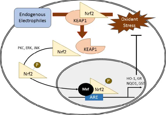
Figure 3: Nrf2-Depdenent Activation of the Antioxidant Response Element. Endogenous electrophilic interaction or exogenous oxidant stress leads to KEAP1 release of Nrf2. Nrf2 is then phosphorylated via PKC, ERK, or JNK to facilitate translocation to the nucleus where interaction with a Maf protein allows for binding to the antioxidant response element (ARE) to induce gene expression of proteins critical in the antioxidant response.
1.3.1.1: Antioxidant Response Element
The antioxidant response element or ARE is an important transcription factor binding
element that exists in the promoters of many cytoprotective genes including heme oxygenase-1
(HO-1), NAD(P)H: quinone oxidoreductase 1, GR, GSTs, Trx, metallothioneins, and
sulfotransferases (109). All of these genes when translated have active roles in reducing proteins
and peptides or participate in the detoxification of reactive xenobiotics. Of particular importance
in xenobiotic-induced oxidative injury is HO-1. The inducible protein HO-1 is involved in the
16
capabilities by either acting as a non-specific target for ROS or inhibitors of cellular oxidative
processes (110). Increased HO-1 expression in the respiratory system as a result of exposure to
PM leads to improved lung function by improving mucociliary function and inhibiting neutrophil
infiltration that could lead to fibrotic lesions (111). The universal role of HO-1 in responding to
oxidative injury has made it a valuable marker of oxidative stress.
Of the transcription factors that bind to the ARE, the most notable is Nrf2, which
activates ARE and BACH1, a repressor of ARE activity (112). At rest, BACH1 associates with
the ARE and must be displaced in order for ARE activation by Nrf2 (112). Nrf2 is normally
bound to Kelch-like ECH-associated protein 1 (KEAP1) in the cytosol. KEAP1 serves as an
adaptor protein for the cullin 3/ring box 1-E3 ubiquitin ligase complex to target Nrf2 for
degradation by ubiquitination (113). However, during oxidative stress KEAP1 releases Nrf2
which allows the translocation of Nrf2 to the nucleus to induce the antioxidant response (114).
Once Nrf2 reaches the nucleus it forms a heterodimer with a transcription factor of the
musculoaponeurotic fibrosarcoma (Maf) family to bind to the ARE (112).
Cellular oxidant stress leads to a separation of KEAP1 and Nrf2, allowing for Nrf2
activation of ARE-related genes. There are two distinct mechanisms through which this can
occur. The first is through an increase in electrophilic species in the cell including divalent
metals, NO·, and aldehydes which can all target specific cysteine residues on KEAP1, which
compromises its ability to form disulfides with Nrf2 (115). The second mechanism is
phosphorylation of Nrf2 by extracellular signal-regulated kinases (ERK), c-Jun NH2-terminal
kinase (JNK) (Xu 2006) or protein kinase C (PKC) (116). Phosphorylation of Nrf2 leads to its
17
argued by some researchers that Nrf2-induced ARE gene expression requires both electrophilic
attack of KEAP1 and phosphorylation of Nrf2 (117).
1.3.2: Perturbation of Redox-Dependent Processes
Oxidant-induced signaling events such as the activation of the KEAP1/Nrf2 pathway are
likely in response to overt oxidative stress. As discussed previously, there are many other
redox-dependent processes within cellular physiology that are largely controlled by “redox switch”
proteins that can be targeted by H2O2 (118). Many exposures are known to elevate intracellular
ROS including H2O2. Thus it is possible that these exposures could affect these redox-dependent
processes without leading to overt cytotoxicity. Among these processes include activation of
signaling pathways, regulation of bioenergetics, cytoskeletal maintenance, and the regulation of
the cell cycle (119). The focus of this dissertation will be restricted to the effect of toxicological
exposures on signaling and bioenergetics via redox-dependent mechanisms.
1.3.2.1: Activation of Signaling Pathways
One of the first recognized redox-dependent mechanisms in physiology was the
inhibition of PTPs (120). PTPs contain a catalytic cysteine that can be sulfenylated by H2O2,
which inhibits the protein’s ability to dephosphorylate other proteins (121,122). This has major
implications for the activation of signaling pathways as PTPs and other phosphatases are more
active than their kinase counter-parts (123). This provides an intracellular imbalance of proteins
that are not phosphorylated, so when H2O2 inhibits PTPs it allows for the stabilization of
18
as that in protein tyrosine phosphatase 1B (PTP1B), is sulfenylated it rapidly interacts with a
nearby amine forming a stable sulfenamide and abrogates PTP activity (121).
It has been shown through previous work that PTPs can be inhibited by targeting the
catalytic cysteine (125). The thiolate can be directly attacked by electrophiles, which
compromises the protein’s ability to dephosphorylate targets (45). More recently, work in our lab
has demonstrated that reducing intracellular H2O2 levels specifically modulated zinc- and
1,2-NQ-induced gene expression (56). It is likely that xenobiotic-induced H2O2 enhances signaling
through the sulfenylation or possibly hyperoxidaiton of PTPs.
The most prominent PTPs in physiology as well as pathology is PTP1B. PTP1B is
expressed in may tissues and is known to interact with many important targets including
epidermal growth factor receptor (EGFR), insulin-like growth factor receptor, c-Src, and Janus
kinase 2 (126). Because of the widespread physiological role of PTP1B, it is a prominent
candidate for toxicological targeting of redox-dependent processes. It is known that ROS and
electrophilic compounds, including zinc (46) and 1,2-NQ (54), target PTP1B’s catalytic cysteine.
However, it is not known whether ROS contribute to zinc and 1,2-NQ’s ability to target PTP1B,
since it is also known that zinc and 1,2-NQ can also elevate H2O2 levels.
1.3.2.2: Bioenergetics
Another major cellular process that is regulated by redox-dependent proteins is
bioenergetics. Bioenergetics is the ability of cells to actively provide, maintain, and utilize
energy. This process is arguably the most responsive cellular process, reacting within seconds or
minutes to environmental factors such as pO2 and pH (127). David Wilson illustrates this best
19
bioenergetic processes are the programs of the cell (128). Bioenergetics is functionally localized
to the mitochondria through oxidative phosphorylation (129). Although oxidative
phosphorylation can be considered the central processing unit that regulates all the “programs of
the cell” (128), oxidative phosphorylation is regulated by the citric acid cycle to produce the
needed energy to drive ATP synthase. Although citric acid cycle funnels energy to maintain
oxidative phosphorylation, the citric acid cycle is fine-tuned and regulated by glycolysis, which
ultimately supplies the cell with energy by converting glucose to pyruvate.
Although there is a variety of proteins involved in bioenergetics, one of the most
important is glyceraldehyde 3-phosphate dehydrogenase (GAPDH). GAPDH functions as a
critical step in glycolysis to utilize glucose for energy, and it is essential for cellular viability
(130). Furthermore, GAPDH is expressed constitutively at high levels in the cytosol. Due to the
role of GAPDH in the cells it is frequently referred to as a housekeeping protein and in many
studies is utilized as an experimental control. Although GAPDH has an essential role in
glycolysis, in recently published work GAPDH has been demonstrated to have other
“moonlighting” functions such as the regulation of nuclear proteins, stabilization of mRNA, and
even as a receptor (130).
A regulating factor of GAPDH activity is the reversible oxidation of a cysteine in one of
its glyceraldehyde 3-phosphate binding domains. Sulfenylation of GAPDH leads to its inability
to maintain its glycolytic function (131). It is hypothesized that this confers improved antioxidant
potential to the cell as this ultimately leads to an increase in NADPH generation, which is
essential for the glutathione/Grx and Trx systems to provide antioxidant protection (132).
Additionally, the cysteine that regulates GAPDH activity has a substantially higher reactivity
20
together suggest that GAPDH could also serve an antioxidant function as well. However, the
important consideration in regards to toxicological exposure is that elevation of ROS perturbs
basic glycolytic function by inhibiting GAPDH through sulfenylation of its active site cysteine,
even if it is at the cost of reducing elevated ROS by providing energy through the pentose
phosphate pathway.
1.4: Approaches to Study Redox Toxicology
Despite the need to better understand redox biology and the interface of redox biology
and environmental health, there have been few techniques available to reliably obtain
information. There are two distinct issues at hand that complicate the ability to study redox
toxicology. The first is that the initiating events for redox-dependent processes are rapid and
frequently localized, while the spatiotemporal resolution of most techniques is inadequate to
provide insightful data (133,134). In the case of techniques that are able to detect a changes in
the redox status of cells, such as the case in electron spin resonance, the resources are technically
and cost prohibitive for most experiments, especially once applied to toxicological experiments
(135). The other disadvantage of traditional techniques in redox biology is that interventions
utilized to decipher the effect of ROS use many “antioxidants” that provide unspecific targeting
of redox species and processes, as is the case with N-acetylcysteine (NAC) which not only up
regulates the synthesis of glutathione, but it itself can react with free radicals and ROS, thus
providing a weak causal link in the physiology (136). Another issue with developing such causal
arguments is that that the antioxidant used could become an oxidant itself, further complicating
21
Although the last few decades have provided considerable technical advancements in the
field of redox biology, which will be discussed in this section, there are still challenges in
implementing these techniques in toxicology (137,138). The primary concern is that most of
these cutting-edge technologies are founded on the homeostatic redox environment (133,139).
For redox biology this characteristic is advantageous; however, toxicological exposures have the
potential to dramatically alter physiological processes that may compromise the functionality of
these tools. Thus it is essential to implement controls that not only test the experimental
conditions but also technical conditions to ensure the exposure does not impact the readout of the
technique (140).
1.4.1: Small Molecule Redox Sensors
Small molecule redox sensors are chemicals, sometimes in the form of dyes, which
change characteristic properties upon interaction with ROS or antioxidants. The first generation
of these sensors lacked appropriate specificity for rigorous testing. For instance,
2′,7′-di-chlorofluorescein (DCF) is a small chemical molecule that was reported to increase fluorescence
upon interaction with H2O2, yet after increased scrutiny it was shown to react with multiple ROS
including HO·, O
2·-, and NO· (141). Additionally, DCF has been demonstrated to induce
oxidative stress itself by redox cycling, which makes it a poor sensor to understand the actual
role of redox-dependent processes (142). More specific, selective sensors have been developed
including Peroxy green-1 (PG-1), Peroxy orange-1 (PO-1), Peroxy yellow-1 (PY-1), and
Peroxyfluor-6-acetoxymethyl ester (PF6-AM). These sensors use a boronate ion in their structure
that interacts with H2O2, specifically, and have not been demonstrated to effect redox-dependent
22
their utility, they also have their weaknesses (142). Amongst these weaknesses is that they are
unable to reversibly interact with their target ROS, and as such cannot capture the dynamic
nature of redox-dependent cellular processes.
1.4.2: Genetically Encoded Redox Sensors
The discovery of green fluorescent protein (GFP) opened many avenues for biological
and biomedical research (144), and further expanded with modifications to GFP changing its
fluorescent color, including yellow fluorescent protein (YFP) (145). One of the most important
characteristics of GFP-based molecules is that they can be expressed in subcellular regions in
specific cell types (146). Furthermore, GFP expression can be conditionally controlled, which
can be a valuable characteristic in certain experiments. These reasons led to the development of
redox-based sensors based on GFP and its variants.
1.4.2.1: HyPer
Through the insertion of circularly permutated YFP (cpYFP) into the H2O2 sensing
protein OxyR1A, the Belousov group developed a fluorogenic genetically encoded sensor,
named HyPer, that responds to intracellular H2O2 levels (147). HyPer can be excited at dual
wavelengths (404 and 488 nm) and emits in the green at ~520 nm. The excitation maximum is
different for each wavelength based on the redox status of the sensor. Specifically, the 404 nm
excitation reaches maximum excitation when reduced, and upon oxidation loses fluorescence.
The opposite holds true for HyPer’s 488 nm excitation as it reaches maximum excitation in the
presence of high levels of intracellular H2O2, and loses fluorescence under reducing conditions.
23
excitations can be used to calculate a ratio, which controls for many common issues with
fluorescence microscopy including variable sensor distribution in the cell and photobleaching.
This sensor’s experimental value was quickly ascertained by targeting it to a variety of
subcellular organelles including the mitochondria (148), and successful expression in the widely
used small-vertebrate model, zebrafish, revealed the role of H2O2 in development and wound
healing (149,150). HyPer has also been chimerically linked to the H2O2 producing protein EGFR
allowing for the measurement of micro-domain increases in H2O2 (151). Importantly,
experimental results using HyPer have demonstrated that xenobiotic induction of H2O2 rarely is
globally elevated and in most cases leads to an increase in specific subcellular compartments
(56,148)
HyPer is sensitive to pH at a physiologically relevant range (147). Thus it is imperative
to monitor HyPer concurrently using pH sensors such as pHred (152). One can also use
side-by-side experiments such as pHluorin or pHluorin2 (153). However, the ideal pH control to utilize
in HyPer experiments is SypHer, which is structurally a point mutation of HyPer that makes the
sensor insensitive to H2O2 while retaining sensitivity to changes in pH (154). A newer version of
SypHer, SypHer2 has been developed with improved experimental responsiveness (155).
1.4.2.2: rxYFP, Oba-Q Proteins and roGFP
YFP was made redox sensitive through two point mutations leading to the development
of rxYFP (156). rxYFP monitors the EGSH metabolically through the interaction of
glutaredoxin-1 (Grxglutaredoxin-1). Grxglutaredoxin-1 reduces oxidized proteins through transfer of electrons between glutathione.
This attribute was utilized to construct a chimeric protein linking rxYFP to Grx1 that resulted in
24
of Grx1 (138). Expression of rxYFP could be targeted to a variety of subcellular compartments;
however, both the parent sensor and its chimeric form are highly sensitive to pH changes
(157,158). This sensitivity to pH is further complicated by its characteristic excitation at only
one wavelength, making it a technically difficult sensor to use in live cell imaging experiments
because of the extensive need of controls. Despite this disadvantage rxYFP has been
successfully used in redox-based immunoblots (159). A group of proteins similar to rxYFP have
recently been developed known as oxidation balance sensed quenching (Oba-Q) proteins (160).
Although these sensors are only excitable at one wavelength, they are all based on variant
chromophores of GFP including Sirius (Oba-Qs), CFP’ (Oba-Qc), and BFP (Oba-Qb) allowing
for expression of multiple sensors in the same cell without overlap of fluorescent emission
spectra.
The redox sensitive version of GFP (roGFP) was developed using two point mutations
(161). The family of roGFPs have been developed each with its distinct midpoint potential and
dynamic range (133) that are functional in a variety of subcellular compartments (Figure 4). Of
the available family members, roGFP1 and roGFP2 are currently the most used. roGFP1 is not
sensitive to pH while roGFP2 is, but has a significantly larger dynamic range (161). Both
roGFP1 and roGFP2 have dual excitation wavelengths (404 and 488 nm) and emit in the green,
affording the benefit of ratiometric analysis. However, unlike HyPer the roGFP sensors
maximum fluorescence emission from 404 and 488 nm are reversed, where 488 nm reaches
maximum fluorescence under reducing conditions and the 404 nm wavelength reaches strongest
fluorescence under oxidizing conditions. Additionally, one group has utilized the same point
25
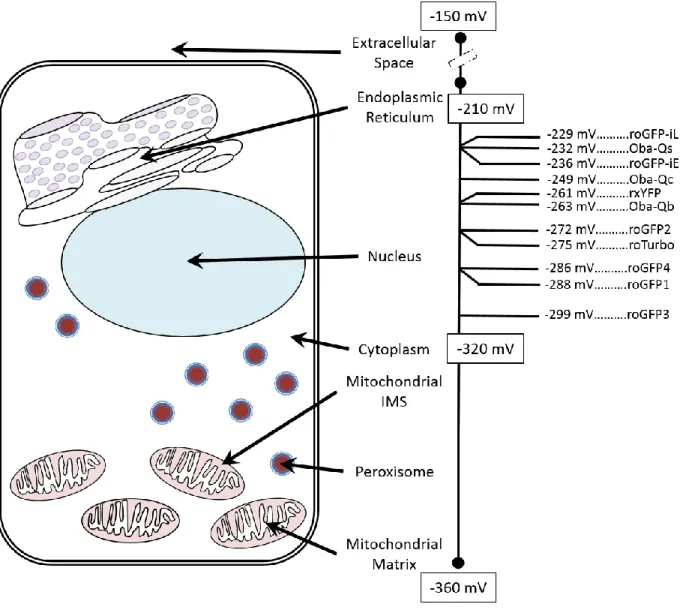
Figure 4: Midpoint Potentials for EGSH Sensors. The average redox potentials for subcellular
compartment are shown with the mitochondrial matrix as the most reducing compartment (-360 mV) and the extracellular space the most oxidizing compartment (-150 mV). Of the available EGSH sensors only a few have a high enough midpoint potential to be functional in the endoplasmic reticulum (roGFP-iL, roGFP-iE (163), Oba-Qs (160)). There are many sensors that can be effectively targeted to the nucleus, cytoplasm, mitochondrial intermembrane space (IMS), peroxisome, or mitochondrial matrix (Oba-Qc, Oba-Qb (160), rxYFP (156), roGFP1-4 (161), roTurbo (162)). The roGFP3 (161) will have improved accuracy in highly reducing compartments such as the mitochondrial matrix.
Although roGFP is a highly efficient sensor, it is limited by its slow equilibration with the
cellular environment as it is like rxYFP and the Oba-Q proteins in that it metabolically monitors
26
chimeric protein linking roGFP2 to Grx1 (roGFP2-Grx1), which dramatically improved sensor
kinetics (164). The same research group further improved the subcellular localization of the
sensor as well as responsiveness to changes in EGSH by switching the order of the chimeric
protein (Grx1-roGFP2) (165,166). It is likely that Grx1-roGFP2 had improved sensor
characteristics compared to roGFP2-grx1 due to the elimination of steric hindrances improving
the kinetic reaction between roGFP2 and Grx1. Another key study is the development of the
chimeric protein that linked roGFP2 to mrx1, the analog of Grx1 in mycobacterium. This
chimeric protein (roGFP2-mrx1) was able to successfully monitor the EMSH of mycobacterium as
a readout for an effect on the host as well as assess the efficacy of antibiotics (167). This
development established the potential to use roGFP as a way to monitor the redox metabolism of
prokaryotic models as well.
1.4.3: Dimedone-Based Chemical Probes
Detecting ROS and antioxidants directly is a powerful method to interrogate the role of
redox toxicology in adverse health outcomes; however, elucidating the identity of those targets
as well as how those outcomes are propagated can be just as important. Altered protein
sulfenylation is a good candidate to characterize the effect of oxidant events, especially if
intracellular H2O2 levels are affected. However, as mentioned previously, sulfenylation is a
rapid, short-lived modification and due to the scarcity of cysteines available to be sulfenylated, it
is physiologically rare (168). To detect sulfenylation one must have a highly specific and
sensitive method of detection (169). Many of these experiments utilize dimedone to detect
sulfenic acids (170). Dimedone is a small cell permeable molecule that is tolerated by cells that
27
all other cysteine-based PTMs (137). This allows for the detection of sulfenic acids by mass
spectrometric or immunoblot methods targeted to identify the dimedone-thioether bond on the
protein.
1.4.3.1: DYn-2 and DAz-2
Dimedone-based approaches to detect sulfenic acids have beneficially impacted the field
of redox biology, yet many of these approaches have a low signal-to-noise ratio. In response,
Kate Carrol’s group developed DYn-2 and DAz-2, which have chemical structures that have the
dimedone warhead with either an alkynyl or azide tail, respectively (171). These probes are
slightly more reactive than dimedone, but their real utility is that they can be coupled by
copper-catalyzed alkynyl-azo cycloaddition (CuAAC) to attach biotin to the DYn-2 or DAz-2 labeled
sulfenic acids (172-174). This allows one to take advantage of the biotin-avidin binding to
dramatically improve sensitivity. This approach, rather than directly linking dimedone to biotin,
is preferred, since biotin cannot cross the cell membrane (175). However, a drawback to using
CuAAC in biological samples is that it can damage the proteins that are to be detected (176,177).
1.4.4: In Vitro Models
Although many of these techniques can be implemented in vivo, in vitro cell culture
models currently provide the highest spatial and temporal resolution to mechanistically link
oxidant events to a specific adverse outcome. When researching the effects of air pollution on
cell toxicity, it is vital to utilize the most appropriate cell type. It has already been discussed that
air pollution has the capacity to affect multiple organs and tissue types, but the initial target of air
28
model for such studies are primary human airway epithelial cultures grown at an air-liquid
interface (178). However, primary cell cultures are notoriously difficult to use in mechanistic
studies as they are resistant to gene induction techniques and due to varying genotypes lead to
low signal-to-noise results.
Even though immortalized cell lines exist, many researchers are hesitant to utilize them in
toxicological studies due to concerns of relevance to clinical scenarios. These concerns are
certainly warranted, especially since established cell lines have neoplastic characteristics and
may be considered to have progressed towards transformation away from normal, healthy cells.
Relevant to redox biology, transformed cells have markedly different oxidant processes as
compared to their respective background, normal cells (179). Thus, it is important to validate
whether an immortalized cell line used in redox studies have a similar oxidant capacity as their
respective tissue found in intact, normal individuals. For instance the established human
bronchial epithelial cell line, BEAS-2B (180,181) has been demonstrated to maintain a similar
oxidative and antioxidative capacity as primary human bronchial epithelial cells (182). With
these considerations in mind, the BEAS-2B cell line are a powerful model to elucidate the effect
of air pollution on the human bronchial epithelia.
1.5: Summary
Air pollution is recognized as an increasing global public health problem. A mechanistic
contributor to air pollutant-induced adverse human health outcomes is oxidative stress. Oxidative
stress can be defined as a pathological increase in ROS, such as H2O2, that leads to impaired
cellular functions. Of particular interest is H2O2-induced sulfenylation of the regulatory proteins
29
is known that many air pollutants can lead to an increase in H2O2, but the role of air
pollutant-induced H2O2 has yet to be thoroughly studied. Thus, a hypothesis was formulated that exposure
to air pollution alters cellular redox homeostasis through the elevation of H2O2 resulting in
oxidant-dependent activation of adaptive and signaling pathways. To test this hypothesis three
goals were undertaken: 1) determine the role of pollutant-induced H2O2 in inflammatory and
adaptive gene expression, 2) examine the role of protein sulfenylation in air pollutant-induced
signaling, and 3) explore the application of H2O2-induced oxidant events as translational
30
CHAPTER 2: Role of H2O2 in the Oxidative Effects of Zinc Exposure in Human Airway
Epithelial Cells1
2.1: Introduction
Human exposure to ambient particulate matter (PM) is a public health concern of global
proportions. Observational studies demonstrate an association between exposure to PM and
elevated rates of cardiovascular morbidity and mortality (183-187). Despite the association
between these adverse health effects and ambient PM levels, the constituents in PM responsible
for its toxicity and the underlying mechanisms remain largely unknown. Epidemiological (36)
and toxicological (35,188) studies have specifically implicated the particle-associated transition
metal zinc (Zn2+) as a contributor to PM health effects. Although zinc is an essential nutrient and
vital to many physiological processes, inhalational exposure to zincis associated with a number
of adverse health outcomes (41).
The health effects of zincinhalation are modeled by metal fume fever, an occupational
disease characterized by a self-limited febrile flu-like condition with airway inflammation
resulting from inhalation of ZnO particles generated during welding (38). The mechanisms
responsible for the pathophysiological effects of Zn2+ inhalation have been investigated in
cultured human airway epithelial cells (HAEC) by our laboratory (48,189,190) and by other
groups utilizing diverse in vitro models (191-194). Observations from these studies show that
1 This chapter previously appeared as an article in the Journal of Redox Biology. The original citation is as follows:
31
Zn2+ induces inflammatory and adaptive gene expression through processes that involve the
deregulation of signaling cascades. Specifically, Zn2+ is thought to perturb multiple signaling
pathways by direct interaction with thiol groups on key regulatory proteins, including protein
tyrosine phosphatases (PTP) (44-46). Zn2+ is a known mediator in signaling pathways, including
the Keap1/Nrf2/ARE pathway (115,195).
Unlike other transition metals associated with PM (e.g., Fe, Ni, Cu, V), Zn2+ lacks two
adjacent valence states and, therefore, does not support single electron transfers to produce
reactive oxygen species (ROS), meaning that ROS generated during Zn2+ exposure are derived
from cellular metabolism. Zn2+ interferes with mitochondrial respiration at multiple points (41)
and consistent with this, we recently reported that exposure of HAEC to Zn2+ results in increased
intracellular generation of H2O2 of mitochondrial origin (196). Physiologically, H2O2 serves as a
second messenger that plays pivotal roles in the reversible inactivation of regulatory proteins,
most notably PTP (78,81,82,197). Thus, there is evidence that toxicological Zn2+ exposure can
induce gene expression through signaling mechanisms by direct interaction as well as through
the generation of H2O2.
In order to determine the dependence of Zn2+-induced responses on H2O2, the present
study expanded our previous live-cell imaging approach to monitor oxidative changes in the
cytosol and mitochondria of HAEC exposed to Zn2+ (56,140). We utilized cytosolic
overexpression or ectopic mitochondrial expression of the H2O2 scavenging enzyme catalase in
BEAS-2B cells bearing the genetically-encoded fluorogenic ratiometric sensors HyPer or
roGFP2, which report on H2O2 and the glutathione redox potential (EGSH), respectively
(133,147,198). In this study we examined the link between oxidative events associated with Zn2+


![Crystal structure of 6 (4 chlorophenyl) 6a nitro 6a,6b,8,9,10,12a hexahydro 6H,7H spiro[chromeno[3,4 a]indolizine 12,11′ indeno[1,2 b]quinoxaline]](data:image/gif;base64,R0lGODlhAQABAIAAAP///wAAACH5BAEAAAAALAAAAAABAAEAAAICRAEAOw==)