Function and tissue focus of daf-18/PTEN in
maintaining blast cell multipotency and quiescence in
Caenorhabditis elegans dauer larvae
Claudia Tenen
Submitted in partial fulfillment of the
requirements for the degree of
Doctor of Philosophy
under the Executive Committee
of the Graduate School of Arts and Sciences
Columbia University
© 2019
Claudia Tenen
All rights reserved
Abstract
Function and tissue focus of daf-18/PTEN in maintaining blast cell multipotency
and quiescence in
Caenorhabditis elegans
dauer larvae
Claudia Tenen
Cellular quiescence, a reversible state of cell-cycle exit, and developmental potential, the
ability to differentiate into appropriate cell types, are properties essential for normal development
and stem cell function (reviewed in (Cheung and Rando, 2013; Fiore et al., 2018; Mihaylova et
al., 2014)). Understanding the mechanisms by which cells maintain quiescence has important
implications for developmental biology, as this reversible state of cell-cycle exit is a key attribute
of stem cells, as well as for cancer biology, as quiescence plays a key role in tumor dormancy
and metastasis. Environmental conditions are key in regulating whether stem cells maintain
quiescence or exit to resume divisions and developmentally progress. I aim to investigate how
the properties of quiescence and developmental potential are retained over long periods of time
and how they are appropriately regulated by external environmental inputs. The nematode
Caenorhabditis elegans is an excellent model for investigating both of these questions because it
is capable of entering and maintaining a developmentally arrested state for an unusually long
time compared to the normal lifetime of the worm, and because the decision to enter this arrest is
regulated entirely by external environmental inputs (Cassada and Russell, 1975).
Upon encountering conditions unfavorable for growth, C. elegans enters an alternative,
developmentally arrested state called dauer diapause in which precursor cells remain quiescent
for months – a period many times the lifespan of a worm grown under favorable conditions
(Cassada and Russell, 1975). Maintaining precursors in this arrested state is important in order
for the worms to develop normally once conditions improve and requires components of the
conserved Insulin/Insulin-like (IIS) signaling pathway (Karp and Greenwald 2013 and this
work); of note, the IIS pathway also regulates mammalian quiescence (Eijkelenboom and
Burgering, 2013). Canonical regulation of dauer diapause includes IIS, TGFß, and dafachronic
acid (DA)/nuclear hormone receptor (NHR) signaling (reviewed in (Murphy and Hu, 2013a)).
Here, I investigate how DAF-18, the sole C. elegans ortholog of the tumor suppressor
PTEN (Phosphatase and tensin homolog) (Gil et al., 1999; Mihaylova et al., 1999; Ogg and
Ruvkun, 1998; Rouault et al., 1999), maintains quiescence in dauer through regulation of these
conserved signaling pathways using the C. elegans gonad as a model. The gonad is composed of
somatic cells and the germline. Both the somatic gonad and germline develop
post-embryonically from precursor cells present when dauer arrest occurs, and these precursor cells
remain quiescent for the duration of dauer diapause (Cassada and Russell, 1975; Hong et al.,
1998; Narbonne and Roy, 2006). After exit from dauer, division and differentiation resume.
DAF-18/PTEN is required for germline quiescence during dauer diapause (Narbonne and Roy,
2006), and my results implicate DAF-18/PTEN in the control of quiescence of the somatic
tissues as well, including the somatic gonad. In this role, DAF-18/PTEN activity in the somatic
gonad non-autonomously coordinates both germline stem cell (GSC) and somatic gonad blast
(SGB) quiescence. I have demonstrated this somatic gonad focus through mosaic analysis,
tissue-specific rescue, and tissue-specific excision mosaics. We propose that DAF-18/PTEN
mediates production of a signal promoting quiescence from the somatic gonad to the SGBs and
GSCs and that this signal does not absolutely require or solely target the IIS, TGFß, or DA/NHR
signaling pathways normally implicated in regulation of dauer diapause.
Table of Contents
List of Figures …………...……… iv
Acknowledgements ……….…………. vi
Chapter 1. Introduction……… 1
1. Introduction to
Caenorhabditis elegans
dauer larvae………. 1
2. Development of the somatic gonad and germline in
C. elegans
……… 2
2.1 Gonadogenesis in
C. elegans
continuous development ……… 2
2.2 Gonadogenesis in
C. elegans
with dauer life history ………… 4
2.3 The role of LIN-12/Notch signaling in somatic gonad
development ………... 5
3. Vulval development in continuous development and dauer life history.. 8
4. Regulation of dauer diapause ……….. 10
4.1 Insulin/insulin-like signaling (IIS)………. 11
4.2 TGFß ………. 13
4.3 DA/NHR ………. 14
4.4 Guanylyl cyclase signaling ………... 15
5.
daf-18/
PTEN in dauer ………. 16
6.
daf-18/
PTEN in L1 arrest ……… 18
6.1 Germline precursor quiescence in L1 arrest ……….. 19
6.2 Somatic precursor quiescence in L1 arrest ……… 20
7. Chapter 1 Figures ….………...…. 22
7.1 Figure 1 ………..… 23
7.2 Figure 3 ……… 25
7.2 Figure 4 ………...…………. 26
7.2 Figure 5 ………..……….. 27
7.2 Figure 6 ………..…….. 28
7.2 Figure 7 ………..….. 29
Chapter 2. daf-18/PTEN acts in the somatic gonad to coordinate somatic gonad and
germline development in C. elegans dauer larvae……….. 30
1. Summary ………... 31
2. Results ……… 32
3. Discussion ………... 39
5. Figure legends ………...…. 42
6. Methods ………... 47
7. Figures ………... 65
7.1 Figure 1 ……….…...……. 66
7.2 Figure 2……….…...……. 67
7.3 Figure 3 ……….……… 68
7.4 Figure 4 ……….……… 69
7.5 Supplemental Figure 1 …………..………...…….… 70
7.6 Supplemental Figure 2 …………..………...…….… 71
7.7 Supplemental Figure 3 ………..………...…….… 72
7.8 Supplemental Figure 4 …………..………...…….… 73
Chapter 3 General Discussion ………..………..……... 74
3.1 Signal regulated by DAF-18/PTEN to regulate SGB and/or GSC
quiescence ……….. 75
3.1.1 Nature of the DAF-18/PTEN-mediated and somatic
gonad-derived signal . ………75
3.1.2 Tissue focus for daf-18/PTEN: cellular redundancy within the
somatic gonad ……….………78
3.1.3 Screening for clues to the identity of the signal …..………….. 81
3.1.4 Secreted PTEN ……….………..………... 82
3.1.5 Other signal mechanism candidates ………..… 84
3.1.6 Soma-germline crosstalk: sid-1 and small RNA signaling …... 85
3.2 DAF-18/PTEN activity autonomous to quiescence-regulating cells …... 86
3.2.1 DAF-18/PTEN is not likely to inhibit Insulin/insulin-like
signaling (IIS) to non-autonomously maintain cellular quiescence ...87
3.2.2 PI3K signaling and non-IIS pathways .………….……… 89
3.3 Concluding remarks ..……….……….. 91
3.4 Chapter 3 Figures ..………... 93
3.1.1 Figure 1 .……… 94
3.1.2 Figure 2 .……… 95
3.1.3 Figure 3 ………. 96
References
……….……… 97
List of Figures
Chapter 1
Figure 1. Continuous development and dauer formation life cycle in
Caenorhabditis elegans
………...… 23
Figure 2. Somatic gonad and germline development .……… 24
Figure 3. VPC fate patterning in continuous development ……… 25
Figure 4. Integration of signaling pathways regulating the decision to form
dauers ………. 26
Figure 5. Insulin/insulin-like signaling pathway ………..…. 27
Figure 6. The
daf-7/
TGF-
β
signaling pathway ……….. 28
Figure 7. DAF-9-mediated DA signaling through DAF-12/NHR .………… 29
Chapter 2
Figure 1. Developmental progression of Somatic Gonad Blast cells (SGBs)
and Germline Stem Cells (GSCs) in
daf-18(0)
dauers ……….…….. 66
Figure 2.
daf-18
acts in the somatic gonad primordium to maintain SGB and
GSC quiescence in dauer ………... 67
Figure 3.
daf-18
has a diffuse cellular focus within the somatic gonad …… 68
Figure 4. Evidence for a novel signal regulated by
daf-18
/PTEN and model 69
Figure S1. Time course of SGB progression in
daf-18(0)
dauers ………… 70
Figure S2. Additional tissue-specific
daf-18(+)
rescue experiments ……... 71
Figure S3. Genetic ablation of the DTCs and AC by
hlh-2(RNAi)
………... 72
Fig. S4. Genetic mosaics lacking daf-18 activity in defined lineages …….. 73
Chapter 3
Figure 1. Tissue-specific expression of
daf-18(+)
in the somatic gonad rescues
somatic gonad and germline quiescence in dauer .…….…….…….……. ….94
Figure 2. The somatic gonad primordium envelops both germline arms in
dauer ……….. 95
Figure 3. In dauers, the transgene
qIs90[ceh-22b::yfp]
is brightly expressed in
the DTCs (Z1.aa and Z4.pp) and the two SS cells that are DTC lineal sisters
(Z1.ap and Z4.pa) . …….…….…….…….…….…….…….…….…….…… 96
Acknowledgements
Foremost, I owe immense thanks to my mentor and advisor Dr. Iva Greenwald, who
provided an incredible amount of guidance throughout my time in her lab, both personally and
professionally. Dr. Greenwald is passionate about teaching as well as scientific discovery, and
that combination afforded me an invaluable and productive experience. The care she applies to
both her work and her students has not only been instructive for my scientific and professional
development but also inspirational to me. I am very grateful for all the time, effort, and patience
she has applied to my education and for her tremendous kindness. I aspire to her level of
scientific rigor and mentoring skills in my own career in the future.
I am also very grateful to my Thesis Committee members Dr. Oliver Hobert, Dr. Mimi
Shirasu-Hiza, and Dr. Daniel Kalderon, as well as Dr. Lori Sussel who kindly participated as a
member for a significant portion of my graduate studies. Their insightful comments and critiques
contributed substantially to my thesis.
My additional committee member Dr. Xantha Karp deserves thanks not only for her help
and guidance as a member of my defense committee, but also for her guidance, scientific
discussion, and feedback throughout my time as a graduate student. Her work has formed the
basis of many projects, including my own.
I additionally thank both Dr. Oliver Hobert as well as the entire Hobert lab for scientific
and technical help throughout my graduate studies; this aid came in the form of weekly lab
meetings as well as every day interactions and included the sharing of reagents and equipment.
Overall, Dr. Hobert and his lab greatly contributed to my education during graduate school.
I am grateful to the Columbia MSTP program including especially Dr. Patrice Spitalnik,
who has helped me with various steps of the program, even including providing advice before I
joined. I also thank the Integrated Program in Cellular, Molecular and Biomedical Studies
(CMBS), including Zaia Sivo, for guiding me through my graduate studies.
I owe many, many thanks to the entirety of the Greenwald lab (including both past and
present members) for their support and many contributions. This lab has been a constant source
of scientific discussions, aid, and friendship throughout my time here. I additionally thank
Gleniza Gomez for her special brand of encouragement and her technical assistance; Dr. Daniel
Shaye for scientific advice and aid; Dr. Yuting Deng for extensive late-night discussions and also
for late-night technical/scientific help; Dr. Ryan Underwood for inspiring scientific curiosity and
Frozen-themed ziplock bags; Justin Benavidez for his level-headed debates/discussions, advice,
and sense of humor; Katherine Luo for indulging me in many debates and discussions, generally
less level-headed and excessively fun; and I thank Jessica Chan for her continuous friendship,
scientific discussions, and introductions to many new places to eat. I am very grateful to my
rotation students, all of whom required no mentorship to excel and made my lab life both easier
and more pleasant: Hana Littleford, Catherine O’Keeffe, Justin Shaffer, and Bryan Wang. I owe
thanks to Dr. Maria Sallee for her help and inspiration in the lab, personal support, great sense of
humor, and a little bit of pop culture education. I thank Dr. Claire de la Cova both for her initial
mentorship during my rotation and for her continued mentorship and friendship afterward, and
also Dr. Robert Townley for help, good musical taste, and tough questions. I also thank Dr.
Michelle Attner for both guidance and friendship. Finally, I thank our newest lab member, Dr.
Julia Wittes, for being a great help even in the short time we overlapped.
I also thank my family for their unceasing support, including my father, Dr. Daniel Tenen
(“don’t stress!”); my sister Nicole, and my sister Danielle, who fielded many phone calls and
never once lost patience with me. Finally, I give particular thanks to two people whose incredible
support has been essential for completing this work: my partner, Tony Xing, and my mother, Dr.
Linda K. Clayton. Tony has supported, encouraged, and helped me pursue my academic
endeavors for over a decade now. His support has been integral to my ability to pursue this
project. My mother, Linda, has been incredibly supportive of me for my entire life, but also has
been constantly available and helpful throughout my graduate studies. In addition, Linda has
provided scientific feedback and has helped immensely in critiquing and editing my scientific
writing. This body of work was truly made possible for me by their support and efforts.
Chapter 1. Introduction
1. Introduction to
Caenorhabditis elegans
dauer larvae:
A fundamental question in developmental biology with strong implications for human
health and disease is how cells remain quiescent and multipotent for extended lengths of time.
The
C. elegans
dauer larva, described further below, is an outstanding model for studying
conserved mechanisms of cellular quiescence and the maintenance of multipotency: in both
dauer larvae and mammals, the conserved IIS pathway components 18/PTEN and
DAF-16/FoxO regulate these properties and directly regulate genes that control cell cycle progression
(Baugh and Sternberg, 2006; Dijkers et al., 2000; Escoté and Fajas, 2015; Mellough et al., 2015;
Rafalski and Brunet, 2011). Genetic studies of dauer larvae have also contributed fundamentally
to our understanding of the insulin signaling pathway. Of particular relevance to this thesis,
studies in
C. elegans
clarified the role of the protein and lipid phosphatase DAF-18/PTEN
(Phosphatase and tensin homolog) as the major negative regulator of insulin signaling (Gil et al.,
1999; Mihaylova et al., 1999; Ogg and Ruvkun, 1998; Rouault et al., 1999; Worby and Dixon,
2014), and first showed that the transcription factor DAF-16/FoxO (Forkhead box class O) is a
major target negatively regulated by insulin signaling (Ogg et al., 1997). Both DAF-18/PTEN
and DAF-16/FoxO have critical roles in human disease: PTEN is an important tumor suppressor,
while FoxO can promote or suppress tumorigenesis depending on cell context (Eijkelenboom
and Burgering, 2013; Milella et al., 2015; Worby and Dixon, 2014). Both PTEN and FoxO have
roles in other diseases as well (Burgering and Medema, 2003; Cantley and Neel, 1999; Ogg et
al., 1997). Dysregulation of PTEN or FoxO contributes to disease in part because both regulate
cellular quiescence: for example, PTEN is required to maintain quiescence in primordial
follicles, the source of mammalian ova ((Adhikari et al., 2010; Eijkelenboom and Burgering,
2013; Milella et al., 2015). These roles in quiescence are shared by their
C. elegans
orthologs
during larval development (Fukuyama et al., 2006, 2012; Karp and Greenwald, 2013; Narbonne
and Roy, 2006; Wolf et al., 2014; Zheng et al., 2018a). We therefore expect that our studies of
how DAF-18/PTEN maintains quiescence in dauer will reveal new conserved mechanisms
directly relevant to the maintenance of stem cell quiescence in mammalian systems.
C. elegans
is also an outstanding and proven model for identifying interactions between
different signaling pathways in development. Investigating cellular quiescence and relevant
signaling pathways in
C. elegans
dauer larvae has the potential to impact not only therapy for
human cancers but also for infectious disease: the dauer larva is analogous to the infective stage
of parasitic nematodes as it is long-lived, non-feeding, and capable of forming a reproductive
adult (Ashton et al., 1999; O’Halloran and Burnell, 2003). In
C. elegans
dauer larvae, failure to
maintain quiescence and block signaling pathways such as LIN-12/Notch prevents dauers from
forming normal, reproductive adults upon recovery (Colella et al., 2016; Karp and Greenwald,
2013), this work); therefore, studies of quiescence and signaling in dauer may identify novel
therapeutic targets for parasitic infections. Thus, as a proven model system amenable to rapid
genetic screens, the
C. elegans
dauer is likely to elucidate therapeutically relevant mechanisms in
quiescence that can then be verified in and applied to mammalian contexts.
This thesis will be concerned with the maintenance of quiescence during dauer arrest by
DAF-18/PTEN in the somatic gonad and germline in dauer.
2. Development of the somatic gonad and germline in
C. elegans
Under favorable conditions, the nematode
Caenorhabditis elegans
progresses through
four larval stages (L1-L4) before molting into an adult (Figure 1).
C. elegans
hatch with 4 gonad
precursors: Z1 and Z4 form the entire somatic gonad, and Z2 and Z3 the germline (Hubbard and
Greenstein, 2005; Kimble and Hirsh, 1979). The somatic gonad then develops in two distinct
phases (Figure 2A). In the first stage, Z1 and Z4 divide in L1 to form the somatic gonad
primordium, which consists of twelve cells that do not divide during L2 (Kimble and Hirsh,
1979; Kimble and White, 1981). By the end of L2, three of the twelve cells are terminally
differentiated, the anchor cell (AC) and two distal tip cells (DTCs), such that there are nine
somatic gonad blast (SGB) cells and three specified cells (Hubbard and Greenstein, 2005;
Kimble and Hirsh, 1979; Kimble and White, 1981). The SGBs include three ventral uterine
(VU), four spermathecal/sheath (SS), and two dorsal uterine (DU) precursor cells (Newman et
al., 1996). The SGBs and specified cells (twelve cells total) together form the somatic gonad
primordium.
In the second stage of somatic gonad development, starting in L3, the SGBs divide and
their descendants undergo fate specification to form the structures of the adult somatic gonad
(Figure 2A).
The AC and DTCs do not divide but are essential for development of the adult
reproductive system. The AC induces vulval development and development of the uterine-vulval
connection, including the uterine-seam syncytium cell (the utse), which is formed from the
fusion of the AC with a syncytium of VU descendants (Kimble and Hirsh, 1979; Kimble and
White, 1981; Newman et al., 1996). The DTCs are required for promoting mitotic divisions in
the germline stem cells (GSCs) and extension and flexion of the germline arms.
In the presence of food, the germline precursors Z2 and Z3 start dividing in L1 and the
GSCs continue to divide mitotically thereafter, maintaining a pool of GSCs in the adult (Hubbard
and Greenstein, 2005; Korta and Hubbard, 2010). Meiotic divisions do not begin until the L3
stage. The descendants of the first ~70 GSCs that differentiate by meiotic divisions become
sperm (four per GSC), and thereafter the meiotic progeny either form oocytes or undergo
apoptosis. The GSCs therefore proliferate continuously in all stages, in contrast to somatic gonad
development.
The initial GSC divisions are GLP-1/Notch-independent (Austin and Kimble, 1987), and
continued GSC proliferation requires activation of GLP-1/Notch in the germline precursors by
the ligand LAG-2, which is produced by the DTC of each arm (reviewed in (Hubbard and
Greenstein, 2005)). Notch signaling is also essential for somatic gonad development:
LIN-12/Notch signaling in the somatic gonad is required for normal fate specification in somatic
gonad precursors and their descendants in both stages of somatic gonad development (both
described in more detail below).
Under starvation conditions, dauer entry occurs between the two phases of somatic gonad
development; dauer entry additionally interrupts the continuous proliferation of germline
precursors (Cassada and Russell, 1975; Hong et al., 1998), Figure 2B, described below).
2.2 Gonadogenesis in
C. elegans
with dauer life history:
In replete conditions,
C. elegans
develops rapidly and continuously through larval stages
before molting into a reproductive adult with a lifespan of approximately two weeks. In contrast,
under unfavorable conditions, the nematode deviates from continuous development and instead
enters a larval stage of interrupted development and prolonged quiescence, dauer diapause
(Figure 1) (Cassada and Russell, 1975). After the L1 stage, the larvae enter the L2d stage, an
extended alternative to L2; if conditions do not improve, they undergo an extended L2d-dauer
molt to form dauers (Fielenbach and Antebi, 2008). Dauers can survive for many months (in
contrast to the normal 2-3-week lifespan) without food and display increased resistance to harsh
environmental conditions such as desiccation or detergents (Cassada and Russell, 1975). If
conditions improve, dauers recover and resume development as post-dauer L3 (pdL3) larvae and
become reproductive adults overtly indistinguishable from those without dauer life history
(Cassada and Russell, 1975). However, there are some anatomical differences, including
supernumerary muscle arms and changes in IL2 neuron morphology (Dixon et al., 2008;
Schroeder et al., 2013), as well as differences in gene expression profiles (Hall et al., 2010).
Dauer arrest interrupts both somatic gonad and germline development: somatic gonad
development is interrupted after the first stage, and the mitotic divisions of the germline that are
normally continuous throughout the larval stages are halted (Figure 2B (Cassada and Russell,
1975; Hong et al., 1998)). The L2d somatic gonad and germline are similar to that of L2: the
somatic gonad primordium still consists of twelve cells, nine precursors and the differentiating
AC and DTCs (Colella et al., 2016; Narbonne and Roy, 2006). Germline development ceases
upon dauer formation with approximately 40 GSCs in the wild type reference strain N2
(Narbonne and Roy, 2006). For the duration of dauer diapause, the nine somatic gonad precursor
cells and all GSCs from L2/L2d remain quiescent until dauer recovery.
2.3 The role of LIN-12/Notch signaling in somatic gonad development:
Notch signaling is an intercellular transduction pathway that mediates cell-cell
identified many components of the Notch pathway that are conserved in other systems, including
mammals, and provided key insights that helped elucidate the mechanism of Notch signal
transduction (Greenwald, 2012).
C. elegans
has two Notch orthologs,
lin-12
and
glp-1
, and both
are required for proper somatic gonad and germline development:
lin-12
is required for
development of the somatic gonad, vulva, and gonad-vulval connection, while
glp-1
is required
for GSC mitotic proliferation (Austin and Kimble, 1987; Ferguson and Horvitz, 1985; Ferguson
et al., 1987; Greenwald et al., 1983; Newman et al., 1995).
LIN-12/Notch signaling is required for several distinct events during development of the normal
somatic gonad:
i. AC-VU decision through lateral specification: early in gonadal development, four cells
in the somatic gonad primordium have the potential to become the AC, termed the
α
cells and ß
cells. The two ß cells lose this potential early and eventually become VUs. The two
α
cells retain
AC potential longer and, through lateral specification by LIN-12/Notch signaling, small
stochastic differences in initial levels of LIN-12/Notch are amplified through a feedback
mechanism. Finally, the
α
cell with relatively lower LIN-12/Notch activity is specified as the
AC, and the other
α
with higher LIN-12/Notch becomes the VU (Greenwald et al., 1983; Kimble
and Hirsh, 1979; Kimble and White, 1981; Seydoux and Greenwald, 1989; Wilkinson et al.,
1994). In LIN-12/Notch mutants, AC and VU fate specification does not occur normally: loss of
LIN-12/Notch function results in specification of multiple ACs over the VU fate, while in
activated dominant mutants all cells with AC potential are instead specified as VUs (Greenwald
et al., 1983). The fluorescent reporter and integrated transgene
arIs107[mir-61::yfp]
is expressed
in
the
α
and ß cells, and this expression becomes restricted to the VUs after AC-VU fate
specification and therefore can be used as a marker of VU cells after this decision (Yoo and
Greenwald, 2005).
ii. Pi cell (defined here) fate induction and utse formation: The VUs begin dividing in L3
and their descendants adopt a variety of cell fates required for development of the adult somatic
gonad. Six of the VU granddaughters adopt what is termed a
π
cell fate, and the others adopt the
ρ
cell fate (Newman et al., 1995). The AC induces
π
cell fate specification in these VU
descendants by activating 12/Notch signaling in these cells through expression of the
LIN-12/Notch ligand, LAG-2.
π
cell daughters later fuse to form a syncytium, and after inducing vulval development
(see below), the AC fuses with this syncytium (Ghosh and Sternberg, 2014; Newman and
Sternberg, 1996; Newman et al., 1996). Once fusion with the AC has occurred, nuclei migrate
outwards from the proximal gonad to form a mature utse. Formation of the utse requires the
π
cells and therefore also requires LIN-12/Notch signaling for specifying the
π
cell fate.
The adoption of the
π
cell fate requires cell-cell interactions and indicates cell fate
determination, as
π
cell fate is determined once the VU granddaughters are born (Newman et al.,
1995): ablation of the AC when the VUs have not divided or have only divided once results in
failure to specify
π
cell fates in a majority of larvae, and only the
ρ
cell fate is specified.
However,
π
cell fates are specified normally with ablation of the AC after the VU
granddaughters are born, six of which will become the
π
cells (Newman et al., 1995).
Subsequent utse formation by
π
cell daughters represents terminal differentiation of these cells.
Both
π
cell fate specification and utse formation can be visualized with multicopy
integrated transgenes.
syIs80[lin-11::gfp]
and
kuIs29[egl-13::gfp]
are direct transcriptional
targets of LIN-12/Notch and markers of
π
cell and
π
cell daughters and are expressed once they
are specified in the late L3 (Cinar et al., 2003; Gupta et al., 2003; Hanna-Rose and Han, 1999;
Oommen and Newman, 2007).
arIs51[cdh-3::gfp]
is expressed in the differentiated anchor cell
by the end of the L2 larval stage and its expression later expands to the entire utse syncytium
when the AC fuses with the immature syncytium of
π
cell daughters in the early-to-mid L4 larval
stage (Karp and Greenwald, 2003; Pettitt et al., 1996) also see (Ghosh and Sternberg, 2014).
iii. Vulval induction: in the four
π
cell daughters that do not contribute to utse formation,
an alternative uv1 cell fate is specified through EGF signaling from the vulval precursor cell
descendant vulF cells (Chang et al., 1999). The uv1 cells are required for proper establishment of
the vulval-uterine connection and extend processes connecting to the vulval vulF cells (Newman
et al., 1996). Therefore, signaling from vulval cells is required for normal somatic gonad
development, and proper vulval fate specification requires LIN-12/Notch signaling: the six
vulval precursor cells (VPCs) remain quiescent during the L1 and L2 stages and are induced to
divide and specify a precise pattern of cell fates by the AC during the L3 (reviewed in
(Greenwald and Kovall, 2013) and (Sternberg, 2005). This patterning is primarily mediated by
crosstalk between the LIN-12/Notch and EGFR signaling pathways and requires the AC. Vulval
development and regulation of VPC in dauer are described in further detail below.
3. Vulval development in continuous development and dauer life history
Though I have focused on the somatic gonad, one well-characterized example of the
maintenance of quiescence and multipotential in somatic blast cells during dauer diapause is the
vulval precursor VPCs. The VPCs remain quiescent and retain VPC potential throughout dauer
diapause, and the IIS signaling target
daf-16/
FoxO is required for both (Euling and Ambros,
1996; Karp and Greenwald, 2013). Additionally, in this context, blocks to both the
LIN-12/Notch and EGFR signaling pathways are required for normal post-dauer development. Here I
briefly review VPC development in continuous development and in dauer.
In the L1, there are six VPCs, numbered P3.p-P8.p. Each VPC is multipotent and has the
potential to adopt the 1°, 2°, or 3° fate. In the early L3 stage, an EGF-like inductive signal from
the AC of the gonad activates EGFR signaling in P6.p specifying 1° fate, and P6.p in turn
produces a lateral signal that activates LIN-12/Notch in its neighbors P5.p and P7.p, causing
them to adopt a 2° fate (Figure 3, adapted from (Karp and Greenwald, 2013)) (Reviewed in
(Sternberg, 2005)). P3.p, P4.p, and P8.p, which do not receive either of these patterning signals,
adopt a 3° fate and each divides once to produce two daughters that fuse with the hypodermis.
Fluorescent reporters for transcriptional targets of the EGFR or LIN-12/Notch pathways mark
these fates: for example, as a result of P6.p adopting 1° fate, the LIN-12 activating ligand
lag-2
is
expressed and can be visualized with a fluorescent marker driven by the
lag-2
promoter (Chen
and Greenwald, 2004; Zhang and Greenwald, 2011). Likewise, 2° fate normally adopted by P5.p
and P7.p in continuous development can be marked using a reporter for the lateral signaling
target of LIN-12, lst-5 (Choi, 2009; Karp and Greenwald, 2013; Yoo and Greenwald, 2005).
The potential of VPCs to adopt 1°, 2°, or 3° fates – their multipotency – appears to
involve mechanisms for blocking the activity of the EGFR and LIN-12/Notch pathways (Karp
and Greenwald, 2013). Expression of LIN-12(intra
Δ
P), a constitutively active form of LIN-12
which is stable and nuclear in dauer VPCs, induces a 2° fate marker in up to 75% of all VPCs in
L2d, but this expression is completely blocked by the L2d-dauer molt such that none of these
markers are expressed in dauer.
The IIS target
daf-16
/FoxO is required for the blocks to signal transduction of activated
EGFR and LIN-12/Notch during dauer diapause, maintaining VPC quiescence and multipotency
(Karp and Greenwald, 2013).
daf-16
encodes the ortholog of mammalian FoxO, a transcription
factor important for longevity, stress resistance, and dauer formation (Larsen et al., 1995; Lin et
al., 1997; Vowels and Thomas, 1992) In
daf-16(0)
dauer larvae, VPCs adopt 1° and 2° fates and
often divide as well, indicating that DAF-16 activity is necessary for maintaining not just VPC
multipotency but also quiescence during dauer diapause (Karp and Greenwald, 2013). The role
of DAF-16 in maintaining VPC multipotency and quiescence is likely autonomous to the VPCs:
VPC-specific RNAi against
daf-16
in dauers results in 1° fate adoption in P6.p.
4. Regulation of dauer diapause
The cellular basis for the regulation of dauer entry is complex. Three major signaling
pathways regulate dauer entry: Insulin/Insulin-like growth factor signaling (IIS), TGFß, and
DA-DAF-12/steroid hormone signaling (Antebi, 2015; Gumienny and Savage-Dunn, 2013; Hu,
2007; Murphy and Hu, 2013a) Environmental conditions detected by sensory neurons regulate
the production of insulin-like growth factors, DAF-7/ TGFß, and steroid hormone ligands
(Dafachronic Acids, DA); these inputs are integrated to control the decision to proceed to
reproductive adulthood or enter dauer diapause (Gerisch et al., 2004; Mak and Ruvkun, 2004).
Active IIS and TGFß signaling converge on the nuclear hormone receptor (NHR) homolog
DAF-12, which promotes continuous development when bound by DA ligand (Figure 4, from (Karp
and Greenwald, 2013)). When IIS and TGFß signaling are inactive, DAF-12 ligand is absent and
DAF-12 instead binds to the SHARP corepressor homolog and DAF-12 interacting protein
DIN-1 to promote dauer formation (Ludewig et al., 2004). cGMP signaling also regulates dauer
formation, generally upstream of the IIS and TGF
b
pathways (discussed further below and
reviewed in (Fielenbach and Antebi, 2008)).
The roles of some components of these pathways in regulating germline and/or somatic
gonad quiescence during dauer development (L2d and the L2d-dauer molt) have been identified,
though less is known about the maintenance of their quiescence during the dauer larval stage
itself; these are described below.
4.1 Insulin/insulin-like signaling (IIS):
Apart from regulating the dauer entry decision and dauer formation, the IIS signaling
pathway regulates many processes and aspects of development in
C. elegans,
including aging, fat
metabolism, stress resistance, behavior and learning, germline proliferation, and non-dauer
developmental arrest (reviewed in (Murphy and Hu, 2013a)). Active IIS promotes continuous
development, while reduced IIS permits dauer entry. Loss of function of the sole
C. elegans
insulin receptor ortholog
daf-2
(Kimura et al., 1997; Ruvkun and Hobert, 1998) forces
constitutive dauer formation (Gottlieb and Ruvkun, 1994). Loss of function in other conserved
components of the IIS pathway, including AGE-1, PDK-1, AKT-1, and AKT-2, also results in
constitutive dauer formation, and dauer formation in these mutants requires the activity of the
FoxO ortholog DAF-16 (Figure 5, reviewed in (Murphy and Hu, 2013a)). As in other systems,
daf-2/
IR is regulated by insulin-like peptides; however, in
C. elegans
it is predicted that there are
approximately 40 insulin-like (INS) molecules and many of these can regulate dauer formation.
Additionally, some of the INS peptides are agonistic to
daf-2
while others antagonize
daf-2
activity (Cornils et al., 2011; Murphy et al., 2007; Pierce et al., 2001). The
ins
genes are mainly
expressed and function in neurons, though some expression and function has been identified in
the intestine as well (Murphy et al., 2007; Pierce et al., 2001).
The lipid and protein phosphatase
daf-18
/PTEN is required for germline quiescence in
dauer (Narbonne and Roy, 2006) and is a conserved inhibitor of IIS signaling, therefore
promoting activity of the canonical IIS target and transcription factor DAF-16/FoxO (Figure 5,
adapted from (Murphy and Hu, 2013a)).
daf-18/
PTEN is described in further detail below.
Activation of the worm ortholog of the insulin receptor, DAF-2, inhibits DAF-16 and promotes
continuous development, so loss of
daf-2
results in active DAF-16 and a
daf-c
phenotype (Larsen
et al., 1995; Lin et al., 1997; Vowels and Thomas, 1992) Conversely, in
C. elegans
,
daf-16
is
required for dauer formation and
daf-16
null mutants are
daf-d. daf-16(0)
mutants fully suppress
dauer formation in
daf-c
mutants of the IIS pathway, indicating that DAF-16/FoxO is the main
target of IIS in regulating dauer formation (Gottlieb and Ruvkun, 1994; Larsen et al., 1995;
Vowels and Thomas, 1992). In dauers formed using the
daf-c
mutant
daf-7(e1370)
(described
below), DAF-16 regulates both quiescence and multipotency in the VPCs, in part by repression
of LIN-12/Notch targets (Karp and Greenwald, 2013).
daf-16
is important for longevity, stress
resistance, and dauer formation. In the presence of food, IIS promotes phosphorylation of
DAF-16, leading to its cytoplasmic sequestration so it cannot activate its nuclear targets (Larsen et al.,
1995; Lee et al., 2001; Lin et al., 1997, 2001; Vowels and Thomas, 1992)
daf-16
is not required
for maintaining quiescence in the dauer germline (Narbonne and Roy, 2006), but it is required in
the VPCs in dauer for maintenance of both quiescence and multipotency, at least in part through
a DAF-16-mediated block to LIN-12/Notch signaling (described above, (Karp and Greenwald,
2013)).
Aside from
daf-18/
PTEN, the PP2A holoenzyme
pptr-1
is also an IIS inhibitor that
regulates dauer entry through regulation of phosphorylation and activity of AKT-1
daf-2
mutants, while knockdown of other PP2A subunits does not. This suppression of dauer
formation is not seen in an alternative pathway
daf-c
mutant,
daf-7/
TGFß.
4.2 TGFß:
When active, the TGFß signaling pathway, promotes continuous development (Ren et al.,
1996; Schackwitz et al., 1996). Of the five TGFß-related genes, a strong role in dauer formation
has only been identified for
daf-7
/TGFß (Gumienny and Savage-Dunn, 2013). DAF-7
signals
through a canonical receptor pathway which, when active, inhibits activity of the Co-Smad
DAF-3 and its partner, the Sno/Ski homolog DAF-5.
daf-7
is expressed specifically in the ASI sensory
neurons, while other components of the pathway are expressed in other tissues (Ren et al., 1996;
Schackwitz et al., 1996).
In addition to regulating dauer formation,
daf-7/
TGFß signaling regulates germline
proliferation, but may have both pro- and anti-proliferative roles.
daf-7
signaling independent of
DAF-3 and DAF-5 appears to negatively regulate GSC proliferation through regulation of DTC
expression of the GLP-1/Notch ligand
lag-2
(Park et al., 2010). However, DAF-7 activity in the
ASI neurons has also been identified as positively regulating GSC proliferation under favorable
conditions through its receptor DAF-1, which is expressed in the DTCs (Dalfó et al., 2012). In
this role,
daf-7/
TGFß signaling likely regulates DAF-3 and DAF-5 activity as in the canonical
pathway; additionally, in this role,
daf-7
appears to promote proliferation independently of
GLP-1/Notch. Finally, this positive regulatory role is also independent of factors that promote dauer
formation in other pathways including DAF-18/PTEN and DAF-16/FoxO (IIS) and DAF-12
(NHR).
In dauer, however,
daf-7
/TGFß is not required for germline progression in
daf-18(0)
dauers and the GSCs arrest appropriately in
daf-7(ts)
dauers (Narbonne and Roy, 2006).
However, it is possible that other TGFß ligands may promote gonad proliferation in
daf-18(0)
dauers. For example, the TGFß ligand
dbl-1
has some role in repressing dauer formation:
dbl-1
null single mutants do not affect dauer formation but do enhance the
daf-c
phenotype of
daf-7(e1372)
at 20
°
C (Morita et al., 1999). Additionally, the biological function of the TGFß
homologs
tig-2
and
tig-3
are unknown and could have unidentified roles in dauer regulation
(Suzuki et al., 1999). Analysis of other TGFß-related genes may reveal unidentified roles in
dauer regulation or functional redundancy with
daf-7.
4.3 DA/NHR:
Nuclear hormone receptor (NHR) signaling acts downstream of both IIS and TGFß
signaling in regulating the dauer entry decision (Antebi, 2015; Gerisch et al., 2004; Mak and
Ruvkun, 2004). Steroids called Dafachronic Acids (DAs) and essential ligands for the NHR
DAF-12 promote continuous development. When unliganded, DAF-12/NHR promotes dauer
entry by interacting with corepressors such as DIN-1S/SHARP (Ludewig et al., 2004), reviewed
in (Fielenbach and Antebi, 2008).
daf-12
ligand binding domain (LBD) mutants which abrogate
ligand-bound DAF-12 activity have up to twice the GSCs observed in wild-type dauers, but no
somatic tissue progression (Antebi et al., 1998). In addition to diminishing ligand binding,
daf-12
LBD mutants may also affect corepressor binding and therefore unliganded DAF-12, thus
forming partially defective dauers which are generally SDS-sensitive (Antebi et al., 1998;
Ludewig et al., 2004). Likewise, the DAF-12 corepressor
din-1s/
SHARP
is required for the
proper establishment of SGB and GSC quiescence during L2d and L2d-dauer molt, and this
function may require DAF-12
activity (see below (Colella et al., 2016; Ludewig et al., 2004).
din-1/SHARP
is
not required for maintenance of SGB or GSC quiescence in dauer (Colella et al.,
2016). Whether DAF-12 activity regulates maintenance of quiescence in dauer in addition to its
establishment is unknown; however, while
daf-12/
NHR
is essential for dauer formation,
din-1s/
SHARP is not, suggesting other transcriptional complexes are likely able to substitute as a
partner for pro-dauer DAF-12 activity (Ludewig et al., 2004).
Production of the DAs that are essential for liganded DAF-12/NHR activity and which
act as ligands for DAF-12 in promoting continuous development requires
daf-9
, a steroidogenic
hydroxylase (Gerisch et al., 2004; Jia et al., 2002; Ludewig et al., 2004).
daf-9(0)
mutants are
daf-c
and fully arrest in dauer unconditionally; this arrest is
daf-12
-dependent (Albert and
Riddle, 1988; Antebi et al., 1998; Gerisch, B., Antebi, 2004; Gerisch et al., 2001; Jia et al., 2002;
Motola et al., 2006). DAF-9 acts non-autonomously to regulate other tissues in the
neuroendocrine XXX cells of the head and the hypodermis, and this activity involves positive
feedback between the XXX cells and the hypodermis;
daf-9
is also expressed in the spermatheca
(Gerisch, B., Antebi, 2004; Mak and Ruvkun, 2004; Schaedel et al., 2012). This positive
feedback loop ensures robust and unified regulation of the decision to bypass or enter dauer in
the entire organism.
4.4 Guanylyl cyclase signaling:
A guanylyl cyclase signaling pathway that regulates dauer formation has been reported to
act upstream of the IIS and TGF-
b
pathways (reviewed in (Fielenbach and Antebi, 2008) and
(Murphy and Hu, 2013a). The
daf-c
mutant
daf-11
encodes a transmembrane guanylyl cyclase
and is expressed in chemosensory neurons. Candidate downstream effectors of this pathway as
well as many components of a dauer-regulating guanylyl cyclase signaling pathway are
expressed in sensory neurons as well (Coburn and Bargmann, 1996; Coburn et al., 1998;
Komatsu et al., 1996).
Guanylyl cycling may also at least partially mediate pheromone-mediated signals for
dauer entry. Dauer pheromone refers to the small molecule ascarosides that mediate population
density signaling in
C. elegans
. Ascarosides have been demonstrated to induce dauer formation,
though some discrepancy exists in the identity of the particular ascarosides that have this activity
(Butcher et al., 2007; Jeong et al., 2005). Subsequently, GTP-binding protein (G protein)–
coupled receptors (GPCRs) have been identified that mediate dauer formation in response to
pheromone signaling, and biosynthetic components of the pheromone-producing pathways have
been identified as well ((Butcher et al., 2009; Kim et al., 2009).
5.
daf-18/
PTEN in dauer
daf-18/
PTEN is the sole lipid and protein phosphatase in
C. elegans
. The lipid
phosphatase activity of
daf-18/
PTEN inhibits IIS by antagonizing the activity of the PI3K
AGE-1, and thereby promoting activity of the IIS target DAF-16/FoxO through nuclear localization
(Figure 5, adapted from (Murphy and Hu, 2013a); green, promoting activity; red,
dauer-inhibiting activity, (Gil et al., 1999; Mihaylova et al., 1999; Ogg and Ruvkun, 1998; Rouault et
al., 1999; Solari et al., 2005).
daf-18
null mutants fully suppress the dauer-constitutive
phenotypes of mutations in the components of the IIS pathway. Based on differential
suppression by a loss-of-function allele
daf-18(e1375),
as well as analysis of gain-of-function IIS
mutants
, daf-18
appears to suppress both
age-1-
dependent and -independent outputs of
daf-2/IR
activity (reviewed in (Murphy and Hu, 2013a)).
daf-18
is required for germline quiescence in dauer, but appears to act independently of
daf-16
in this role (Masse et al., 2005; Narbonne and Roy, 2006).
daf-18
mutant dauers are dauer
defective, but dauers can be obtained in a
daf-7(e1372ts)/
TGFß or
daf-2(e1370ts)
/IR
background; in the presence of the hypomorphic allele
daf-18(e1375),
dauers in these
backgrounds arrest with over twice the number of GSCs compared to control dauers (Narbonne
and Roy, 2006). Additionally,
daf-18(ok480)
null mutant dauers can be obtained in
daf-7
mutants, and these dauers arrest with over four times the number of GSCs than controls. The
requirement for
daf-18
in maintaining arrest of somatic tissues and the question of whether
daf-18
is required during L2d/L2d-dauer molting stages to establish quiescence, or during dauer
arrest to maintain quiescence, have not been tested.
Many aspects of dauer formation and maintenance are regulated non-cell autonomously
from the nervous system, intestine, and hypodermis (as in (Gerisch et al., 2004; Libina et al.,
2003; Masse et al., 2005). Analysis of tissue-specific expression of
daf-18
in the null
daf-18(mg198); daf-2(e1372)
mutants has shown that
daf-18
expression in each of multiple tissues,
including the intestine, nervous system, seam cells, and body wall muscle, can autonomously and
non-autonomously rescue the various dauer defects observed in
daf-18(0)
mutants. Such
daf-18
expression, however, cannot rescue the “gonadal developmental arrest”; gonadal developmental
arrest was only rescued by
daf-18
expression from its own widely-expressed promoter (Masse et
al., 2005). Thus, the tissue focus for regulation of gonadal arrest by
daf-18
has not been
determined.
Also notably, like
din-1s
(described above), the two AMP-activated kinase catalytic
subunits
aak-1
and
aak-2
are required additively for establishing germline arrest in dauer, and so
is the upstream AMPK activator
par-4/
LKB1, but none of these genes affect SGB quiescence in
dauer (Colella et al., 2016; Narbonne and Roy, 2006). Like
din-1s, aak-2
was explicitly found to
have a role in establishing GSC quiescence during L2d/L2d-dauer molt stages but not
maintaining it in dauer.
The only component reported to be required for maintaining quiescence in the somatic
gonad and germline during dauer arrest, rather than establishing quiescence during
L2d/L2d-dauer molt, is
cki-1
, which encodes an ortholog of the cyclin dependent kinase inhibitor p27/Kip
(Hong et al., 1998). Both “ongoing divisions” were observed in the somatic gonad and germline
in
daf-7(e1372); cki-1(RNAi)
dauers.
In summary, little is known about maintenance of somatic gonad and germline
quiescence during dauer arrest, or which tissue (or tissues) regulates this quiescence. Only
cki-1
is definitively reported to have a role in maintenance of quiescence in these tissues (Hong et al.,
1998), though explicit timing has not been tested for most genes with identified roles in
establishing or maintaining somatic gonad and germline quiescence; precise time courses have
been reported for
aak-2
and
din-1s
only (Colella et al., 2016; Narbonne and Roy, 2006). Whether
daf-18/
PTEN is required for establishing quiescence, maintaining it, or both in the germline is
not known.
daf-18/
PTEN is required to maintain GSC quiescence in dauer, but whether it has a
role in somatic gonad quiescence as well has not been tested. Additionally, its tissue focus in this
role has not been investigated.
6.
daf-18/
PTEN in L1 arrest
My thesis is a study of dauer, but there is another well-studied arrested state in
C. elegans
for which
daf-18/
PTEN is required
,
L1 arrest. L1 arrest occurs if eggs hatch into a nutritionally
deficient environment; this arrest occurs because postembryonic development is not initiated
upon the hatching of
C. elegans
eggs, but occurs only when nutrition is supplied (Lewis and
Fleming, 1995). During L1 arrest, both somatic and gonad precursors remain quiescent until
replete conditions are detected at which point developmental progression (Fukuyama et al., 2006;
Hong et al., 1998). The genes required for quiescence of germline precursors in L1 arrest have
been relatively well-characterized, and our understanding of the mechanisms for maintaining
quiescence in somatic precursors has improved significantly in recent years. As described here, it
is clear that
daf-18/
PTEN is required for maintenance of many precursors in L1 arrest, including
the germ and somatic precursors; while other roles for
daf-18/
PTEN have been described,
including for survival of L1 arrested larvae (Fukuyama et al., 2012), here I focus on its
requirement to regulate tissue quiescence and the tissue focus for
daf-18/
PTEN in this role.
6.1 Germline precursor quiescence in L1 arrest
In L1-arrested larvae cultured without food, the germline precursors Z2 and Z3 arrest in a
G2 4N stage and do not divide until L1 arrest is exited (Fukuyama et al., 2003, 2006). In
daf-18(0)
L1-arrested larvae, however, Z2 and Z3 divide, indicating that DAF-18/PTEN is required
for maintenance of quiescence in the germline precursors (Fukuyama et al., 2006). The
AMP-activated protein kinases (AMPKs)
aak-1
and
aak-2
are also required for maintenance of
germline quiescence in L1 arrest, though redundantly. Both
daf-18
and
aak
appear to act by
inhibiting TORC1 (TOR Complex 1), as loss of activity of multiple TORC1 components
suppresses germ cell divisions in L1 arrest (Fukuyama et al., 2003, 2006, 2012).
Notably, the divisions of Z2 and Z3 observed in
daf-18(0)
larvae are suppressed by
mutations in the
age-1
and
akt-1
components that mediate IIS signaling
,
but not by a
loss-of-function
daf-16
mutant (Fukuyama et al., 2006). These findings suggest that DAF-18/PTEN
maintains Z2/Z3 quiescence through inhibition of IIS, and that downstream effector genes other
than
daf-16/
FoxO are involved in this role. Additionally, arrest does not require several other
daf-d
genes known to be required for dauer arrest including
daf-3
or
daf-5
(TGFß),
daf-22
(dauer
pheromone production),
daf-12
(NHR signaling), or
daf-6
(normal amphid and phasmid
morphology and function).
Cell-autonomy of
daf-18
in this context has not been demonstrated. However, an
extrachromosomal array carrying
daf-18(+)
rescues the germ precursor defect (Fukuyama et al.,
2006), suggesting that this function may not be cell-autonomous as extrachromosomal arrays are
often silenced in the germline (William G. Kelly, SiQun Xu, 1997). The tissue focus of
aak
in
maintaining germline quiescence in L1 arrest has also not been determined. Both
daf-18
and
aak
are required for L1 survival, and their cellular focus in this role has been identified: both function
in the intestine, and
daf-18,
additionally, functions in the hypodermis, while
aak
functions in the
neurons (Fukuyama et al., 2012). Therefore, both
daf-18
and
aak
appear to have distinct foci for
their roles in L1 arrest survival vs. maintenance of germline precursor quiescence.
6.2 Somatic precursor quiescence in L1 arrest
It has recently been demonstrated that
daf-18/
PTEN is required to maintain quiescence of
somatic precursors during L1 arrest. Loss of developmental arrest in the form of divisions or
migration is seen in at least three types of post-embryonic somatic precursors, the M mesoblast,
Q neuroblast, and P neuroblast (Fukuyama et al., 2015; Zheng et al., 2018a). The developmental
progression observed is characterized by divisions in the M and Q precursors and by ventral
migration of the P neuroblast. Like the germline precursors, reactivation of M and P cells is
mediated by TOR signaling; however, unlike Z2 and Z3,
daf-16
also has a role, albeit lesser, in
maintaining arrest of somatic precursors (Baugh and Sternberg, 2006; Fukuyama et al., 2015). In
contrast, Q cell divisions in
daf-18(0)
L1 arrest are induced by
mpk-1
signaling, which is
activated through INS gene expression induced by the TGFß ortholog
dbl-1/
BMP (but not
daf-7/
TGFß) (Zheng et al., 2018a). Intriguingly, these divisions are suppressed with loss of PP2A
activity in
pptr-2
and
sur-6
mutants
,
indicating that these PP2As promote Q cell division
downstream of
akt-1
; however, the PP2A encoded by
pptr-1,
which negatively regulates the IIS
pathway through AKT-1 (Padmanabhan et al., 2009), does not suppress Q cell division.
Therefore, some PP2As may promote AKT-1 activity while others inhibit it; alternatively,
pptr-1
is redundant with another PP2A in this context.
The tissue focus of
daf-18/
PTEN in maintaining somatic precursor quiescence is distinct
for different somatic blast cells:
daf-18
is required non-autonomously in the hypodermis to
prevent M cell and P cell progression and also displays some rescue in neurons and intestine, but
not muscle. However,
daf-18
is required autonomously to maintain Q neuroblast quiescence
(Fukuyama et al., 2015; Zheng et al., 2018a).
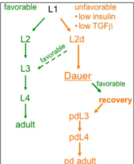
Figure 1. Continuous development and dauer formation life cycle in
Caenorhabditis
elegans.
In favorable conditions (green)
C. elegans
develops through four larval stages, L1-L4,
before molting into a reproductive adult. In unfavorable conditions (orange), TGFß and IIS
signaling are low and L1s molt into an alternative to the L2 called the L2d (Cassada and Russell,
1975). If conditions improve, continuous development can be resumed, but otherwise L2ds
undergo an extended molt to form dauer larvae. Dauers have an extended lifespan and increased
stress resistance and, if conditions improve, recover to continuously develop through post-dauer
L3 and L4 stages to become reproductive adults.
Figure 2.Somatic gonad and germline development. (A) At the start of the L1 stage, the gonad primordium consists of four cells: the somatic gonad precursors Z1 and Z4, and the germline precursors Z2 and Z3 (Kimble and Hirsh, 1979; Kimble and White, 1981). In the first phase, Z1 and Z4 generate twelve cells. Three cells are terminally differentiated: two distal tip cells (DTCs), which promote mitosis in the germline and guide later gonad outgrowth, and the anchor cell (AC), which later organizes uterine and vulval patterning (Kimble and Hirsh, 1979; Kimble and White, 1981; Newman et al., 1996). The other cells are precursors to later structures, including 3 ventral uterine precursor cells (VUs), which generate descendants that will form the ventral uterus in the second phase of somatic gonadal
development. The second stage of somatic gonadal development begins in mid-L3, when the precursor cells divide. At that time, the VUs produce 12 granddaughters; in 6 of these granddaughters, LIN-12/Notch is activated by a LAG-2 signal from the AC via cell-cell interactions, causing them to adopt a fate called “pi” (Newman et al., 1995). The pi cells divide once and eight of their daughters fuse with the AC to form the Uterine Seam (utse) syncytium. (B) In dauer, somatic gonad development is interrupted after the first phase (Hong et al., 1998; Narbonne and Roy, 2006).
germline
somatic gonad
L1
L2
L3
L4
Phase
1
Phase
2
ACGSC
GSC
Z1
Z2
Z3
Z4
DTC
GSC
GSC
Phase 2
π
π
π
π
π
π
GSC
AC utseDTC
Figure 2A
Figure 2B
L1
L2
Phase
1
ACGSC
GSC
Z1
Z2
Z3
Z4
DTC
dauer
L1
L2d
ACGSC
GSC
DTC
Figure 3. VPC fate patterning in continuous development. From (Karp and Greenwald,
2013). “(A) VPC specification in wild-type hermaphrodites. During L3, an EGF-like signal (red)
from the anchor cell (AC) activates Ras signaling in P6.p, causing it to adopt 1° fate and produce
ligands, including LAG-2 and APX-1, which activate LIN-12/Notch in P5.p and P7.p.” Sourced
in Karp and Greenwald 2013 from (Sternberg, 2005).
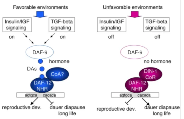
Figure 4
.
Integration of signaling pathways regulating the decision to form dauers.
From
(Karp and Greenwald, 2013). The insulin/insulin-like signaling (IIS) and TGFß signaling
pathways converge on signaling mediated by the nuclear hormone receptor (NHR) DAF-12
(Gerisch, B., Antebi, 2004; Mak and Ruvkun, 2004). In favorable environments, IIS and TGFß
signaling are both active and inhibit activity of downstream targets 16/FoxO and
DAF-3/Smad, DAF-5/Sno/Ski, allowing production of dafachronic acids (DAs), which require activity
of the steroid hydroxylase DAF-9. These DAs act as ligands for DAF-12/NHR which then
promotes continuous development. In unfavorable environments, both IIS and TGFß signaling
are inactive, and DAF-12/NHR unliganded activity promotes dauer formation.
Figure 5. Insulin/insulin-like signaling pathway.
Adapted from (Murphy and Hu, 2013a).
Green, dauer-promoting activity (loss of function: dauer defective). Red, dauer-inhibiting
activity (loss of function: dauer constitutive). Insulin/insulin-like peptides (ILPs) can have
agonistic or antagonistic activity. When agonistic ILPs activate the insulin receptor DAF-2,
signaling through IIS-promoting components (colored red) promote phosphorylate the canonical
IIS target 16/FoxO such that it is sequestered in the cytoplasm. When IIS is inactive,
16/FoxO is localized in the nucleus and can activate its targets. 18/PTEN promotes
DAF-16/FoxO nuclear localization by antagonizing AGE-1/PI3K activity. Not shown are other IIS
targets which include HSF-1 and SKN-1.
Figure 6.
The
daf-7/
TGF-β signaling pathway.
From (Gumienny and Savage-Dunn, 2013).
daf-7 expressed in the ASIs promotes signaling in target cells that ultimately inhibits the
functions of DAF-3/Smad and DAF-5/Sno/Ski. DAF-7/TGFß signaling is active in favorable
conditions and promotes continuous development (opposing dauer development). DAF-7/TGFß
activates receptor composed of two DAF-1 type I receptor and DAF-4 type II receptor subunits
which promote signal transduction through the Smads DAF-8 and DAF-14.
From Gumienny and
Savage-Dunn 2013
Figure 7. DAF-9-mediated DA signaling through DAF-12/NHR. From (Antebi, 2006) with
slight adaptations. In favorable environments, the IIS and TGFß signaling pathways promote
dafachronic acid (DA, blue circles) signaling and therefore ligand-bound DAF-12 activity, while
in unfavorable conditions, ligand is absent and unliganded DAF-12 interacts with corepressors
like DIN-1/SHARP to promote dauer formation. Production of DAF-12-binding DA ligands
requires the steroid hydroxylase DAF-9.
DAs
Chapter 2
daf-18/PTEN acts in the somatic gonad to coordinate somatic gonad and germline development in C. elegans dauer larvae
Claudia C. Tenen1 and Iva Greenwald2,3
1Integrated Program in Cellular, Molecular and Biophysical Studies 2Dept. of Biological Sciences
Columbia University New York, NY 10027 USA