development of type 2 diabetes in mice
Fan-Yan Wei, … , Tsutomu Suzuki, Kazuhito Tomizawa
J Clin Invest. 2011;121(9):3598-3608. https://doi.org/10.1172/JCI58056.
The worldwide prevalence of type 2 diabetes (T2D), which is caused by a combination of environmental and genetic factors, is increasing. With regard to genetic factors, variations in the gene encoding Cdk5 regulatory associated protein 1–like 1 (Cdkal1) have been associated with an impaired insulin response and increased risk of T2D across different ethnic populations, but the molecular function of this protein has not been characterized. Here, we show that Cdkal1 is a mammalian methylthiotransferase that biosynthesizes 2-methylthio-N6-threonylcarbamoyladenosine (ms2t6A) in
tRNALys(UUU) and that it is required for the accurate translation of AAA and AAG codons. Mice with pancreatic β cell– specific KO of Cdkal1 (referred to herein as β cell KO mice) showed pancreatic islet hypertrophy, a decrease in insulin secretion, and impaired blood glucose control. In Cdkal1-deficient β cells, misreading of Lys codon in proinsulin occurred, resulting in a reduction of glucose-stimulated proinsulin synthesis. Moreover, expression of ER stress–related genes was upregulated in these cells, and abnormally structured ER was observed. Further, the β cell KO mice were hypersensitive to high fat diet–induced ER stress. These findings suggest that glucose-stimulated translation of proinsulin may require fully modified tRNALys(UUU), which could potentially explain the molecular pathogenesis of T2D in patients carrying cdkal1 risk alleles.
Research Article Metabolism
Find the latest version:
Research article
Deficit of tRNA
Lys
modification
by Cdkal1 causes the development
of type 2 diabetes in mice
Fan-Yan Wei,1 Takeo Suzuki,2 Sayaka Watanabe,1 Satoshi Kimura,2 Taku Kaitsuka,1
Atsushi Fujimura,3 Hideki Matsui,3 Mohamed Atta,4 Hiroyuki Michiue,3
Marc Fontecave,4 Kazuya Yamagata,5 Tsutomu Suzuki,2 and Kazuhito Tomizawa1
1Department of Molecular Physiology, Faculty of Life Sciences, Kumamoto University, Kumamoto, Japan. 2Department of Chemistry and Biotechnology,
School of Engineering, The University of Tokyo, Tokyo, Japan. 3Department of Physiology, Okayama University Graduate School of Medicine, Dentistry and
Pharmaceutical Sciences, Okayama, Japan. 4Institut de Recherches en Technologie et Sciences pour le Vivant IRTSV-LCBM, UMR 5249, CEA/CNRS/UJF,
CEA-Grenoble, Grenoble, France. 5Department of Medical Biochemistry, Faculty of Life Sciences, Kumamoto University, Kumamoto, Japan.
The worldwide prevalence of type 2 diabetes (T2D), which is caused by a combination of environmental and
genetic factors, is increasing. With regard to genetic factors, variations in the gene encoding Cdk5 regulatory
associated protein 1–like 1 (Cdkal1) have been associated with an impaired insulin response and increased risk
of T2D across different ethnic populations, but the molecular function of this protein has not been
character-ized. Here, we show that Cdkal1 is a mammalian methylthiotransferase that biosynthesizes 2-methylthio-
N
6-threonylcarbamoyladenosine (ms
2t
6A) in tRNA
Lys(UUU) and that it is required for the accurate translation of
AAA and AAG codons. Mice with pancreatic
β
cell–specific KO of Cdkal1 (referred to herein as
β
cell KO mice)
showed pancreatic islet hypertrophy, a decrease in insulin secretion, and impaired blood glucose control. In
Cdkal1-deficient
β
cells, misreading of Lys codon in proinsulin occurred, resulting in a reduction of
glucose-stimulated proinsulin synthesis. Moreover, expression of ER stress–related genes was upregulated in these
cells, and abnormally structured ER was observed. Further, the
β
cell KO mice were hypersensitive to high fat
diet–induced ER stress. These findings suggest that glucose-stimulated translation of proinsulin may require
fully modified tRNA
Lys(UUU), which could potentially explain the molecular pathogenesis of T2D in patients
carrying
cdkal1
risk alleles.
Introduction
Type 2 diabetes (T2D) is caused by a combination of genetic and environmental factors. Recent advances in whole-genome associa-tion studies have identified a number of genetic variaassocia-tions associ-ated with T2D (1–4). The Cdk5 regulatory associassoci-ated protein 1– like 1 (cdkal1) gene is one of the most reproducible risk genes in T2D across different ethnic populations (5). Variations in cdkal1
have been associated with impaired insulin secretion and increased risk of T2D (6–8). Although there is increasing evidence associ-ating single nucleotide polymorphisms in cdkal1 with T2D, the molecular function of Cdkal1 is unknown.
We recently identified Cdkal1 as a member of the methylthio-transferase (MTTase) family, a subfamily of the radical S-adenosyl-methionine (SAM) superfamily (9). The MTTase family utilizes SAM and [4Fe-4S] clusters to catalyze the methylthiolation of various substrates. For instance, MiaB, a bacterial MTTase protein, catalyzes the methylthiolation of N6-isopentenyladenosine (i6A) to generate 2-methylthio-N6-isopentenyladenosine (ms2i6A) at position 37 (A37), 3′ adjacent to the anticodon in some tRNAs (10, 11). This hyper-modification of A37 is essential for the efficient and accurate trans-lation of cognate codons by the ribosome (12, 13). We have shown that Cdkal1 (and its bacterial homolog YqeV) catalyze the methyl-thiolation of N6-threonyl carbamoyl adenosine (t6A) to synthesize
2-methylthio-N6-threonyl carbamoyl adenosine (ms2t6A) for tRNA in bacteria (9). However, the enzymatic characteristics of Cdkal1 in mammalian cells and its relevance to T2D are completely unknown. By using pancreatic β cell–specific Cdkal1 KO mice (referred to here-in as β cell KO mice), we show that Cdkal1 has critical roles in the quality control of protein translation and is relevant to T2D.
Results
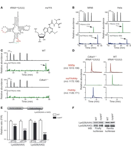
Cdkal1 catalyzes ms2t6A modification of mammalian tRNALys(UUU).
To determine the biochemical function of Cdkal1 in mamma-lian cells and its relevance to T2D, we used mass spectrometric analysis to examine modified bases in total RNA from MIN6 cells, a pancreatic β cell–derived insulinoma cell line, and HeLa cells, a human-derived cell line (Figure 1B). As expected, the proton adduct ms2t6A (m/z 459) could be clearly detected along with t6A (m/z 413) in both cell types (Figure 1B). In addition, we also detected ms2t6A in total RNA from various mouse tis-sues (Supplemental Figure 1; supplemental material available online with this article; doi:10.1172/JCI58056DS1). To investi-gate whether Cdkal1 was involved in the ms2t6A modification, we examined total RNA isolated from the pancreas of WT and
Cdkal1–/– mice. The ms2t6A modification was detected only in the WT mice but not in the Cdkal1–/– mice (Figure 1C). These
results suggest that Cdkal1 only catalyzes the ms2t6A modifica-tion in mammalian cells. Because ms2t6A is present at position 37 of tRNALys in Bacillus subtilis (14, 15), we isolated 2 species of tRNALys (tRNALys[UUU] [Figure 1A] and tRNALys[CUU] [Supple-mental Figure 2A]) from mouse livers and performed an RNA
Authorship note: Takeo Suzuki and Sayaka Watanabe contributed equally to this work.
Conflict of interest: The authors have declared that no conflict of interest exists.
fragment analysis. Ms2t6A was specifically found at position 37 of tRNALys(UUU) in WT liver (Figure 1D), whereas tRNALys(CUU) bore t6A at position 37 (Supplemental Figure 2B). As no frag-ment containing t6A was detected in tRNALys(UUU) of WT liver, the methylthio modification appeared to be introduced univer-sally (Figure 1D). When the nucleosides from the flow-through fraction after the isolation of tRNALys(UUU) were analyzed, no ms2t6A could be detected (Supplemental Figure 3), suggesting that ms2t6A is a modification specific to tRNALys(UUU). In con-trast, the ms2t6A-containing fragment (m/z 1172.16) was com-pletely replaced with a t6A-containing fragment (m/z 1126.17) in tRNALys(UUU) isolated from livers of Cdkal1–/– mice (Figure 1D).
These results demonstrate that mouse Cdkal1 is a methylthiolase that converts t6A to ms2t6A in tRNALys(UUU).
The ms2t6A modification is required for decoding fidelity. The
2-methyl-thio modification ms2i6A is important for preventing the misread-ing and frame-shiftmisread-ing of cognate codons durmisread-ing protein translation in bacteria (12–14). These observations prompted us to speculate that the 2-methylthio modification ms2t6A in tRNALys(UUU) is also required for translational accuracy. To examine whether the ms2t6A modification prevents either the frame-shifting or misreading of tRNALys(UUU)’s cognate codons (AAA and AAG), we utilized a dual luciferase-based reporter assay in WT B. subtilis and yqeV-deficient
B. subtilis (ΔyqeV), which lacks the ms2t6A modification (Supple-mental Figure 4A, Figure 1E, and ref. 16). Because Lys529 in firefly luciferase is essential for enzymatic activity, the misreading or frameshifting of this codon would result in a loss of firefly lucifer-ase activity (17, 18). Two constructs in which Lys529 is encoded by AAA or AAG codons were introduced into WT and ΔyqeV strains, and relative firefly luciferase activity was measured (Figure 1E). In the ΔyqeV strain under noninducible conditions (–IPTG), a spe-cific reduction in firefly luciferase activity was observed with the AAA construct, but not with the AAG construct (Figure 1E). Under inducible conditions (+IPTG), a marked reduction in firefly lucifer-ase activity was observed with both constructs in the ΔyqeV strain, and an even greater reduction in activity was observed with the AAG construct (Figure 1E), although the IPTG-induced protein level of the renilla-firefly fusion protein was the same in the WT and ΔyqeV
strains (Figure 1F). We next determined whether the 2-methylthio modification ms2t6A is involved in the reading frame maintenance of the relevant codons. We employed constructs that fused Renilla
and firefly luciferases separated by a short sequence containing a +1 frameshift site (Supplemental Figure 4B). We observed no sig-nificant frameshift activity of either construct in the ΔyqeV strain as compared with the WT strain. These results suggest that the 2-methylthio modification ms2t6A in tRNALys(UUU) is important for preventing the misreading of its cognate codons, especially when the rate of translation is relatively high.
Cdkal1 is an ER-localizing protein that is functionally dissociated with Cdk5/p35. Cdkal1 was ubiquitously expressed in mouse tissues through all the developmental stages and was especially abun-dant in the heart, kidney, and pancreas (Supplemental Figure 5). To investigate the subcellular distribution of Cdkal1, HEK293 and MIN6 cells were transfected with EGFP-Cdkal1 and ER-tracker. EGFP-Cdkal1 colocalized with ER-tracker (Figure 2A). Moreover, Cdkal1 was also colocalized with endogenous Bip, an ER protein (Figure 2B). Cdkal1 has 3 unique domains, a radical SAM domain, a TRAM domain, and a hydrophobic domain (Figure 2C). Both the radical SAM domain (a catalytic domain) and the TRAM domain (a potential tRNA-binding domain) are conserved among mammals
and bacteria (9). In contrast, the hydrophobic domain at the C terminus exists only in mammalian Cdkal1 (9). This hydrophobic domain was determined to carry the ER-localization signal because deletion of this domain disrupted ER localization (Figure 2D). Fur-thermore, endogenous Cdkal1 was detected in the rough ER frac-tion purified from mouse liver (Supplemental Figure 6). The ER localization was finally confirmed by immunoelectron microscopic examination in EGFP-Cdkal1–transfected MIN6 cells (Figure 2E).
We previously reported that Cdk5 regulates insulin secretion in pancreatic β cells (19). Cdkal1 may function through interac-tion with a Cdk5 regulatory subunit, p35, as Cdk5rap1, an amino acid homolog of Cdkal1, interacts with p35 and inhibits Cdk5 activity (20, 21). However, Cdkal1 neither interacted with p35 in HEK293 cells overexpressing p35 nor inhibited Cdk5 activity in vitro (Supplemental Figure 7), suggesting that the molecular function of Cdkal1 in β cells is independent of the pathway in which Cdk5/p35 participates.
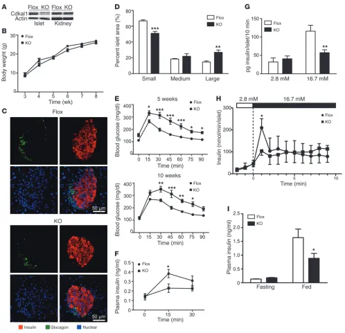
Cdkal1 deficiency in β cells causes glucose intolerance. To investigate the physiological functions of Cdkal1 in pancreatic β cells, β cell– specific Cdkal1-deficient mice (β cell KO) were generated by cross-ing transgenic mice in which exon 5 of cdkal1 was floxed by the LoxP sequence with transgenic mice in which Cre recombinase was regulated under the control of the rat insulin promoter (Supple-mental Figure 8A). Exon 5 of cdkal1 was deleted in the pancreatic islets of β cell KO mice, but not the other tissues of the β cell KO mice, (Supplemental Figure 8B). Cdkal1 protein expression was faint in the islets of β cell KO mice compared with that in the islets of littermate control mice (Flox) (Figure 3A). In contrast, the same level of Cdkal1 was observed in kidney of Flox mice and β cell KO mice (Figure 3A). The β cell KO mice showed normal development (Figure 3B). Immunohistochemical analyses revealed no obvi-ous morphological abnormalities in α or β cells in the pancreatic islets of β cell KO mice relative to Flox mice (Figure 3C). How-ever, we noticed that KO islets were larger than Flox islets, and we performed a detailed analysis to investigate islet area. We divided the islets into 3 groups: small islets (0–5,000 μm2), medium islets (5,001–10,000 μm2), and large islets (>10,000 μm2), and we calcu-lated the relative abundance of each group. In β cell KO mice, the number of small islets was significantly lower than in Flox mice, and the number of large islets was significantly greater (Figure 3D). Because there was no difference in total islet number between β cell KO and Flox mice (data not shown), pancreatic islets in β cell KO mice may be able to lapse into a hypertrophic condition.
Because insulin secretion is impaired in patients with variants of the cdkal1 gene (6–8), the mice were given an intraperitoneal glu-cose tolerance test (IPGTT). The β cell KO mice showed glucose intolerance compared with the Flox mice at 5 and 10 weeks after birth (Figure 3E). Moreover, plasma insulin levels 15 minutes after the glucose challenge were significantly lower in the β cell KO mice (Figure 3F). We also investigated insulin secretion in islets isolated from Flox and β cell KO mice. After 16.7 mM glucose stimulation, the insulin level was significantly lower in β cell KO mice than in Flox mice (Figure 3G). Because patients with variants of the cdkal1
research article
investigated insulin secretion in Flox and β cell KO mice under nor-mal feeding conditions. The mice were fasted overnight and then re-fed for 1.5 hours. Plasma insulin levels in the fasting condition and postprandial condition were determined (Figure 3I). There was
[image:4.585.65.514.63.569.2]a significant decrease in postprandial insulin secretion in β cell KO mice when compared with Flox mice. These results suggest that Cdkal1 deficiency in pancreatic β cells impairs glucose-stimulated insulin secretion and thus induces glucose intolerance.
Figure 1
Methylthiolation of tRNALys(UUU) by Cdkal1 controls the decoding accuracy of the lysine codon. (A) The molecular structure of tRNALys(UUU) and
ms2t6A. (B) Results of a mass spectrometric analysis of the ms2t6A modification of tRNA in MIN6 and HeLa cells. The upper panels show the UV trace,
and the middle and lower panels show the mass chromatograms for detecting t6A (m/z 413, arrow) and ms2t6A (m/z 459, arrow), respectively. (C)
Results of a mass spectrometric analysis of the ms2t6A modification of tRNA isolated from the pancreas of Cdkal1–/– and WT mice. The arrow indicates
ms2t6A (m/z 459). (D) Modification of tRNALys(UUU) isolated from the liver of Cdkal1–/– and WT mice. The upper panels show mass chromatograms
of GGDp fragments in tRNALys(UUU). The middle and lower panels show mass chromatograms of ms2t6AAΨp fragments and t6AAΨp fragments,
respectively. (E) WT and ΔyqeV cells were transformed with a reporter plasmid in which both Renilla renilla and firefly luciferases are cloned with the lac promoter (upper panel). Relative activity was determined by normalizing firefly luciferase intensity to renilla luciferase intensity (F/R, lower panel). Data are presented as the mean ± SEM, and asterisks indicate statistical significance determined by Student’s t test. ***P < 0.001; n = 4. (F) The expression
Cdkal1 deficiency induces aberrant proinsulin synthesis. Given the molec-ular function of Cdkal1 in B. subtilis (Figure 1), we speculated that Cdkal1 deficiency in pancreatic β cells decreases the decoding fideli-ty in lysine codon due to insufficient modification in tRNALys(UUU). The Lys residue is particularly important for processing proinsulin to generate mature insulin and C-peptide because 1 of the 2 Lys resi-dues in human proinsulin is located at the cleavage site between the C-peptide and A chain of insulin. Thus, misreading of Lys codon in proinsulin by insufficiently modified tRNALys(UUU) might result in aberrant processing of (pro)insulin and subsequent glucose intoler-ance. To investigate misreading of Lys codon in Cdkal1-deficient β cells, pancreatic islets isolated from both β cell KO and Flox mice were labeled with both 14C-lysine and 3H-leucine. If misreading of Lys codon occurs in Cdkal1-deficient β cells, we would observe a change in the ratio of incorporation of 14C-lysine to 3H-leucine in
(pro)insulin when compared with the incorporation ratio in Flox β cells. As expected, there was a significant decrease of relative incor-poration of 14C-lysine in (pro)insulin in Cdkal1-deficient β cells when compared with Flox β cells (KO: 0.82 ± 0.007 versus Flox: 1.0 ± 0.01, P = 0.0004; Figure 4A). Since misreading of Lys codon in proinsulin might cause aberrant processing, we investigated the C-peptide content, which indicates proper processing of proinsulin, in the pancreas of β cell KO and Flox mice. The C-peptide content in β cell KO pancreas was significantly lower than C-peptide content in Flox pancreas (β cell KO: 12.4 ± 1.41 ng/mg protein versus Flox: 22.4 ± 3.29 ng/ mg protein, P = 0.0429; Figure 4B). Accordingly, plas-ma C-peptide levels were significantly lower in β cell KO mice than in Flox mice (Figure 4C). Pancreatic sections were also immunostained with anti–C-peptide antibodies. Consistent with the reduction in C-peptide levels in β cell KO mice, the intensity of C-peptide
stain-Figure 2
Cdkal1 localizes on ER through its hydrophobic domain. (A) Colocalization of overexpressed Cdkal1-EGFP (green) and ER-tracker (red) on ER
in HEK293 cells and MIN6 cell. Scale bars: 10 μm. (B) Colocalization of overexpressed Cdkal1-EGFP (green) with endogenous Bip in HEK293
cells. Scale bar: 10 μm. (C) The domain structure of Cdkal1 protein. (D) EGFP-tagged full-length Cdkal1 or Cdkal1 with truncation of C terminus
hydrophobic domain (Cdkal1ΔC) was transfected in HeLa cells together with ER-tracker. Localization of full-length Cdkal1 or Cdkal1ΔC was visualized using confocal microscope. Scale bars: 10 μm. (E) MIN6 cells were transfected with Cdkal1-EGFP, and the localization of Cdkal1 was
[image:5.585.51.536.78.479.2]research article
ing was apparently reduced in KO islets compared with Flox islets (Supplemental Figure 9A). Previous study using pancreatic β cell– specific transgenic mice overexpressing mutant elF2α has shown that aberrant protein translation could impair the correct targeting
[image:6.585.45.543.79.560.2]of proinsulin (22). Therefore, subcellular localization of proinsulin was investigated in β cell KO mice. High magnification revealed that proinsulin was mainly confined to the perinuclear area and colocalized with C-peptide in the β cells of Flox mice. In contrast,
Figure 3
Conditional deletion of the Cdkal1 gene causes glucose intolerance. (A) Conditional deletion of Cdkal1 in pancreatic islets in β cell KO (KO) mouse. (B) Comparison of the body weights of β cell KO and Flox mice. (C) Pancreatic sections obtained from β cell KO and Flox mice at 5 weeks of age were immunostained with anti-insulin (red) and anti-glucagon (green) antibodies. Nuclei were counterstained with DAPI. (D)
Compari-son of relative islet area in pancreas of β cell KO and Flox mice. Area of 529 islets from 3 Flox mice and 572 islets from 3 β cell KO mice were examined and classified into small, medium, and large islet area. The relative distribution of each islet area was compared between β cell KO and Flox. (E) Blood glucose during glucose tolerance test at 5 weeks (upper) and 10 weeks (lower). n = 4–7. (F) Plasma insulin levels during a
glucose tolerance test at 15 weeks. n = 10–11. (G) Glucose-stimulated insulin secretion in islets (n = 8) isolated from β cell KO or Flox mice was determined. (H) Glucose-stimulated insulin secretion in perfused islets of Flox and β cell KO mice. n = 4–5. (I) Plasma insulin levels in Flox or
there were large aggregates of proinsulin-positive granules, which were not colocalized with C-peptide–positive granules in islets of β cell KO mice (Supplemental Figure 9B). These results suggest that Cdkal1 deficiency may cause abnormal proinsulin translation, which in turn leads to the impairment of both the processing and targeting of proinsulin.
In addition, we investigated the total protein synthesis level and proinsulin synthesis level in islets of β cell KO and Flox mice. Total protein synthesis in KO islets was not changed under either low- or high-glucose conditions (Figure 4D). There was no dif-ference in proinsulin levels between KO islets and control islets under low-glucose conditions (Figure 4E). However, a significant decrease in proinsulin synthesis was observed in KO islets stimu-lated with high glucose compared with Flox islets (Figure 4E). A decrease in insulin synthesis was also observed in MIN6 cells transfected with siRNA targeting Cdkal1 (Supplemental Figure 10). To investigate whether Cdkal1 deficiency had any effect on the synthesis of other crucial β cell proteins, we examined the protein levels of Kir6.2, SUR1, and Pdx1. There were no obvious differences in protein levels between KO islets and Flox islets (Figure 4F). These results suggest that tRNA modification by Cdkal1 is crucial for translation fidelity and efficiency of proin-sulin in pancreatic β cells.
Cdkal1 deficiency induces ER stress in β cells. Accumulation of unfolded or misfolded proteins in the ER lumen triggers stress response, which has been proposed to cause the dysfunction of pancreatic β cells (22–25). Notably, temporary or chronic imbal-ance in the protein synthesis environment can induce ER stress
in β cells and subsequent glucose intolerance in vivo (22, 26, 27). To investigate whether aberrant proinsulin synthesis caused by Cdkal1 deficiency triggers stress responses in β cells, we exam-ined the expression levels of a variety of genes essential for β cell function. There were no differences in the levels of insulin 1 or 2 mRNAs between KO and Flox islets (Figure 5A). Among pancre-atic β cell marker genes, the mRNA levels of glucose transporter 2 (Glut2) were significantly reduced in KO islets (Figure 5B). More-over, Glut2 was distributed diffusely in the cytoplasm of β cells in Cdkal1-deficient islets (Figure 5C). The decreased expression and abnormal localization of Glut2 correlate with ER stress (22). Therefore, the relative expression of ER stress–related genes was then examined in the pancreatic islets of β cell KO and Flox mice. Among all the stress-related genes, only the expression of spliced
Xbp1 was found to be elevated (Figure 5D). In addition, phospho-EIF2 levels were higher in KO islets than in Flox islets (Supple-mental Figure 11). Furthermore, electron microscopy revealed distended ER, which indicates ER stress (27, 28), in pancreatic β cells from β cell KO mice but not from Flox mice (Figure 5E). These results suggest that cdkal1 gene deficiency may induce an ER stress response and glucose intolerance.
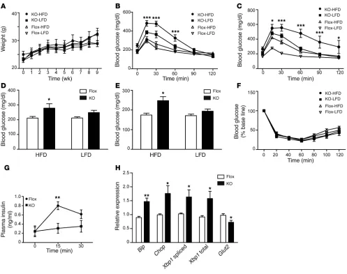
[image:7.585.50.540.79.325.2]Cdkal1 deficiency enhances susceptibility to high-fat diet stress. Environ-mental stress such as a high-fat diet (HFD) has a great impact on glucose metabolism. Interestingly, recent study has found the asso-ciation of polymorphism in the cdkal1 gene with the prevalence of metabolic syndrome in Japanese men (29). We therefore speculated that a HFD might induce profound glucose intolerance in β cell KO mice. To investigate the effect of an HFD, β cell KO and Flox mice
Figure 4
Aberrant insulin synthesis in the pancreatic β cells of β cell KO mice. (A) Relative incorporation of 14C-lysine to 3H-leucine in immunoprecipitated
(pro)insulin in islets of β cell KO or Flox mice in KRB buffer containing 16.7 mM glucose for 1 hour. (B) Pancreatic C-peptide content of β cell KO or Flox mice was measured by ELISA, and value was normalized to total protein concentration. n = 5–8; *P < 0.05 by Student’s t test. (C) Plasma
C-peptide concentrations in Flox and β cell KO mice fasted for 7 hours. n = 10. ***P < 0.001 by Student’s t test. (D) Relative total protein synthesis
under basal condition (2.8 mM) or stimulated condition (16.7 mM) was determined by normalizing 35S incorporation to the total protein concentra-tion. n = 4; *P < 0.05 by Student’s t test. (E) Proinsulin synthesis in KO or Flox islets under basal condition (2.8 mM) or stimulated condition (16.7 mM)
research article
were fed either an HFD or a low-fat diet (LFD) for up to 8 weeks. There was no difference in weight gain between β cell KO and Flox mice during the experimental period (Figure 6A). Islet hypertro-phy was observed in both β cell KO and Flox mice fed an HFD for 8 weeks as a compensatory effect of the diet (Supplemental Figure 12). However, significant glucose intolerance developed in β cell KO mice fed an HFD for 3 weeks, whereas Flox mice fed an HFD showed normal glucose tolerance compared with Flox mice fed an LFD (Figure 6B). Blood glucose levels at 15, 30, and 60 minutes after the intraperitoneal injection were higher in β cell KO mice than in Flox mice. After 8 weeks on an HFD, glucose intolerance was more severe in the β cell KO mice (Figure 6C). Blood glucose concentrations in β cell KO mice were continuously higher than control levels and increased 2 hours after a glucose injection (Fig-ure 6C). In addition, both nonfasting blood glucose levels (Fig(Fig-ure 6D) and 7 hour–fasting blood glucose levels (Figure 6E) were sig-nificantly higher in β cell KO mice than Flox mice after 3 weeks on an HFD, whereas nonfasting and 7 hour–fasting blood glucose
[image:8.585.58.530.77.450.2]levels in LFD-fed β cell KO mice were compatible with those in LFD-fed Flox mice (Figure 6, D and E). To investigate whether insulin sensitivity was affected in β cell KO mice, an insulin toler-ance test was performed in β cell KO and Flox mice fed an HFD or LFD for 7 weeks. There were no differences in the action of insulin between β cell KO and Flox mice fed either diet (Figure 6F). We also investigated whether an HFD had any effect on liver function as well as counter-insulin responses such as glucagon production. To examine gluconeogenesis in the liver, Flox and β cell KO mice fed an HFD for 10 weeks were injected with pyruvate and blood glucose levels were measured. There was no significant difference in gluconeogenesis between Flox and β cell KO mice (Supplemen-tal Figure 13A). Furthermore, we examined plasma glucagon levels in mice fed an HFD for 10 weeks. There was no difference in fast-ing glucagon levels between Flox and β cell KO mice (Supplemen-tal Figure 13B). From these observations, we speculated that the severe glucose intolerance was mainly caused by HFD-induced ER stress and the consequent decrease in insulin secretion in Cdkal1
Figure 5
ER stress response in the pancreatic β cell KO mice. (A) Quantitative analysis of the mRNA expression of insulin, glucagon, and Cdkal1 in isolated islets of β cell KO and Flox mice. *P < 0.05; n = 4. (B) Comparison of the expression of β cell–related genes between β cell KO and Flox mice. *P < 0.05; n = 4. (C) Subcellular distribution of GLUT2 in islets of β cell KO and Flox mice. Scale bars: 50 μm. (D) Quantitative analysis of
ER stress–related genes in β cell KO and Flox mice. *P < 0.05; n = 4. (E) Transmission electron microscopic examination of the ultrastructure
KO β cells. Serum insulin levels after the injection of glucose were measured in β cell KO and Flox mice fed an HFD. Blood insulin levels after the challenge were significantly lower in β cell KO mice than in Flox mice (Figure 6G). Impaired glucose-stimulated insulin secretion was also observed in isolated Cdkal1-deficient pancreatic islets (Supplemental Figure 14). Finally, pancreatic islets were iso-lated from β cell KO mice and Flox mice fed the HFD for 8 weeks, and the expression levels of ER stress–related genes were examined (Figure 6H). We observed a significant increase in the expression of major ER stress–related genes, including the Bip, CHOP, and Xbp1
genes. Our results indicate that Cdkal1 deficiency induces a mas-sive ER stress response, which in turn decreases insulin secretion, causing severe glucose intolerance.
Discussion
[image:9.585.45.542.80.473.2]Chemical modifications of nucleotides surrounding anticodons in tRNAs are believed to be essential for accuracy and efficiency in protein translation (14). The structural basis of the ms2i6A37 mod-ification at A37 of tRNA was recently identified in bacteria (30). The methylthiolation of i6A37 is capable of stabilizing the codon-anticodon interaction through cross-strand stacking with the base of the first nucleotide of the mRNA codon. This stabilization of the codon-anticodon interaction prevents frame shifting and mis-reading during translation. In the present study, we showed that the ms2t6A modification of tRNALys(UUU) by Cdkal1 is required for the accurate translation of AAA and AAG codons. The human insulin gene contains 2 Lys(AAG) codons. One of the Lys residues
Figure 6
β cell KO mice exhibit increased ER stress and glucose intolerance after consuming an HFD. (A) Changes in body weight of β cell KO and Flox mice on an HFD and a LFD starting from 20 weeks old. (B and C) Results of the glucose tolerance test after 3 weeks (B) and 8 weeks (C) of
consuming an HFD or a LFD. Mice were fasted for 7 hours from 8 am and injected with glucose (1 g/kg body weight). *P < 0.05; ***P < 0.001, KO-HFD versus Flox-HFD mice. n = 4–6. (D and E) Nonfasting blood glucose (D) and 7-hour fasting blood glucose (E) levels after 3 weeks on
an HFD or an LFD. *P < 0.05; n = 6. (F) The insulin tolerance test was performed in mice fed an HFD or an LFD for 7 weeks. (G) β cell KO mice and Flox mice were fed an HFD for 8 weeks. Plasma insulin level during IPGTT (1 g/kg body weight) was examined in β cell KO mice and Flox mice fasted for 14 hours. **P < 0.01; n = 6. (H) Relative expression of ER stress–related genes in β cell KO mice and Flox mice fed an HFD for 8 weeks. **P < 0.01; n = 4–5. Significant differences between groups were examined by repeated measure of ANOVA (A–C, F, and G), 2-way
research article
is located at the cleavage site between the C-peptide and A chain of insulin. Misreading of this Lys codon during insulin synthesis by ms2t6A modification–deficient tRNALys(UUU) may cause the mis-folding or miscleavage of proinsulin, which has an impact on glu-cose homeostasis. Indeed, we observed a decreased incorporation of lysine residue in Cdkal1-deficient β cells, as well as decreased C-peptide levels in pancreas of Cdkal1 KO. Interestingly, SNPs in the Cdkal1 gene have been shown to associate with impaired con-version of proinsulin to insulin (31–33), supporting our finding that Cdkal1 deficiency may cause aberrant proinsulin generation.
The main role of pancreatic β cells is the adequate synthesis and release of insulin in response to glucose. To accomplish this task, the cells induce insulin biosynthesis in response to glucose. Proinsu-lin mRNA represents 20% of the total mRNA expression in glucose-stimulated β cells, whereas (pro)insulin biosynthesis approaches 50% of their total protein production (34, 35). It is inevitable that some insulin will be misfolded in such a mass production (36, 37). However, if augmented absolute levels of misfolded proinsulin are above the threshold, the misfolded proinsulin may lead to the inhibition of insulin production, ER stress, and β cell dysfunction. The onset of diabetes caused by misfolded proinsulin has been well studied in mutant INS gene–induced diabetes of youth (MIDY). In
Akita mice, in which a heterozygous proinsulin-C(A7)Y mutation in the mouse Ins2 gene is identical to the heterozygous mutation causing human MIDY, the mutant proinsulin in Akita mice blocks insulin production and activates ER stress in β cells (38, 39). On the other hand, dysregulation of protein synthesis can also lead to the production of misfolded proinsulin and ER stress. For example, a massive increase of protein synthesis by Perk deficiency causes massive proinsulin production, which leads to abnormal folding of proinsulin and ER stress (37). Taken together, these findings sug-gest that the absolute amount of misfolded proinsulin is a criti-cal determinant of onset of ER stress followed by dysfunction of β cells. In β cell KO mice, the Cdkal1 deficiency may cause a certain amount of proinsulin to be mistranslated, which may be misfolded and accumulate in the ER, leading to further inhibition of insulin production and subsequent activation of ER stress.
A recent study showed impaired mitochondrial ATP generation, first-phase insulin exocytosis, and responsiveness of ATP-sensitive K+ channel to glucose in general Cdkal1–/– mice (40). In β cell KO
mice, we also observed impaired first-phase insulin secretion as well as impaired ATP generation after glucose stimulation (Figure 3H and Supplemental Figure 15). Considering the molecular function of Cdkal1, it is not assumed that Cdkal1 directly regulates these functions. These results suggest that aberrant protein translation may occur in the proteins involved in the regulation of mitochon-drial ATP generation and insulin exocytosis in addition to insu-lin in Cdkal1-deficient mice. Although we did not detect obvious changes in the levels of Kir6.2 and SUR1, other proteins involved in mitochondrial functions may be abnormally translated and in turn cause the defect of ATP generation observed in KO islets.
In conclusion, our results suggest that functional loss of Cdkal1 affects the accuracy of protein translation, causing the synthesis of abnormal insulin, which triggers ER stress in β cells. These results pro-vide epro-vidence linking the molecular function of Cdkal1 with T2D.
Methods
Animals. Cdkal1flox/flox (Flox) mice were generated by flanking exon 5 of the
Cdkal1 gene with the loxP sequence (Supplemental Figure 8A). Flox mice were crossed with transgenic mice expressing Cre recombinase under the
control of the rat insulin 2 promoter (RIP-Cre) to obtain pancreatic-spe-cific Cdkal1 KO mice (Cdkal1flox/flox; RIP-Cre/0: β cell KO). To delete Cdkal1 from all tissues, Flox mice were crossed with transgenic mice carrying Cre recombinase under the control of a CAG promoter (CAG-Cre) provided by RIKEN through a national bioresource project of the Ministry of Edu-cation, Culture, Sports, Science and Technology of Japan (MEXT). All mouse strains (Cdkal1flox/flox, RIP-Cre, CAG-Cre) were backcrossed onto the C57BL/6 genetic background for more than 7 generations.
Animals were housed at 25°C with 12-hour light/12-hour dark cycles. High-fat chow (D12451, 45% kcal% fat) and low-fat chow (D12450B, 10% kcal% fat) were purchased from Research Diets. All animal procedures were approved by the Animal Ethics Committee of Kumamoto University (approval ID; A21-103).
Measurement of blood glucose and insulin levels. Mice were fasted for 14 hours (8:00 pm to 10:00 am) or 7 hours (8:00 am to 3:00 pm), followed by intraperitoneal injection of glucose (1 g/kg). Blood glucose was determined by a glucometer (ACCU-CHEK, Aviva; Roche). Plasma insulin or C-peptide levels were determined using an ELISA kit. To measure pancreatic C-peptide levels, whole pancreases were homogenized in an acid-ethanol solution. Pancreatic C-peptide levels were normalized to total protein concentration measured by BCA reagent (Pierce). For the insulin tolerance test, mice were injected with 1 unit/kg of regular human insulin. For pyruvate tolerance test, mice were fasted overnight and injected with sodium pyruvate (2 g/kg).
Morphological examination. For immunohistochemical examination, pan-creatic sections were stained using anti-insulin (Santa Cruz Biotechnol-ogy Inc.), anti-glucagon (Sigma-Aldrich), and anti-GLUT2 (Santa Cruz Biotechnology Inc.) antibodies. Images were obtained using a FV1000 confocal microscope (Olympus). For islet morphological examination, pancreatic sections were examined as described previously (19). Pancreatic sections for transmission electron microscopic examination were prepared as described previously (41).
Gene expression studies. Islets were isolated from β cell KO mice or Flox mice by intraductal collagenase (Liberase TL grade; Roche) digestion fol-lowed by hand picking. Isolation of total RNA from islets was performed using an RNeasy Mini Kit (QIAGEN). A PrimerScript RT Reagent Kit was used to generate cDNA. Quantitative real-time PCRs were performed using either a TaqMan Gene Expression Kit (Applied Biosystems) or SYBR Pre-mix Ex Taq. The results were normalized to the level of GAPDH or β actin. Primer sequences are provided in Supplemental Table 1.
Metabolic labeling experiments. Fifty islets were washed in Krebs-Ringer bicarbonate buffer (115 mM NaCl, 5 mM KCl, 10 mM NaHCO3, 2.5 mM MgCl2, 2.5 mM CaCl2, and 20 mM HEPES, pH 7.4, 0.1% BSA) containing 2.8 mM glucose and incubated in the same buffer for 1 hour at 37°C. The buffer was then changed to incubation buffer (2.8 mM or 16.7 mM glucose) containing 100 μCi [35S]-methionine and cysteine (Tran35S-LABEL; MP Bio-medical Inc.) for 1 hour. The islets were lysed in 100 μl of lysis buffer (50 mM HEPES, pH 7.4, 150 mM NaCl, 1% Triton X-100, 0.1% SDS, protease inhibi-tor cocktail; Roche). Then 5 μl of lysate was taken for a total protein assay using BCA reagent (Pierce), and 5 μl was taken for measurement of total protein synthesis by trichloroacetic acid precipitation on Whatman filter paper. Proinsulin synthesis was measured by immunoprecipitation of 50 μg of islet lysates with anti-insulin antibody (Santa Cruz Biotechnology Inc.) conjugated on protein A–Dynabeads (Invitrogen). Immunoprecipitated proteins were resolved on a Tris-Tricine gel (Invitrogen). The labeled proin-sulin was quantified by FLP2000 (Fuji Film).
in 50 μl of lysis buffer (50 mM HEPES, pH 7.4, 150 mM NaCl, 0.5% Triton X-100, protease inhibitor cocktail; Roche). Lysates were precleared with Dynabeads Protein A for 1 hour to reduce background absorption to Dyna-beads. Lysates were then incubated with guinea pig anti-insulin antibody (AB3440; Millipore) for 3 hours, and (pro)insulin was immunoprecipitated by adding Dynabeads Protein A. Immunoprecipitated proteins was eluted using nondenaturing elution buffer included in the Dynabeads immunoprecipitation kit (Invitrogen), and radioactivity was measured by a liquid scintillation counter (Aloka).
Analysis of ms2t6A modification in tRNA. Purification of total RNA from
mouse tissues or a cultured cell line was performed using a guanidinium thiocyanate/phenol/chloroform method (42). Individual tRNALys(UUU) or tRNALys(CUU) was purified by reciprocal circulating chromatography (RCC) (43). Purified total RNA or individual tRNA was hydrolyzed to obtain nucleosides or digested to obtain oligonucleotides, then subjected to liquid chromatography/mass spectrometry (44).
Reporter assay for detecting frame-shifts in B. subtilis. Reporters for detect-ing translational fidelity were adapted from a luciferase-based reporter as described previously (16). For protein expression in B. subtilis, report-ers were cloned into pHT01 vectors (MoBiTec). WT (trpC2) B. subtilis
and yqeV-deficient (ΔyqeV) B. subtilis were obtained from the National BioResource Project (B. subtilis; NIG). Transformation of B. subtilis with a pHT01 vector containing each construct was performed according to the protocol of Anagnostopoulos and Spizizen (45). Colonies were cultured at 37°C in 2 ml LB medium containing 2.5 μg/ml chloramphenicol until OD600 = 0.5. Isopropyl β-D-1thiogalactopyranoside (IPTG) was added to cultures at a final concentration of 1 mM. After 1 hour of incubation, the cultures were harvested and lysed in lysis buffer (50 mM HEPES, pH 7.4, 100 mM KCl, 10 mM MgCl2, 2 mg/ml lysozyme). Aliquots of 5 μl were used in the luciferase assay using the Dual-Luciferase Reporter Assay System (Promega).
Islet perifusion. Islets were isolated from Flox mice or β cell KO mice and cultured in RPMI medium with 10% FBS overnight. Seventy islets were loaded on a filter (Millipore) and perifused with KRB buffer with constant bubbling of 95% O2 and 5% CO2 for 30 minutes. Islets were then stimulated
with KRB buffer containing 16.7 mM glucose. Islets were perifused with KRB buffer at a flow rate of 1 ml/min. Insulin levels were measured by ELISA as described above.
Biochemical assay. Western blotting was carried out as described elsewhere. The anti-Kir6.2 antibody was purchased from Sigma-Aldrich, anti-SUR1 antibody was from Santa Cruz Biotechnology Inc., and anti-Pdx1 antibody was from Millipore. ATP levels were measured in 25 islets using an ATP Bioluminescent Kit (Roche). Briefly, islets were incubated in KRB buffer containing 2.8 mM glucose for 30 minutes and then stimulated with KRB buffer containing either 2.8 mM glucose or 16.7 mM glucose for 30 min-utes. The extraction and measurement of ATP in islets were performed according to protocols provided.
Statistics. All data are presented as mean ± SEM. Statistical significance of differences between groups was evaluated using 1-way ANOVA, 2-way ANOVA, repeated measure of 2-way ANOVA, 2-tailed Student’s t test, and the Mann-Whitney U test. P < 0.05 was considered significant.
Acknowledgments
We thank E. Araki and T. Kondo for help with the immuno-histochemistry, K. Asai for providing the materials and the tech-nical advice for transformation of B. subtilis, and N. Maeda for technical assistance. This work was supported by a Grant-in-Aid for Scientific Research from the Ministry of Education, Culture, Sports, Science and Technology of Japan, by the Japan Society for the Promotion of Science (JSPS) through its Funding Program for Next Generation World-Leading Researchers, by the Uehara Memorial Foundation, and by the Takeda Science Foundation.
Received for publication March 17, 2011, and accepted in revised form June 8, 2011.
Address correspondence to: Kazuhito Tomizawa, Department of Molecular Physiology, Faculty of Life Sciences, Kumamoto University, 1-1-1 Honjyo, Kumamoto 860-8556, Japan. Phone: 81.96.373.5050; Fax: 81.96.373.5052; E-mail: [email protected].
1. Steinthorsdottir V, et al. Variant in CDKAL1 influ-ences insulin response and risk of type 2 diabetes.
Nat Genet. 2007;39(6):770–775.
2. Saxena R, et al. Genome-wide association analysis identifies loci for type 2 diabetes and triglyceride levels. Science. 2007;316(5829):1331–1336. 3. Scott LJ, et al. A genome-wide association study of
type 2 diabetes in Finns detects multiple suscepti-bility variants. Science. 2007;316(5829):1341–1345. 4. Zeggini E, et al. Replication of genome-wide associ-ation signals in UK samples reveals risk loci for type 2 diabetes. Science. 2007;316(5829):1336–1341. 5. Dehwah MA, Wang M, Huang QY. CDKAL1 and
type 2 diabetes: a globalmeta-analysis. Genet Mol Res. 2010;9(2):1109–1120.
6. Groenewoud MJ, et al. Variants of CDKAL1 and IGF2BP2 affect first-phase insulin secre-tion during hyperglycaemic clamps. Diabetologia. 2008;51(9):1659–1663.
7. Stancáková A, et al. Association of 18 confirmed susceptibility loci for type 2 diabetes with indices of insulin release, proinsulin conversion, and insulin sensitivity in 5,327 nondiabetic Finnish men. Dia-betes. 2009;58(9):2129–2136.
8. Ruchat SM, et al. Association between insulin secre-tion, insulin sensitivity and type 2 diabetes suscep-tibility variants identified in genome-wide associa-tion studies. Acta Diabetol. 2009;46(3):217–226. 9. Arragain S, et al. Identification of eukaryotic
and prokaryotic methylthiotransferase for biosynthesis of
2-methylthio-N6-threonylcar-bamoyladenosine in tRNA. J Biol Chem. 2010; 285(37):28425–28433.
10. Pierrel F, Douki T, Fontecave M, Atta M. MiaB pro-tein is a bifunctional radical-S-adenosylmethionine enzyme involved in thiolation and methylation of tRNA. J Biol Chem. 2004;279(46):47555–47563. 11. Hernandez HL, et al. MiaB, a bifunctional
radi-cal-S-adenosylmethionine enzyme involved in the thiolation and methylation of tRNA, contains two essential [4Fe-4S] clusters. Biochemistry. 2007; 46(17):5140–5147.
12. Urbonavicius J, Qian Q, Durand JM, Hagervall TG, Björk GR. Improvement of reading frame main-tenance is a common function for several tRNA modifications. EMBO J. 2001;20(17):4863–4873. 13. Wilson RK, Roe BA. Presence of the hypermodified
nucleotide N6-(delta2-isopentenyl)-2-methylthio-adenosine prevents codon misreading by Esch-erichiacoli phenylalanyl-transfer RNA. Proc Natl Acad Sci U S A. 1989;86(2):409–413.
14. Agris PF. Decoding the genome: a modified view.
Nucleic Acids Res. 2004;32(1):223–238.
15. Grosjean H, Sprinzl M, Steinberg S. Posttran-scriptionally modified nucleosides in transfer RNA: their locations and frequencies. Biochimie. 1995;77(1–2):139–141.
16. Kimura S, Suzuki T. Fine-tuning of the ribosomal decoding center by conserved methyl-modifica-tions in the Escherichia coli 16S rRNA. Nucleic Acids Res. 2010;38(4):1341–1352.
17. Kramer EB, Vallabhaneni H, Mayer LM, Farabaugh
PJ. A comprehensive analysis of translational mis-sense errors in the yeast Saccharomyces cerevisiae.
RNA. 2010;16(9):1797–1808.
18. Kramer EB, Farabaugh PJ. The frequency of trans-lational misreading errors in E. coli is largely determined by tRNA competition. RNA. 2007; 13(1):87–96.
19. Wei FY, et al. Cdk5-dependent regulation of glu-cose-stimulated insulin secretion. Nat Med. 2005; 11(10):1104–118.
20. Ching YP, Pang AS, Lam WH, Qi RZ, Wang JH. Identification of a neuronal Cdk5 activator-bind-ing protein as Cdk5 inhibitor. J Biol Chem. 2002; 277(18):15237–15240.
21. Wang X, Ching YP, Lam WH, Qi Z, Zhang M, Wang JH. Identification of a common protein association region in the neuronal Cdk5 activator. J Biol Chem. 2000;275(41):31763–31769.
22. Back SH, et al. Translation attenuation through eIF2alpha phosphorylation prevents oxidative stress and maintains the differentiated state in beta cells. Cell Metab. 2009;10(1):13–26.
23. Eizirik DL, Cnop M. ER stress in pancreatic beta cells: the thin red line between adaptation and failure.
Sci Signal. 2010;3(110):pe7.
24. Oslowski CM, Urano F. The binary switch between life and death of endoplasmic reticulum-stressed beta cells. Curr Opin Endocrinol Diabetes Obes. 2010; 17(2):107–112.
research article
with beta-cell failure and diabetes. Endocr Rev. 2008; 29(3):317–333.
26. Han D, et al. IRE1alpha kinase activation modes control alternate endoribonuclease outputs to determine divergent cell fates. Cell. 2009; 138(3):562–575.
27. Scheuner D, et al. Control of mRNA translation preserves endoplasmic reticulum function in beta cells and maintains glucose homeostasis. Nat Med. 2005;11(7):757–764.
28. Sachdeva MM, et al. Pdx1 (MODY4) regulates pan-creatic beta cell susceptibility to ER stress. Proc Natl Acad Sci U S A. 2009;106(45):19090–19095. 29. Miyaki K, et al. Association of a cyclin-dependent
kinase 5 regulatory subunit-associated protein 1-like 1 (CDKAL1) polymorphism with elevated hemoglobin A1(c) levels and the prevalence of
metabolic syndrome in Japanese men: interaction with dietary energy intake. Am J Epidemiol. 2010; 172(9):985–991.
30. Jenner LB, Demeshkina N, Yusupova G, Yusupov M. Structural aspects of messenger RNA reading frame maintenance by the ribosome. Nat Struct Mol Biol. 2010;17(5):555–560.
31. Kirchhoff K, et al. Polymorphisms in the TCF7L2, CDKAL1 and SLC30A8 genes are associated with impaired proinsulin conversion. Diabetologia. 2008;
51(4):597–601.
32. Stancáková A, et al. Association of 18 confirmed susceptibility loci for type 2 diabetes with indices of insulin release, proinsulin conversion, and insu-lin sensitivity in 5,327 nondiabetic Finnish men.
Diabetes. 2009;58(9):2129–213.
33. Haupt A, et al. The risk allele load accelerates the age-dependent decline in beta cell function. Diabe-tologia. 2009;52(3):457–462.
34. Van Lommel L, et al. Probe-independent and direct quantification of insulin mRNA and growth hor-mone mRNA in enriched cell preparations. Diabetes. 2006;55(12):3214–3220.
35. Schuit FC, In’t Veld PA, Pipeleers DG. Glucose stim-ulates proinsulin biosynthesis by a dose-dependent recruitment of pancreatic beta cells. Proc Natl Acad Sci U S A. 1988;85(11):3865–3869.
36. Schubert U, Antón LC, Gibbs J, Norbury CC, Yewd-ell JW, Bennink JR. Rapid degradation of a large fraction of newly synthesized proteins by protea-somes. Nature. 2000;404(6779):770–774. 37. Liu M, Li Y, Cavener D, Arvan P. Proinsulin
disul-fide maturation and misfolding in the endoplasmic reticulum. J Biol Chem. 2005;280(14):13209–13212. 38. Wang J, et al. A mutation in the insulin 2 gene
induces diabetes with severe pancreatic beta-cell dysfunction in the Mody mouse. J Clin Invest. 1999;
103(1):27–37.
39. Yoshioka M, Kayo T, Ikeda T, Koizumi A. A novel locus, Mody4, distal to D7Mit189 on chromosome 7 determines early-onset NIDDM in nonobese C57BL/6 (Akita) mutant mice. Diabetes. 1997; 46(5):887–894.
40. Ohara-Imaizumi M, et al. Deletion of CDKAL1 affects mitochondrial ATP generation and first-phase insulin exocytosis. PLoS One. 2010; 5(12):e15553.
41. Han XJ, et al. CaM kinase I alpha-induced phos-phorylation of Drp1 regulates mitochondrial mor-phology. J Cell Biol. 2008;182(3):573–585. 42. Chomczynski P, Sacchi N. The single-step method
of RNA isolation by acid guanidinium thiocyanate-phenol-chloroform extraction: twenty-something years on. Nat Protoc. 2006;1(2):581–585.
43. Miyauchi K, Ohara T, Suzuki T. Automated parallel isolation of multiple species of non-coding RNAs by the reciprocal circulating chromatography method. Nucleic Acids Res. 2007;35(4):e24. 44. Ikeuchi Y, et al. Agmatine-conjugated cytidine in a
tRNA anticodon is essential for AUA decoding in archaea. Nat Chem Biol. 2010;6(4):277–282. 45. Anagnostopoulos C, Spizizen J. Requirement for