Complement receptor 2–mediated targeting of
complement inhibitors to sites of complement
activation
Hongbin Song, … , V. Michael Holers, Stephen Tomlinson
J Clin Invest.
2003;
111(12)
:1875-1885.
https://doi.org/10.1172/JCI17348
.
In a strategy to specifically target complement inhibitors to sites of complement activation
and disease, recombinant fusion proteins consisting of a complement inhibitor linked to a
C3 binding region of complement receptor (CR) 2 were prepared and characterized. Natural
ligands for CR2 are C3 breakdown products deposited at sites of complement activation.
Fusion proteins were prepared consisting of a human CR2 fragment linked to either the N
terminus or C terminus of soluble forms of the membrane complement inhibitors decay
accelerating factor (DAF) or CD59. The targeted complement inhibitors bound to
C3-opsonized cells, and all were significantly more effective (up to 20-fold) than corresponding
untargeted inhibitors at protecting target cells from complement. CR2 fusion proteins also
inhibited CR3-dependent adhesion of U937 cells to C3 opsonized erythrocytes, indicating a
second potential anti-inflammatory mechanism of CR2 fusion proteins, since CR3 is
involved in endothelial adhesion and diapedesis of leukocytes at inflammatory sites.
Finally, the in vivo validity of the targeting strategy was confirmed by the demonstration that
CR2-DAF, but not soluble DAF, targets to the kidney in mouse models of lupus nephritis
that are associated with renal complement deposition.
Article
Immunology
Find the latest version:
Introduction
Complement is an important component of immu-nity, but inappropriate and excessive activation of the complement system is involved in numerous patho-logical conditions. Complement activation products that mediate tissue injury are generated at various points in the complement pathway. Complement activation on a cell surface results in the cleavage of serum C3 and the covalent attachment of C3 frag-ments that serve as opsonins for immune effector cells. C3 cleavage also results in the generation of C3a, a soluble anaphylatoxic peptide. Later in the pathway, serum C5 is cleaved to release soluble C5a, a potent anaphylatoxin and chemoattractant with a wide range of bioactive properties. Cleavage of C5
initiates formation of the membrane attack complex (MAC), a cytolytic protein complex that assembles in cell membranes (for a detailed description of the complement system and activation pathways, see Rother et al. [ref. 1]).
Various types of complement-inhibitory proteins are currently under investigation for therapy of inflammatory disease and disease states associated with bioincompatibility (2). Two of the best thera-peutically characterized inhibitors of human com-plement are a soluble form of comcom-plement receptor 1 (sCR1) and an anti-C5 monoclonal antibody. These systemically active inhibitory proteins have shown efficacy in various animal models of disease and more recently in clinical trials (3–9). Anti-C5 mAb inhibits the generation of C5a and the MAC, where-as sCR1 is an inhibitor of complement activation and also blocks the generation of C3 activation products. Soluble forms of human decay accelerating factor (DAF) and membrane cofactor protein (MCP), mem-brane inhibitors of complement activation, have also been shown to be protective in animal models of inflammation and bioincompatibility (10–14). CD59 is a membrane inhibitor of complement that blocks assembly of the MAC by binding to C8 and C9 but does not affect generation of complement opsonins or C3a and C5a. Soluble forms of CD59 (sCD59) have been produced, but low functional activity in vitro, particularly in the presence of serum, suggests that unmodified sCD59 will have little or no thera-peutic efficacy (15–18).
Complement receptor 2–mediated targeting of complement
inhibitors to sites of complement activation
Hongbin Song,
1Chun He,
1Christian Knaak,
2Joel M. Guthridge,
3V. Michael Holers,
3and Stephen Tomlinson
11Department of Microbiology and Immunology and
2Department of Cell Biology and Anatomy, Medical University of South Carolina, Charleston, South Carolina, USA 3Departments of Medicine and Immunology, Division of Rheumatology, University of Colorado Health Sciences Center,
Denver, Colorado, USA
In a strategy to specifically target complement inhibitors to sites of complement activation and dis-ease, recombinant fusion proteins consisting of a complement inhibitor linked to a C3 binding region of complement receptor (CR) 2 were prepared and characterized. Natural ligands for CR2 are C3 break-down products deposited at sites of complement activation. Fusion proteins were prepared consist-ing of a human CR2 fragment linked to either the N terminus or C terminus of soluble forms of the membrane complement inhibitors decay accelerating factor (DAF) or CD59. The targeted complement inhibitors bound to C3-opsonized cells, and all were significantly more effective (up to 20-fold) than corresponding untargeted inhibitors at protecting target cells from complement. CR2 fusion proteins also inhibited CR3-dependent adhesion of U937 cells to C3 opsonized erythrocytes, indicating a sec-ond potential anti-inflammatory mechanism of CR2 fusion proteins, since CR3 is involved in endothe-lial adhesion and diapedesis of leukocytes at inflammatory sites. Finally, the in vivo validity of the tar-geting strategy was confirmed by the demonstration that CR2-DAF, but not soluble DAF, targets to the kidney in mouse models of lupus nephritis that are associated with renal complement deposition.
J. Clin. Invest.111:1875–1885 (2003). doi:10.1172/JCI200317348.
Received for publication November 8, 2002, and accepted in revised form April 9, 2003.
Address correspondence to:Stephen Tomlinson, Medical University of South Carolina, Department of Microbiology and Immunology, BSB 201, 173 Ashley Avenue, Charleston, South Carolina 29425, USA. Phone: (843) 792-1450; Fax: (843) 792-0462; E-mail: [email protected].
Conflict of interest:The authors have declared that no conflict of interest exists.
Nonstandard abbreviations used:complement receptor (CR); decay accelerating factor (DAF); membrane attack complex (MAC); soluble form of complement receptor 1 (sCR1); membrane cofactor protein (MCP); soluble form of CD59 (sCD59); sialyl Lewisx(sLex); short consensus repeat (SCR);
normal human serum (NHS); surface plasmon resonance (SPR); antibody-sensitized sheep erythrocytes (EAs); gelatin veronal buffer (GVB++); sheep erythrocytes (SRBCs); soluble DAF (sDAF);
Targeting complement inhibitors to sites of com-plement activation and disease is likely to improve their efficacy. Since complement plays an important role in host defense and immune complex catabolism, targeted complement inhibitors may also reduce potentially serious side effects resulting from systemic complement inhibition, particularly long-term com-plement inhibition. Recently, a modified form of sCR1 decorated with sialyl Lewisx(sLex) was prepared and shown to bind to endothelial cells expressing P and E selectin. sCR1sLex was shown to be a more potent therapeutic agent than sCR1 in rodent models of inflammatory disease (19–21). Specific targeting of complement inhibitors to a cell surface has been achieved by linking complement inhibitors to anti-body fragments containing an antigen binding site. In feasibility studies, DAF (22) and antibody-CD59 (23) fusion proteins were more effective in vitro at protecting targeted cells than untargeted cells from complement. Nonspecific membrane targeting of recombinant complement inhibitors has also been achieved by coupling inhibitors to membrane-insert-ing peptides (24, 25). Here, we describe a novel means to target complement-inhibitory proteins that may have much broader therapeutic potential than previ-ously described targeting strategies.
C3 activation fragments are abundant complement opsonins found at sites of complement activation, and they serve as ligands for various C3 receptors. One such receptor, complement receptor 2 (CR2), plays an important role in humoral immunity by way of its expression on mature B cells and follicular dendritic cells (26, 27). CR2 is a member of the C3 binding pro-tein family and consists of 15 or 16 short consensus repeat (SCR) domains, structural units that are char-acteristic of these proteins (28, 29). Natural ligands for CR2 are iC3b, C3dg, and C3d, cell-bound breakdown fragments of C3 that bind to the two N-terminal SCR domains of CR2 (30, 31). Cleavage of C3 results initial-ly in the generation and deposition of C3b on the acti-vating cell surface. The C3b fragment is involved in the generation of enzymatic complexes that amplify the complement cascade. On a cell surface, C3b is rapidly converted to inactive iC3b, particularly when deposit-ed on a host surface containing regulators of comple-ment activation (i.e., most host tissue). Even in the absence of membrane-bound complement regulators, substantial levels of iC3b are formed because of the action of serum factor H. iC3b is subsequently digest-ed to the membrane-bound fragments C3dg and then C3d by factor I and other proteases, but this process is relatively slow (32, 33). Thus, the C3 ligands for CR2 are relatively long lived once they are generated and will be present in high concentrations at sites of comple-ment activation. For example, these C3 activation frag-ments have been shown to be present in the glomeru-lus in several forms of human glomerulonephritis (34). Because of the presence of these ligands, we considered soluble CR2 a rational choice as a targeting moiety for
delivery of complement inhibitors to sites of comple-ment-associated disease. We demonstrate herein that CR2-targeted DAF and CD59 bind to C3-coated targets and are significantly more potent than their untarget-ed counterparts at providing protection from comple-ment. We corroborate previous data showing that sCD59 is not an effective inhibitor of complement-mediated lysis but show that CR2-complement-mediated targeting of CD59 significantly increases its complement-inhibitory activity. Data are also presented that indicate CR2 fusion proteins act as antagonists of CR3 (Mac-1, CD11b/CD18), a leukocyte receptor involved in leuko-cyte adhesion and activation. Finally, using a mouse model of lupus nephritis that is associated with renal complement deposition, we show that a CR2-linked inhibitor is preferentially targeted to the kidney.
Methods
Cell lines and DNA. All DNA manipulations were car-ried out in the mammalian expression vector PBM, derived from p118-mIgG1 (35) by deletion of mouse IgG1 Fc coding region. CHO cells used for protein expression were maintained in DMEM (GIBCO; Invit-rogen Corp., Carlsbad, California, USA) supplement-ed with 10% FCS. Stably transfectsupplement-ed CHO cell clones were cultivated in the presence of G418, and for recombinant protein expression, cells were cultured in suspension in serum-free medium for growth of CHO cells in suspension culture (CHO-S-SFM II) without FCS (Invitrogen Corp.). U937 cells were cul-tured in RPMI (Invitrogen Corp.) and 10% FCS.
Antibodies, reagents, and serum. Rabbit antiserum to CHO cell membranes, human DAF, and CD59 were prepared by standard techniques (36). Mouse anti-DAF mAbs 1H4 (37) and 1A10 (38), rat anti-CD59 mAb YTH53.1 (39), and mouse anti human CR2 mAb 171 (binds to SCR 1-2) (40) are described. Anti-sheep eryth-rocyte IgM was from Research Diagnostic Inc. (Flan-ders, New Jersey, USA). All secondary antibodies were purchased from Sigma-Aldrich (St. Louis, Missouri, USA). Purified recombinant sCD59 was a gift from B.P. Morgan (University of Wales, Cardiff, United King-dom). C6-depleted human serum was purchased from Quidel (San Diego, California, USA) and normal human serum (NHS) was obtained from the blood of healthy volunteers in the laboratory.
encoding SS(GGGGS)3 and (GGGS)2 were used for fusion proteins containing CR2 at the C terminus and N terminus, respectively. Gene constructs were pre-pared by standard PCR methods (41). All cloning steps were performed in the PBM vector that was also used for protein expression (35). For expression, plasmids were transfected into CHO cells using lipofectamine according to the manufacturer’s instructions (Invitro-gen Corp.). Stably transfected clones were selected by limiting dilution as described (35), and protein expres-sion of clones was quantitated by ELISA.
ELISA and protein assays. Detection of recombinant proteins and determination of relative protein concen-tration in culture supernatants was achieved using a standard ELISA technique (36). Depending on which type of recombinant protein was being assayed, the cap-ture antibody was either DAF mAb 1H4 or anti-CD59 mAb YTH53.1. Primary detection antibodies were either anti-DAF or anti-CD59 rabbit polyclonal antibody. In some ELISAs, anti-CR2 mAb 171 was also used as primary detection antibody, and although the assay was less sensitive with this antibody, similar data were obtained. The protein concentration of purified recombinant proteins was determined using a bicin-choninic acid protein assay kit (Pierce Chemical Co., Rockford, Illinois, USA).
Protein purification. Recombinant proteins were purified from culture supernatant by affinity chromatography. Affinity columns were prepared by coupling either anti-DAF 1H4 mAb or anti-CD59 YTH53.1 mAb to HiTrap NHS-activated affinity columns (Pharmacia Biotech, Piscataway, New Jersey, USA) according to the manufac-turer’s instructions. Culture supernatants containing recombinant proteins were adjusted to pH 8.0 and applied to affinity columns at a flow rate of 0.5 ml per minute. The column was washed with 6–8 vol of PBS, and recombinant proteins were eluted with 2–3 column vol of 0.1 M glycine (pH 2.4). The fractions containing fusion protein were collected into tubes containing 1 M Tris buffer (pH 8.0) and dialyzed against PBS.
SDS-PAGE and Western blotting. Purified recombinant proteins were separated in SDS-PAGE 10% acrylamide gels (Bio-Rad Life Science, Hercules, California, USA) under nonreducing conditions. Gels were stained with Coomassie blue. For Western blotting, standard proce-dures were followed (36). Briefly, separated proteins were transferred to a polyvinylidene fluoride membrane, and the transferred proteins were detected by means of either anti-DAF mAb 1H4 or anti-CD59 mAb YTH53.1. Mem-branes were developed with an ECL detection kit (Amer-sham Biosciences, Piscataway, New Jersey, USA). CR2-CD59 was also analyzed by SDS-PAGE after glycanase treatment. CR2-CD59 (2 µg) was heated at 95°C for 3 minutes in 15 mM sodium phosphate buffer (pH 7.5) containing 0.1% SDS, 10 mM 2-mercaptoethanol, and 5 mM EDTA. After cooling, CR2-CD59 was incubated with 3 U of Flavobacterium meningosepticumN-glycanase (EC 3.5.1.52, Sigma-Aldrich) for 20 hours at 37°C in the presence of 1% Nonidet P40 and 0.3 mM PMSF.
Flow cytometry. Binding of recombinant fusion pro-teins to C3-opsonized cells was determined by flow cytometry. CHO cells were incubated in 10% anti-CHO antiserum (30 minutes at 4°C), washed, and opsonized with C3 by incubation in 10% C6-depleted NHS (45 minutes at 37°C). The C3-opsonized cells were then washed and incubated with 1 µM recombinant protein (60 minutes at 4°C). After washing, cells were incubat-ed with 10 µg/ml of either DAF mAb 1H4 or anti-CD59 mAb YTH53.1 as appropriate (30 minutes at 4°C), followed by FITC-conjugated secondary antibody (1:100, 30 minutes at 4°C). Cells were then washed, fixed with 2% paraformaldehyde in PBS, and analyzed using a FACScan flow cytometer (Becton Dickinson Immunocytometry Systems, San Jose, California, USA). All incubations and washes were performed in DMEM.
[image:4.576.347.514.527.708.2]Analysis of CR2 fusion protein binding to C3 ligand. Kinet-ic analysis of the interaction of the CR2 fusion proteins with C3dg-biotin was performed using surface plasmon resonance (SPR) measurements made on a BIAcore 3000 instrument (Biacore AB, Uppsala, Sweden). Human C3dg-biotin, prepared as described (42), was bound to the surface of BIAcore streptavidin sensor chips by injecting C3dg-biotin at 50 µg/ml over the sur-face of one flow cell of the chip at 2 µl per minute for 20 minutes. The flow buffer was 0.5×PBS (pH 7.4) plus 0.05% Tween 20. The SPR signal from captured C3dg generated BIAcore response units ranging from 250 to 500. Control streptavidin-coated flow cells were run in the absence of protein. Binding was evaluated over a range of CR2 fusion protein concentrations (15.6–500 nM) in 0.5×PBS and 0.05% Tween 20 at 25°C at a flow rate of 25 µl per minute. CR2 fusion protein samples were injected in 50 µl aliquots using the kinject com-mand. Association of the fusion proteins with the lig-and was monitored for 120 seconds, after which the complex was allowed to dissociate in the presence of buffer only for an additional 120 seconds. The binding surface was regenerated between analyses of different fusion protein concentrations by a 10-second pulse of
Figure 1
200 mM sodium carbonate (pH 9.5) at 50 µl per minute. Binding of CR2 fusion protein fragments to C3dg-immobilized flow cells was corrected for binding to con-trol flow cells. Binding data were fitted to a 1:1 Lang-muir binding model using BIAevaluation Version 3.1 software (BIAcore) and evaluated for best fit by low residual and χ2values. The kinetic dissociation profiles obtained were used to calculate on and off rates (kaand kd) and affinity constants (KD) using the BIAevaluation Version 3.1 program. Between experiments, the strepta-vidin surface was regenerated with a 60-second pulse of 50 mM sodium hydroxide (pH 12.5) at 50 µl per minute, and C3dg-biotin was reapplied as described above.
Complement lysis assays. CHO cells at 60–80% confluence were detached with versene (Invitrogen Corp.), washed twice, and resuspended to 106cells per milliliter in DMEM. Cells were sensitized to complement by adding 10% rabbit anti-CHO cell membrane antiserum to cells (30 minutes at 4°C). Antiserum was then removed, and cells were resuspended in NHS diluted in DMEM. Final assay volumes were either 50 or 100 µl. After 60 min-utes at 37°C, cell viability was determined either by try-pan blue exclusion (both live and dead cells counted) or 51Cr release (43). Both assays gave similar results. To assay complement-inhibitory activity of recombinant proteins, the proteins were diluted in DMEM and added to NHS before addition to CHO cells. A final concentration of 10% NHS was used that resulted in approximately 90% lysis of unprotected antibody-sen-sitized CHO cells. Inhibition of complement-mediated hemolysis was determined using antibody-sensitized sheep erythrocytes (EAs) (Advanced Research Tech-nologies, San Diego, California, USA). Hemolytic assays were carried out in gelatin veronal buffer (GVB++) (Advanced Research Technologies) in a final volume of 300 µl containing 2.5 ×10⁷EAs, NHS at a final dilution of 1:300, and incremental concentrations
of fusion protein. Reaction mixtures were incubated at 37°C for 60 minutes, and reactions were stopped by addition of 300 µl of PBS containing 10 mM EDTA. Cells were removed by centrifugation, and cell lysis was assayed by spectrophotometric quantitation of hemo-globin in the supernatant at 413 nm.
Adhesion of U937 cells to erythrocytes. Assays of CR3-dependent adhesion to C3-opsonized erythrocytes were performed essentially as described (44). Briefly, fresh sheep erythrocytes (SRBCs) were sensitized with a predetermined subagglutinating amount of rabbit anti-SRBC IgM for 30 minutes at 37°C in GVB (Advanced Research Technologies). After washing twice, C3b-opsonized SRBCs were prepared by incu-bating IgM-sensitized SRBCs with an equal volume of a 1:2 dilution of C6-deficient human serum in GVB++ (120 minutes at 37°C). Cells were washed twice and pellets resuspended in GVB. Most of the C3 bound to erythrocytes after this treatment is in the form of iC3b or C3d degradation products (CR2 lig-ands) because of the short half-life of C3b in serum. U937 cells (4 ×10⁵cells in 200 µl) were added to 50
µl of C3-opsonized SRBCs (2 × 10⁶cells), and the mixture was centrifuged (4 minutes per 40 g) and left at room temperature for 90 minutes. Cells were then examined by phase-contrast microscopy, and the number of U937 cells adherent to erythrocytes was determined. At least 100 erythrocytes were scored per sample, and the average number of U937 cells bound per erythrocyte was calculated. Triplicate determina-tions were made for each experiment performed. In some experiments, U937 cells were cultured for 3 days in the presence of 50 ng/ml PMA before harvest, a treatment that results in upregulation of CR3 (45, 46). Cells incubated with IgM-coated SRBCs alone or SRBCs incubated directly with C6-deficient human serum were used as controls.
Biodistribution studies. Standard procedures for deter-mining tissue distribution of injected radiolabeled pro-teins were followed (47, 48). Briefly, 1.7 µg of 125I-labeled CR2-DAF or soluble DAF (sDAF) was injected into the tail vein of 34-week-old (CR2-DAF at 4.20 ×10⁶cpm/µg and sDAF at 4.84 ×10⁶cpm/µg) or 8-week-old (CR2-DAF at 3.75 ×10⁶cpm/µg and sDAF at 3.58 ×10⁶ cpm/µg) female NZB/NZW F1 mice (Jackson Labs, Bar Harbor, Maine, USA). Twenty-four or forty-eight hours after injection, mice were sacrificed, a blood sample was taken, and major organs were removed, shredded, and washed in PBS containing 10 mM EDTA and then weighed and counted. Targeting speci-ficity was evaluated as percent injected dose per gram of tissue. Proteins were iodinated using the iodogen method according to the manufacturer’s instructions (Pierce Chemical Co.).
[image:5.576.44.290.54.219.2]Immunofluorescence microscopy. CR2-DAF or sDAF (270 µg) was injected into the tail vein of 24-week-old MRL/lpr mice. Twenty-four hours later, kidneys were removed and snap frozen. Cryostat sections (5 µM) prepared from frozen kidneys were fixed in acetone
Figure 2
and processed for indirect immunofluorescence microscopy. An equimolar mixture of mouse anti-human DAF 1A10 and 1H6 mAbs were used as pri-mary detection antibodies (final concentration, 10
µg/ml) with an anti-mouse IgG Fc-specific FITC-con-jugated secondary antibody (F4143, Sigma-Aldrich). Standard procedures were followed (49), except that to reduce background staining, most likely caused by deposited immune complexes in the mouse kidney, the secondary FITC-labeled antibody was diluted 1:800 (10 times the recommended dilution). Digital images were acquired and optimized with Adobe Pho-toshop using identical settings.
Results
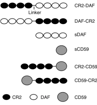
Construct design, expression, and purification. Recombinant fusion proteins contained the four N-terminal SCR units of human CR2 linked to either the N terminus or the C terminus of soluble forms of human CD59 or DAF (constructs depict-ed in Figure 1). Recombinant proteins were purified from the culture super-natant of stably transfected CHO cell clones with yields of between 100 and 200 µg/l. Analysis of purified recombi-nant proteins by SDS-PAGE and West-ern blot revealed proteins within the expected molecular weight range (Figure 2), and except for CR2-CD59, all pro-teins migrated as a single band. The two bands seen for CR2-CD59 were due to differences in glycosylation, since CR2-CD59 migrated as a single band after glycanase treatment (data not shown).
Targeting of fusion proteins to complement opsonized cells. C3 ligand for CR2 was deposited on CHO cells by incubation of CHO cells with complement-activat-ing antibody and C6-depleted serum
(to prevent MAC formation and cell lysis). C3 opsonization was confirmed by flow cytometric analy-sis using an antibody specific for human iC3b (50). All CR2-containing fusion proteins, but not sCD59 or sDAF, bound to C3-coated CHO cells (Figure 3).
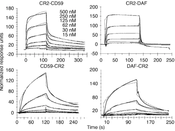
[image:6.576.69.527.47.184.2]Kinetic analysis of interaction between fusion proteins and C3dg ligand. A comparison of the affinity of the differ-ent recombinant fusion proteins for the CR2 ligand C3dg was determined by SPR measurements. The experiments were performed by passing varying con-centrations of the fusion proteins over Biacore strepta-vidin chips containing captured C3dg-biotin. Kinetic analysis of the data showed the best fit to a 1:1 (Lang-muir) binding interaction model using global fitting parameters (Figure 4). Both of the fusion proteins with
Figure 3
Binding of recombinant fusion proteins to C3-opsonized CHO cells. Antibody-sensitized CHO cells were incubated in C6-deficient human serum, washed, and incubated with soluble complement inhibitor (thin black trace) or fusion protein with CR2 at the N ter-minus (light gray trace) or C terter-minus (dark gray trace) at 20 µg/ml. Cell binding of recombinant proteins was detected by flow cytom-etry using anti-DAF or anti-CD59 mAbs followed by a FITC-labeled secondary antibody. Incubation of CHO cells with PBS instead of complement inhibitor is shown in the right-hand panel. Data are representative of three separate experiments.
Figure 4
[image:6.576.251.537.469.683.2]CR2 at the N terminus (CR2-DAF and CR2-CD59) showed similar binding profiles, with fast association and dissociation rates. In contrast, binding of fusion proteins with CR2 at the C terminus (DAF-CR2 and CD59-CR2) showed slow association and dissociation rates (Figure 4 and Table 1). The N-terminal CR2 fusion proteins, however, bound with the highest affin-ity (Table 1). CD59 fusion proteins bound with a high-er affinity than DAF fusion proteins. Affinity constants of the CR2 fusion proteins for C3dg were between the ranges previously published for the interaction between CR2 and C3dg (42, 51). Soluble DAF and sCD59 did not bind to immobilized C3dg (data not shown). Although N-terminal CR2 fusion proteins bind C3dg with a higher affinity than C-terminal coun-terparts, flow cytometry indicated that a larger amount of the C-terminal CR2 fusion proteins bound to C3-opsonized CHO cells than did the corresponding N ter-minus CR2 fusion proteins (Figure 3). The reason for this may be due to different geometries and accessible surface domains of bound fusion proteins that may affect the interaction of anti-DAF or anti-CD59 detec-tion antibodies used for flow cytometry or, alternative-ly, to differences in the form(s) of C3 activation frag-ments on the cell surface as compared with C3dg on Biacore chips. Further analysis is needed to resolve this issue, but the results of functional studies reported below support the former interpretation.
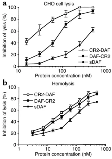
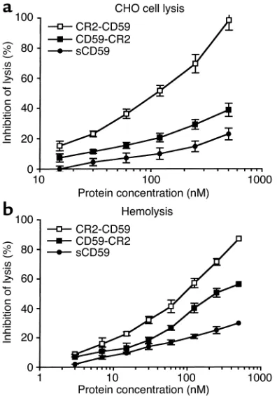
Complement-inhibitory activity of fusion proteins. Comple-ment-inhibitory activity of the targeted and untargeted complement inhibitors was analyzed by measuring their effect on complement-mediated lysis of both CHO cells and erythrocytes. In these experiments, antibody-sensi-tized cells and recombinant proteins were incubated in human serum at a concentration that resulted in 90–100% lysis of unprotected cells. For both cell types, the targeted complement inhibitors were significantly
more effective than their respective untargeted proteins at inhibiting complement-mediated lysis. Targeted DAF proteins were more effective inhibitors than targeted CD59 (Figures 5 and 6). Fusion proteins containing CR2 linked to the N terminus of either DAF or CD59 were more effective inhibitors than C-terminal CR2 fusion proteins. The most potent inhibitor of comple-ment lysis was CR2-DAF, requiring a concentration of 18 nM for 50% inhibition of CHO cell lysis. In contrast, untargeted sDAF required a concentration of 375 nM for 50% inhibition of CHO cell lysis, a 20-fold difference (Figure 5a). sCD59 was a particularly poor inhibitor of complement and provided only 25% protection from CHO cell lysis at 500 nM, the highest concentration tested. CR2-CD59, however, provided 50% inhibition of CHO cell lysis at 102 nM and was more effective than untargeted sDAF (Figure 6a). Table 2 compares the inhibitory activities of the various recombinant com-plement inhibitors. The higher comcom-plement-inhibitory activity of the N-terminal CR2 fusion proteins correlat-ed with the higher affinity these proteins exhibitcorrelat-ed for C3dg ligand (Table 1).
There were some differences between the relative effectiveness of the complement inhibitors at protect-ing CHO cells and erythrocytes from complement-mediated lysis. This was particularly true for the DAF inhibitors; sDAF was significantly more effective at protecting erythrocytes than CHO cells from comple-ment, although targeted DAF was still more effective. There was also little difference in the inhibitory activi-ty of CR2-DAF and DAF-CR2 when erythrocytes were the target cells for complement lysis.
[image:7.576.57.288.88.154.2]Effect of CR2 fusion proteins on cell adhesion. CR3 is a leukocyte receptor involved in endothelial adhe-sion, diapedesis, and the activation of cell cytolytic
Table 1
Kinetic parameters for recombinant fusion protein binding to immo-bilized C3dg-biotin
Analyte ka(1/Ms) kd(1/s) KD(nM)
CR2-CD59 3.45 ×10⁴ 0.169 49 ± 14
CD59-CR2 5.85 ×10² 0.012 205 ± 83.8
CR2-DAF 2.07 ×10⁴ 0.119 575 ± 123
DAF-CR2 2.30 ×10² 0.023 999 ± 26.1
Kinetic values were determined from the average of at least two experiments. Ms, mole-seconds.
Figure 5
[image:7.576.324.510.471.735.2]mechanisms (phagocytosis and degranulation). Since CR2 and CR3 share the same iC3b complement lig-and, we determined whether CR2 fusion proteins interfered with CR3-mediated cell binding. For these experiments, we used U937, a well-characterized promonocytic cell line (CR2–, CR3+) that binds to iC3b-coated erythrocytes in a CR3-dependent mech-anism (46). All of the CR2 fusion proteins, but not sDAF or sCD59, significantly inhibited the binding of U937 cells to C3-opsonized SRBCs (P< 0.01). For this in vitro feasibility experiment, we used a relatively high 500-nM concentration of CR2 fusion protein, and each fusion protein inhibited U937 binding to a similar extent (Figure 7). Similar data were obtained in an experiment using U937 cells that were stimulat-ed with PMA (data not shown), a treatment that results in upregulation of CR3 (45, 46). For comple-ment opsonization of erythrocytes, IgM was used to activate complement, since IgG deposited on the ery-throcytes would engage Fc-γreceptors expressed on U937 cells. In control experiments, U937 cells did not bind to SRBC preincubated with either IgM or C6-deficient human serum alone (data not shown). U937
cells also express CR4 (p150, 95, CD11c/CD18), a third complement receptor recognizing the iC3b lig-and. However, binding of U937 cells to C3-opsonized erythrocytes is CR4-independent and appears to be related to the association of CR4 with the cytoskele-ton and its immobility in the membrane (46).
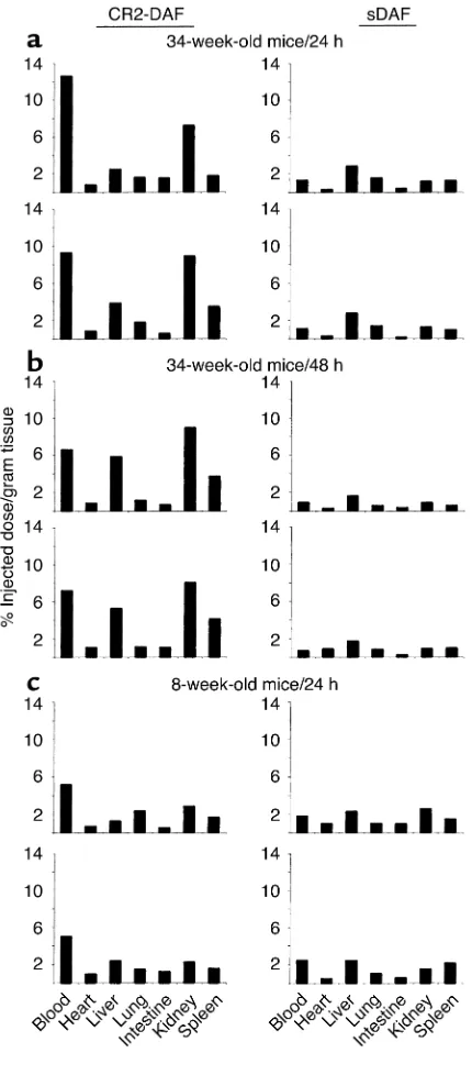
[image:8.576.79.277.54.337.2]Targeting of CR2-DAF to the kidneys of nephritic mice. To determine whether a CR2 fusion protein will target a site of complement activation and disease in vivo, a biodistribution study of CR2-DAF and sDAF in female NZB/W F1 mice was performed. NZB/W F1 mice devel-op a spontaneous autoimmune disease that is very sim-ilar to human systemic lupus erythematosus (SLE), with the production of autoantibodies and the devel-opment of severe immune complex–mediated glomeru-lonephritis that is associated with complement deposi-tion from 26 to 28 weeks of age (4, 52). Biodistribudeposi-tion of [125I]CR2-DAF and [125I]sDAF in 34-week-old NZB/W F1 mice was determined at 24 hours and 48 hours after injection. Twenty-four hours after tail-vein injection of [125I]CR2-DAF, a significantly higher pro-portion of radioactivity was localized to the kidney than to the other organs that were examined (Figure 8a). At 48 hours after injection of [125I]CR2-DAF, there was a similar level of radioactivity in the kidney as at 24 hours, but radioactivity in the liver and spleen was increased and blood radioactivity decreased (Figure 8b). The liver and spleen are sites of immune complex clearance and likely account for increased targeting of [125I]CR2-DAF to these organs at the later time point. [125I]sDAF showed no preferential binding in the kid-ney or any other organ (Figure 8, a and b). In 8-week-old prenephritic NZB/W F1 mice, there was no evi-dence of [125I]CR2-DAF targeting to the kidney (Figure 8c). Of further interest, [125I]sDAF was cleared much more rapidly from the circulation than [125I]CR2-DAF, suggesting that the CR2 moiety is functioning to pro-long the circulatory half-life of the fusion protein. However, the level of [125I]CR2-DAF in the blood of younger mice at 24 hours was about half that recorded in the older mice, and the long circulatory half-life of [125I]CR2-DAF may be a consequence, at least in part, of it binding to circulating immune complexes. Tar-geting of CR2-DAF to complement deposited in the kidney was also examined in another murine model of SLE by direct examination of kidney sections. Similar
Figure 6
[image:8.576.304.540.637.722.2]Inhibition of complement-mediated lysis by recombinant sCD59 and CD59 fusion proteins. Antibody-sensitized CHO cells (a) or SRBCs (b) were incubated with recombinant protein and 10% human serum (CHO cells) or 0.33% human serum (erythrocytes). These concentra-tions resulted in approximately 90% lysis of unprotected cells. Lysis was determined after 60 minutes at 37°C. Background lysis determined by incubating cells in heat-inactivated serum was less than 5% and was subtracted (mean ± SD, n= 4).
Table 2
Concentration of complement inhibitor providing 50% inhibition of lysis
Recombinant protein CHO cell lysis (nM) Erythrocyte lysis (nM)
CR2-DAF 18 21
DAF-CR2 74 33
sDAF 375 90
CR2-CD59 102 91
CD59-CR2 NA 225
sCD59 NA NA
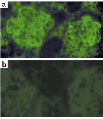
to female NZB/W F1 mice, MRL/lpr mice develop severe proliferative glomerulonephritis with the depo-sition of complement in association with glomerular immune deposits by 24 weeks of age (53). CR2-DAF and sDAF were injected into the tail vein of 24-week-old MRL/lpr mice, and kidney sections were analyzed 24 hours later for human DAF immunoreactivity by fluo-rescence microscopy. Kidney sections from a mouse injected with CR2-DAF displayed a high level of DAF staining, with preferential localization in glomeruli in a pattern identical to that seen for immune complexes. No DAF staining was evident in glomeruli from a mouse injected with sDAF (Figure 9).
Discussion
This study describes the generation and characteriza-tion of soluble human DAF and CD59-containing pro-teins that are targeted to a site of complement activa-tion. Targeting of CD59 and DAF was achieved by linking the inhibitors to a fragment of human CR2 that binds complement C3 activation products. The targeted proteins were significantly more potent than their untargeted counterparts at inhibiting comple-ment. The C3 ligands for CR2 are relatively long lived and are covalently bound, often in large quantities, at sites of complement activation. Thus, CR2-mediated targeting of complement inhibition has the potential to be of therapeutic benefit for numerous complement-associated diseases or disease states. Consistent with this hypothesis, CR2-DAF was shown to target to the kidneys of nephritic NZB/W F1 mice. These mice pro-duce autoantibodies with consequent formation and deposition of immune complexes in the kidney, result-ing in complement activation and deposition (4, 52). Human CR2 binds human and mouse C3 ligands with
[image:9.576.66.278.52.226.2]similar affinities (54), and the biodistribution and microscopy studies establish that a CR2 fusion protein retains targeting function in vivo. However, human CD59 and soluble human DAF are poor inhibitors of rodent complement relative to their activity against human complement (with the exception of human DAF activity against the rat alternative pathway of acti-vation) (43, 55, 56), and so the complement-inhibitory
Figure 7
Effect of recombinant fusion proteins on U937 cell adhesion. SRBCs were sensitized with IgM antibody and incubated in C6-deficient serum. C3-opsonized erythrocytes were coincubated with U937 cells in the presence of 500 nM recombinant fusion protein or PBS. After incubation, the average number of U937 cells bound per erythrocyte was determined by microscopy (mean ± SD, n= 3).
Figure 8
Biodistribution of [125I]CR2-DAF and [125I]sDAF in nephritic
[image:9.576.310.525.254.743.2]function of these proteins was not evaluated in a mouse model. Meaningful validation of the use of CR2-targeted complement inhibitors in a rodent model of disease will require the generation and char-acterization of rodent CR2 complement-inhibitor fusion proteins. The targeting approach is also likely to be effective for other inhibitors of complement such as soluble CR1, an inhibitor currently in clinical trials.
It is tempting to speculate that differences between the complement-inhibitory activity of the N- and C-terminal fusion proteins are due to the different C3dg ligand binding characteristics of the proteins. N-ter-minal CR2 fusion proteins were the most potent com-plement inhibitors, and these proteins bound C3dg with a higher affinity and with faster association and dissociation rates than C-terminal CR2 fusions. How-ever, the difference in C3dg ligand affinity between the two CD59 fusion proteins and between the two DAF proteins was not great. One conceivable reason that a fast on-off rate may be advantageous could be related to cell surface “mobility” or the frequent release of the complement inhibitor from a fixed orientation, allow-ing frequent opportunities for more favorable posi-tioning of the inhibitor for interaction with C3b or nas-cent MAC ligand (depending on the type of inhibitor). For CD59 in particular, previous data suggest that positioning relative to the site of MAC formation is important for its function (23, 57). Other possible rea-sons for the differences in the complement-inhibitory activity of N- and C-terminal CR2 fusion proteins include different positioning of the inhibitor moiety relative to its ligand at the cell surface and steric hin-drance of the inhibitor’s ligand binding site. The rela-tive affinities of the different CR2 fusion proteins for C3dg are reminiscent of the affinities of CR2-SCR 1–2
and CR2 SCR 1–15 for C3dg. The KDvalues for CR2 SCR1–2 and CR2 SCR 1–15 interactions with C3dg were similar, but CR2 SCR 1–2 associated and dissoci-ated much faster, suggesting a contribution of the additional SCR domains to overall affinity (42). Analy-sis of the solution structure of another SCR-contain-ing protein, factor H, indicated that SCR domains are folded back on themselves and interactions between SCR domains may modulate C3 ligand binding char-acteristics (58). Conformational variability between SCR domains is predicted to result from different (native) linker lengths, with longer linkers providing greater conformational flexibility. In this context, the CR2 and DAF SCR domains are linked with a relative-ly long Ser-Grelative-ly linker, and this may permit the fusion partners to fold back on one another, resulting in SCR-SCR interactions that may modulate CR2 binding affinity. This is a possible explanation for the lower KD of the DAF fusion proteins for C3dg as compared with the CD59 fusion proteins (CD59 is a single-domain compact globular protein with no SCR homology).
Complement-mediated lysis assays were performed using antibody-sensitized CHO cells or SRBCs as tar-gets. There were marked differences in the relative activities of some of the complement inhibitors for protecting the different cells from complement-medi-ated lysis. sDAF, DAF-CR2, and CD59-CR2 were sig-nificantly more effective at protecting sheep erythro-cytes than CHO cells from complement-mediated lysis. The reason for these differences is not clear, but it may be related to the multihit characteristics of nucleated cell lysis. Unlike erythrocytes, complement-mediated lysis of nucleated cells is not entirely due to colloid osmotic deregulation, and the deposition of multiple MACs in the plasma membrane is required (59–61). Most of the previous studies investigating the inhibitory activity of soluble (untargeted) complement inhibitors have been performed using erythrocytes as target cells for complement-mediated lysis. However, CHO cells most likely represent a more physiological-ly relevant target for in vitro experiments. Human cells were not used as targets in our experiments, since the expression of endogenous complement-inhibitory proteins would make interpretation of data difficult, and human erythrocytes and most human cell lines are resistant to lysis by human complement in vitro unless the function of endogenously expressed com-plement inhibitors is blocked.
[image:10.576.92.263.53.249.2]Various mechanisms of complement-mediated damage are implicated in different disease condi-tions, and different diseases will most likely benefit from inhibition strategies acting at various points in the complement pathway. For example, if applicable for the disease, a particular benefit of blocking com-plement at a late step in the pathway would be that host defense functions and immune homeostasis mechanisms of complement would remain intact. Thus, a CD59-based inhibitor would provide advan-tages over inhibitors of complement activation (such
Figure 9
as DAF and sCR1) in diseases in which the terminal cytolytic pathway is primarily implicated in patho-genesis. Soluble CD59 is unlikely to have therapeutic benefit because of its very poor activity in vitro, but we have shown that CR2-mediated targeting of CD59 significantly increased its complement-inhibitory activity. In fact, CR2-CD59 was more effective at inhibiting complement-mediated lysis than sDAF, and sDAF has shown therapeutic efficacy in vivo (11). Rodent analogues of CR2-CD59 may also be useful tools for dissecting the relative roles of early comple-ment activation products versus MAC formation in disease pathogenesis. The relative contributions of the various complement activation products to tissue injury in many disease states is poorly understood and controversial.
The CR2 fusion proteins inhibited the binding of U937 cells to C3-opsonized erythrocytes. CR2 and CR3 both bind iC3b, and our data indicate that CR2 fusion proteins act as CR3 antagonists, since U937 binding to C3-opsonized erythrocytes is CR3 dependent (46). As an adhesion molecule, CR3 mediates endothelial adhe-sion and diapedesis at sites of inflammation through its high-affinity interaction with ICAM-1. As a com-plement receptor, CR3 promotes and enhances phago-cytosis and degranulation through its interaction with iC3b. Both ICAM-1 and iC3b bind to overlapping epi-topes on CR3 (62). CR3 can thus be an important determinant in promoting cell-mediated tissue damage at sites of inflammation, and antibodies that block CR3 have shown effectiveness in several inflammatory conditions (62). The antagonistic effect of CR2 on CR3 binding therefore indicates a second potential anti-inflammatory mechanism of action of the CR2 com-plement-inhibitor fusion proteins that may act syner-gistically with complement inhibition. Of further relevance, soluble CR2 has been shown to suppress antibody responses in mice (54), and CR2 complement-inhibitor fusion proteins may have additional immu-nologic effects that will need to be investigated.
In summary, targeting complement inhibitors to sites of complement activation and disease is likely to con-siderably enhance their efficacy. Indeed, the data indi-cate that for disease states that would benefit from CD59-based therapy, the targeting of CD59 to the site of complement activation will be a requirement. An advantage of CR2-mediated targeting over other tar-geting approaches, such as antibody-mediated target-ing or sLex-mediated targeting to adhesion molecules, is that the CR2 moiety will target any accessible site of complement activation and will have broad therapeutic potential. CR2 fusion proteins can also act as CR3 (and CR2) antagonists, and this may represent a second and potentially important therapeutic benefit. Human CR2 complement-inhibitor fusion proteins are also less like-ly to be immunogenic than recombinant inhibitors con-taining antibody variable regions. The predicted ability of targeted inhibitors of complement activation to pro-vide an effective local concentration with low levels of
systemic inhibition also diminishes the possibility of compromising host defense mechanisms, particularly with long-term systemic complement inhibition (this is a less important consideration for CD59-based in-hibitors). CR2-targeted inhibitors may also target infec-tious agents that activate complement, but whether this would seriously interfere with complement-dependent defense pathways is difficult to predict. This would depend on such considerations as the rate of pathogen multiplication relative to effective inhibitor levels both in the circulation and retained on host tissue at sites of complement activation.
Acknowledgments
We thank Masaki Imai for assistance with some exper-imental procedures. This work was supported by grants from the S.L.E. Foundation Inc., the American Heart Association, and the NIH (AI 34451 to S. Tomlinson and CA53615 to V.M. Holers).
1. Rother, K., Till, G.O., and Haensch, G.M., editors. 1998. The complement system. Springer-Verlag. Berlin, Germany. 564 pp.
2. Lambris, J.D., and Holers, V.M. 2000. Therapeutic interventions in the com-plement system. Humana Press. Totowa, New Jersey, USA. 258 pp. 3. Wang, Y., Rollins, S.A., Madri, J.A., and Matis, L.A. 1995. Anti-C5
mono-clonal antibody therapy prevents collagen-induced arthritis and ame-liorates established disease. Proc. Natl. Acad. Sci. U. S. A.92:8955–8959. 4. Wang, Y., Hu, et al. 1996. Amelioration of lupus-like autoimmune dis-ease in NZB/W F1 mice after treatment with a blocking monoclonal antibody specific for complement component C5. Proc. Natl. Acad. Sci. U. S. A.93:8563–8568.
5. Kaplan, M. 2002. Eculizumab (Alexion). Curr. Opin. Investig. Drugs.
3:1017–1023.
6. Whiss, P.A. 2002. Pexelizumab Alexion. Curr. Opin. Investig. Drugs.
3:870–877.
7. Weisman, H.F., et al. 1990. Soluble human complement receptor type 1: in vivo inhibitor of complement suppressing post-ischemic myocardial inflammation and necrosis. Science.249:146–151.
8. Rioux, P. 2001. TP-10 (AVANT Immunotherapeutics). Curr. Opin. Investig. Drugs.2:364–371.
9. Zamora, M.R., et al. 1999. Complement inhibition attenuates human lung transplant reperfusion injury: a multicenter trial. Chest.
116(Suppl. 1):46S.
10. Higgins, P.J., et al. 1997. A soluble chimeric complement activating inhibitory protein that possesses both decay-accelerating and factor I cofactor activities. J. Immunol.158:2872–2881.
11. Moran, P., et al. 1992. Human recombinant soluble decay accelerating factor inhibits complement activation in vitro and in vivo. J. Immunol.
149:1736–1743.
12. Christiansen, D., Milland, J., Thorley, B.R., McKenzie, I.F., and Loveland, B.E. 1996. A functional analysis of recombinant soluble CD46 in vivo and a comparison with recombinant soluble forms of CD55 and CD35 in vitro. Eur. J. Immunol.26:578–585.
13. Salerno, C.T., et al. 2002. A soluble chimeric inhibitor of C3 and C5 con-vertases, complement activation blocker-2, prolongs graft survival in pig-to-rhesus monkey heart transplantation. Xenotransplantation.9:125–134. 14. Kroshus, T.J., et al. 2000. A recombinant soluble chimeric comple-ment inhibitor composed of human CD46 and CD55 reduces acute cardiac tissue injury in models of pig-to-human heart transplanta-tion. Transplantation.69:2282–2289.
15. Rushmere, N.K., Van Den Berg, C.W., and Morgan, B.P. 2000. Produc-tion and funcProduc-tional characterizaProduc-tion of a soluble recombinant form of mouse CD59. Immunology.99:326–332.
16. Quigg, R.J., He, C., Hack, B.K., Alexander, J.J., and Morgan, B.P. 2000. Production and functional analysis of rat CD59 and chimeric CD59-Crry as active soluble proteins in Pichia pastoris. Immunology.99:46–53. 17. Sugita, Y., et al. 1994. Recombinant soluble CD59 inhibits reactive
haemolysis with complement. Immunology.82:34–41.
18. Meri, S., Lehto, T., Sutton, C.W., Tyynela, J., and Baumann, M. 1996. Structural composition and functional characterization of soluble CD59: heterogeneity of the oligosaccharide and glycophosphoinositol (GPI) anchor revealed by laser-desorption mass spectrometric analysis. Biochem. J.316:923–935.
anti-inflammatory effects of complement inhibitors possessing sialyl lewisX moieties. J. Immunol.162:4952–4959.
20. Ritterhaus, C.W., et al. 1999. Recombinant glycoproteins that inhibit complement activation and also bind the selectin adhesion molecules. J. Biol. Chem.274:11237–11244.
21. Huang, J., et al. 1999. Neuronal protection in stroke by an sLex-glycosy-lated complement inhibitory protein. Science.285:595–599.
22. Zhang, H., Lu, S., Morrison, S.L., and Tomlinson, S. 2001. Targeting of functional antibody-decay accelerating factor (DAF) fusion proteins to a cell surface. J. Biol. Chem.276:27290–27295.
23. Zhang, H.-F., Yu, J., Bajwa, E., Morrison, S.L., and Tomlinson, S. 1999. Targeting of functional antibody-CD59 fusion proteins to a cell surface. J. Clin. Invest.103:55–66.
24. Linton, S.M., et al. 2000. Therapeutic efficacy of a novel membrane-tar-geted complement regulator in antigen-induced arthritis in the rat. Arthritis Rheum.43:2590–2597.
25. Smith, G.P., and Smith, R.A. 2001. Membrane-targeted complement inhibitors. Mol. Immunol.38:249–255.
26. Carroll, M.C. 1998. The role of complement and complement receptors in induction and regulation of immunity. Annu. Rev. Immunol.16:545–568. 27. Carroll, M.C. 2000. The role of complement in B cell activation and
tol-erance. Adv. Immunol.74:61–88.
28. Holers, V.M. 1989. Complement receptors. Year Immunol.4:231–240. 29. Dierich, M.P., Schulz, T.F., Eigentler, A., Huemer, H., and Schwable, W. 1988. Structural and functional relationships among receptors and reg-ulators of the complement system. Mol. Immunol.25:1043–1051. 30. Lowell, C.A., et al. 1989. Mapping of the Epstein-Barr virus and C3dg
binding sites to a common domain on complement receptor type 2. J. Exp. Med.170:1931–1946.
31. Szakonyi, G., et al. 2001. Structure of complement receptor 2 in complex with its C3d ligand. Science.292:1725–1728.
32. Law, S.K., Fearon, D.T., and Levine, R.P. 1979. Action of the C3b-inacti-vator on the cell-bound C3b. J. Immunol.122:759–765.
33. Seya, T., and Nagasawa, S. 1985. Limited proteolysis of complement protein C3b by regulatory enzyme C3b inactivator: isolation and characterization of a biologically active fragment, C3d,g. J. Biochem. (Tokyo)97:373–382. 34. Holers, V.M. 2001. Complement in clinical immunology. Mosby.
Philadel-phia, Pennsylvania, USA. 289 pp.
35. Quigg, R.A., et al. 1998. Blockade of antibody-induced glomeru-lonephritis with Crry-Ig, a soluble murine complement inhibitor. J. Immunol.160:4553–4560.
36. Harlow, E., and Lane, D. 1988. Antibodies: a laboratory manual. Cold Spring Harbor Laboratory. Cold Spring Harbor, New York, USA. 726 pp. 37. Coyne, K.E., et al. 1992. Mapping of epitopes, glycosylation sites, and
complement regulatory domains in human decay accelerating factor. J. Immunol.149:2906–2913.
38. Kinoshita, T., Medof, M.E., Silber, R., and Nussenzweig, V. 1985. Distri-bution of decay-accelerating factor in peripheral blood of normal indi-viduals and patients with paroxysmal nocturnal hemoglobinuria. J. Exp. Med.162:75–92.
39. Davies, A., et al. 1989. CD59, an LY-6-like protein expressed in human lymphoid cells, regulates the action of the complement membrane attack complex of homologous cells. J. Exp. Med.170:637–654. 40. Guthridge, J.M., et al. 2001. Epitope mapping using the X-ray
crystallo-graphic structure of complement receptor type 2 (CR2)/CD21: identifi-cation of a highly inhibitory monoclonal antibody that directly recog-nizes the CR2-C3d interface. J. Immunol.167:5758–5766.
41. Dieffenbach, C.W., and Dveksler, G.S., editors. 1995. PCR primer: a lab-oratory manual. Cold Spring Harbor Laboratory Press. Cold Spring Harbor, New York, USA. 714 pp.
42. Guthridge, J.M., et al. 2001. Structural studies in solution of the recom-binant N-terminal pair of short consensus/complement repeat domains of complement receptor type 2 (CR2/CD21) and interactions with its lig-and C3dg. Biochemistry.40:5931–5941.
43. Yu, J., et al. 1999. Protection of human breast cancer cells from comple-ment-mediated lysis by expression of heterologous CD59. Clin. Exp. Immunol.115:13–18.
44. Duits, A.J., Jainandunsing, S.M., van de Winkel, J.G., and Capel, P.J. 1991. Selective enhancement of Leu-CAM expression by interleukin 6 during differentiation of human promonocytic U937 cells. Scand. J. Immunol.
33:151–159.
45. Rothlein, R., and Springer, T.A. 1986. The requirement for lymphocyte function-associated antigen 1 in homotypic leukocyte adhesion stimu-lated by phorbol ester. J. Exp. Med.163:1132–1149.
46. Ross, G.D., Reed, W., Dalzell, J.G., Becker, S.E., and Hogg, N. 1992. Macrophage cytoskeleton association with CR3 and CR4 regulates receptor mobility and phagocytosis of iC3b-opsonized erythrocytes. J. Leukoc. Biol.51:109–117.
47. Sharkey, R.M., Motta-Hennessy, C., Pawlyk, D., Siegel, J.A., and Golden-berg, D.M. 1990. Biodistribution and dose estimates for yttrium- and iodine-labeled monoclonal antibody IgG and fragments in nude mice. Cancer Res.50:2330–2336.
48. Sharkey, R.M., Natale, A., Goldenberg, D.M., and Mattes, M.J. 1991. Rapid blood clearance of immunoglobulin G2a and immunoglobulin G2b in nude mice. Cancer Res.51:3102–3107.
49. Coligan, J.E., Kruisbeek, A.M., Margulies, D.H., Shevach, E.M., and Strober, W., editors. 2002. Current protocols in immunology[book on CD-ROM]. John Wiley and Sons Inc. Hoboken, New Jersey, USA. 50. Iida, K., Mitomo, K., Fujita, K., and Tamura, N. 1987. Characterization
of three monoclonal antibodies against C3 with selective specificities. Immunology.62:413–417.
51. Sarrias, M.R., et al. 2001. Kinetic analysis of the interactions of comple-ment receptor 2 (CR2, CD21) with its ligands C3d, iC3b, and the EBV glycoprotein gp350/220. J. Immunol.167:1490–1499.
52. Andrews, B.S., et al. 1978. Spontaneous murine lupus-like syndromes. Clinical and immunopathological manifestations in several strains. J. Exp. Med.148:1198–1215.
53. Biesecker, G., Katz, S., and Koffler, D. 1981. Renal localization of the membrane attack complex in systemic lupus erythematosus nephritis. J. Exp. Med.154:1779–1794.
54. Hebell, T., Ahearn, J.M., and Fearon, D.T. 1991. Suppression of the immune response by a soluble complement receptor of B lymphocytes. Science.254:102–105.
55. Harris, C.L., Spiller, O.B., and Morgan, B.P. 2000. Human and rodent decay-accelerating factors (CD55) are not species restricted in their com-plement-inhibiting activities. Immunology.100:462–470.
56. Yu, J., et al. 1997. Mapping the regions of the complement inhibitor CD59 responsible for its species selectivity. Biochemistry.36:9423–9428. 57. Fodor, W.L., Rollins, S.A., Guilmette, E.R., Setter, E., and Squinto, S.P. 1995. A novel bifunctional chimeric complement inhibitor that regu-lates C3 convertase and formation of the membrane attack complex. J. Immunol.155:4135–4138.
58. Aslam, M., and Perkins, S.J. 2001. Folded-back solution structure of monomeric factor H of human complement by synchrotron X-ray and neutron scattering, analytical ultracentrifugation and constrained molecular modelling. J. Mol. Biol.309:1117–1138.
59. Koski, C.L., Ramm, L.E., Hammer, C.H., Mayer, M.M., and Shin, M.L. 1983. Cytolysis of nucleated cells by complement: cell death displays multi-hit characteristics. Proc. Natl. Acad. Sci. U. S. A.80:3816–3820. 60. Ramm, L.E., Whitlow, M.B., and Mayer, M.M. 1982. Transmembrane
channel formation by complement: functional analysis of the number of C5b6, C7, C8, and C9 molecules required for a single channel. Proc. Natl. Acad. Sci. U. S. A.79:4751–4755.
61. Takeda, J., et al. 1986. Number of hits necessary for complement-medi-ated hemolysis. Microbiol. Immunol.30:461–468.
62. Ross, G.D. 2000. Regulation of the adhesion versus cytotoxic functions of the Mac-1/CR3/αMβ2-integrin glycoprotein. Crit. Rev. Immunol.