
Original Article
HOXC6 expression is associated with a poor prognosis
in early-stage cervical squamous cell carcinoma
Jianhong Shang1*, Hongwei Shen2*, Jun Liu2, Yalan Deng2, Shuzhong Yao2, Shanyang He2
1Department of Ultrasonic Medicine, Fetal Medical Center, 2Department of Obstetrics and Gynecology, The First
Affiliated Hospital, Sun Yat-sen University, Guangzhou, P. R. China. *Equal contributors.
Received February 17, 2017; Accepted March 20, 2017; Epub May 1, 2017; Published May 15, 2017
Abstract: Homeobox C6 (HOXC6) is involved in malignant progression in certain cancers. However, the expression profile of HOXC6 and its clinical/prognostic significance are unclear in early-stage cervical cancer. The mRNA and protein expression levels of HOXC6 were analyzed in five cervical cancer cell lines and four corresponding cervical cancer tumors by real-time PCR and Western blotting, respectively. Immunohistochemistry was performed to detect the expression of HOXC6 in paraffin-embedded tissues from 90 early-stage cervical cancer patients, 60 cervical intraepithelial neoplasia (CIN) patients and 19 normal cervical tissues (NCTs). Statistical analyses were used to evaluate the clinicopathological significance of HOXC6 expression. In this study, HOXC6 expression was differen -tially increased in cervical cancer cell lines and tissues than in normal tissues and ANTs. Moreover, its expression was significantly up-regulated at protein level in early-stage cervical cancer, compared to CIN and NCTs (P=0.004). Statistical analysis showed that HOXC6 expression was significantly correlated with tumor size (P=0.044), type of tumor growth (P=0.009), stromal invasion (P=0.030), lymph node metastasis (P=0.027), positive surgical margins (P=0.038), vaginal involvement (P=0.038), postoperative adjuvant therapy (P=0.012), recurrence (P=0.044) and survival (P=0.033). Kaplan-Meier analysis indicated that patients with higher HOXC6 expression had shorter overall survival duration than patients with lower HOXC6 expression. Cox-regression analysis demonstrated that HOXC6 expression was a factor significantly associated with the 5-year DFS and OS rates for 90 SCC patients. Our findings suggest that HOXC6 expression was associated with a poor prognosis in early-stage SCC, and it may serve as a prognostic biomarker.
Keywords: Cervical cancer, prognosis, HOXC6
Introduction
Cancer statistics indicated that an estimated
527,600 new cases and 265,700 deaths of
cervical cancer would occur in the world annu-ally [1], despite its decreasing incidence and mortality in several Western countries
associ-ated with wide implementation of Papanicolaou testing and vaccines [2-4]. And nearly 90% of
cervical cancer occurred in developing coun-tries, so that it remains a critical issue in China with 98,900 new cases and 30,500 deaths in
2015 [5]. The treatment strategy for cervical
cancer depends on the FIGO stage and several traditional clinical pathological variables [6]. However, traditional pathological variables are
not sufficiently reliable for predicting clinical outcomes or for guiding optimal treatment strategies. Therefore, there is still an urgent
need for additional research to identify novel biomarkers to supply practical information for
patient prognosis and suitable therapeutic options. It can provide the earliest and the most
accurate forecast information and guide clinical
individualized treatment.
The homeobox (HOX) genes, key factors in the regulation of embryogenesis, encode a group of transcription factors and regulate the expres
-sion of downstream target genes via specific DNA binding [7]. They are also involved in sev -eral processes such as cellular morphogenesis
and differentiation [8-11]. There are 39 differ
-ent human HOX genes clustered into four differ -ent groups (HOXA, HOXB, HOXC and HOXD) [8,
9]. Recent studies have found that the expres
Table 1. Correlation between HOXC6 expression and clinicopathologic features of early-stage cervical
cancer (N=90)
Variable N Percentage None or low (N=22) High (N=68)HOXC6 expression Chi-square value p Value FIGO stage
Ia1 5 5.56 2 3 5.58 0.409
Ia2 7 7.78 2 5
Ib1 33 36.67 11 22
Ib2 21 23.33 2 19
IIa1 12 13.33 3 9
IIa2 12 13.33 2 10
Age (years)
≤40 35 38.89 9 26 0.050 0.823
>40 55 61.11 13 26
Tumor size (cm)
<4 53 58.89 17 38 4.065 0.044
≥4 37 41.11 5 32
Types of tumor growth
Unclear 8 8.89 4 4 9.448 0.009a
Endophytic 25 27.78 10 15
Exophytic 57 63.33 8 43
Differentiation grade
G1 3 3.33 0 3 1.370 0.504a
G2 31 34.45 9 22
G3 56 62.22 13 43
Stromal invasion
<1/2 40 44.44 15 25 7.046 0.030
≥1/2 50 55.56 7 43
Lymphovascular space invasion (LVSI)
Yes 15 16.67 2 13 1.203 0.273
No 75 83.33 20 55
Pelvic lymph node metastasis (PLNM)
Yes 43 47.78 6 37 4.907 0.027
No 47 52.22 16 31
Positive surgical margins
Yes 33 36.67 4 29 4.284 0.038
No 57 63.33 18 39
Parametrial infiltration
Yes 7 7.78 2 5 0.070 0.791
No 83 92.22 20 63
Vaginal involvement
Yes 33 36.67 4 29 4.284 0.038
No 57 63.33 18 39
Postoperative adjuvant therapy
Yes (total) 53 58.89 18 35 6.323 0.012
No 37 41.11 4 33
Recurrence
Yes 37 41.11 5 32 4.065 0.044
No 53 58.89 17 36
Vital status at follow-up
Alive 52 57.78 17 35 4.536 0.033
Death from 38 42.22 5 33
lungs, and ovaries, oral squamous cell carcino-ma and esophageal squamous cell carcinocarcino-ma [10-17]. In a recent study, HOXC6 was shown to play an important role in several cellular events
through the regulation of its functional biologi -cal targets such as Bcl-2 and JunD [18, 19].
Although HOXC6 is critical for various regulated
cellular processes and is correlated with
can-cer progression, the function of HOXC6 in SCC
is largely unknown.
In the present study, we investigated the
clini-cal relationship between expression pattern of
HOXC6 and early-stage cervical cancer
pro-gression. We found that the HOXC6 expression was significantly increased in cervical cancer
cells and tissues, and was closely associated
with tumor size, type of tumor growth, stromal
invasion, lymph node metastasis, positive sur-gical margins, vaginal involvement, postopera-tive adjuvant therapy, recurrence and survival. In addition, using Cox-regression analysis, the
effectiveness of HOXC6 as a factor significantly
associated with the 5-year DFS and OS rates
for SCC patients was assessed. Collectively,
HOXC6 may be a prognostic biomarker in early-stage cervical cancer.
Materials and methods
Samples and patients
The current retrospective study enrolled 90
patients diagnosed with early-stage cervical squamous cell carcinoma (SCC) who under-went radical hysterectomy and
lymphadenec-tomy in the Department of Obstetrics and Gynecology, the First Affiliated Hospital, Sun Yat-sen University from January 2006 to
De-cember 2009. All enrolled patients were in Ia2-IIa2 stage without preoperative radiotherapy or
chemotherapy and with available clinical follow-up data. The mean patient age was 42.90±7.815 (ranging from 27 to 68). Clinical stages were
determined according to the International
Federation of Obstetrics and Gynecology, 2009 (FIGO). The last follow-up was carried out in
December 2015, with the mean observation
period of 56 months (6-92 months), and there were 38 cancer-related deaths. 19 samples of normal cervix from patients undergoing simple hysterectomy because of uterine leiomyomata
were obtained as controls. In addition, samples
from 20 patients with CINI and 17 with CINII
undergoing biopsy, and 23 with CINIII
undergo-ing hysterectomy were also selected as
con-trols. Four fresh cervical SCC and their corre -sponding adjacent noncancerous tissues
(ANTs) were collected from the cervical cancer
patients without preoperative radiotherapy or
chemotherapy, which were used for quantita -tive polymerase chain reaction (qPCR) and Western blotting analysis. Prior written consent
of the patients for the use of these clinical materials for research purposes, and approval from the Institutional Ethical Board (IRB) in the First Affiliated Hospital of Sun Yat-sen University were obtained. The clinical information is sum -marized in Table 1.
Cell lines
The cervical cancer cell lines, including HeLa
high), SiHa, C33A and HCC94 (HOXC6-low), were cultured in Eagle’s minimum essen-tial medium (Gibco BRL, Rockville, MD) and Caski cells were cultured in RPMI-1640 medi-um (Gibco BRL). Media were supplemented with 10% fetal bovine serum (HyClone Labora-tories, Logan, UT) and 1% antibiotics (100 U/ml
penicillin and 100 μg/ml streptomycin) in a 5% CO2-humidified atmosphere at 37°C. These five
cell lines were purchased from American Type Culture Collection (ATCC, MD, USA).
RNA extraction and quantitative RT-PCR Total RNA from cultured cells and fresh tissues were extracted using the Trizol reagent
(Invi-trogen, Carlsbad, CA, USA) according to
manu-facturer’s instructions. 2 μg RNA from each sample was used for cDNA synthesis using ran -dom hexamer primers. qPCR was used to
quan-tify HOXC6 mRNA level. Each sample was test -ed in triplicate. Expression data were nor-
malized to the geometric mean with reference to the housekeeping gene GAPDH. The HOXC6 primer sequences were 5’-AAGAACTCCATCCG-CCACAAC-3’ (forward) and 5’-GCTTAAACACCT-GGTCCAATGTC-3’ (reverse). Primers for GAPDH were 5’-ACCACAGTCCATGCCATCAC-3’ (forward) and 5’-TCCACCACCCTGTTGCTGTA-3’ (reverse).
Western blotting
Cell lysates from cell lines and fresh tissue were obtained using cold RIPA buffer. Total pro
-teins (50 μg) were fractionated using sodium dodecyl sulfate-polyacrylamide gel electropho
-resis (SDS-PAGE) and transferred onto nitrocel
described previously. Anti-HOXC6 (Santa Cruz Biotechnology, Inc., Santa Cruz, CA, USA) anti-body was used.
Immunohistochemical assay
IHC analysis was used to study altered protein expression in 90 human cervical cancer tis-sues, 60 cervical intraepithelial neoplasia pa-
tients and 19 normal cervical tissues. Briefly, 4-lm-thick paraffin sections of the cervical can
-cer tissue from the patient were baked at 65°C for 30 min and then deparaffinized with xylene
and rehydrated. Submerging the sections into
EDTA antigenic retrieval buffer and then micro
-waving were used for antigen retrieval. The
samples were then treated with 3% hydrogen peroxide in methanol to quench endogenous
peroxidase activity, followed by incubation with 1% bovine serum albumin to block nonspecific
binding. Sections were then incubated with anti-HOXC6 antibody (Santa Cruz Biotechnolo-
gy, Inc., Santa Cruz, CA, USA) overnight at 4°C.
Normal rabbit serum was used as a negative
control. After washing, the tissue sections were then incubated with a biotinylated anti-mouse
secondary antibody (Abcam), followed by fur -ther incubation with streptavidin-horseradish
peroxidase complex (Abcam). The tissue sec -tions were immersed in 3-amino-9-ethyl carba-zole and counterstained with 10% Mayer’s he- matoxylin, dehydrated and mounted in Crystal
Mount. Two observers who were blinded to the
histopathological features and patient data of the samples evaluated the degree of immunos
-taining of formalin-fixed, paraffin embedded sections independently. The scores given by
the two independent investigators were
aver-aged and were based on both the proportion of
positively stained tumor cells and the intensity
of staining. The staining results were scored based on the following criteria: (i) percentage of
positive tumor cells in the tumor tissue: 0 (0%), 1 (1-10%), 2 (11-50%), 3 (51-70%) and 4 (71-100%); (ii) staining intensity: 0 (none), 1 (weak),
2 (moderate), 3 (strong). The staining index was
calculated as: staining intensity score ×
propor-tion of positive tumor cells (range from 0 to 12). A final score ≥7 was considered high
expres-sion.
Statistical analysis
Statistical analyses were carried out using the
[image:4.612.89.287.70.264.2]SPSS 13.0 software package (SPSS Inc., Chi-cago, IL, USA). The relationship between HOXC6
Figure 1. Overexpression of HOXC6 mRNA and pro -tein in cervical cancer cell lines. qPCR (A) and West-ern blotting (B) examined the expression of HOXC6 mRNA and protein in cervical cancer cell lines (Hela, SiHa, CasKi, C33A, HCC94) and NCT. Expression lev -els were normalized against GAPDH. Error bars rep-resent the standard deviation of the mean (SD) cal -culated from three parallel experiments. *P<0.05.
[image:4.612.91.284.380.605.2]expression and clinicopathological characteris-tics was assessed using the chi-square test and Fisher’s exact test. Bivariate correlations between study variables were calculated by
Spearman’s rank correlation coefficients.
Sur-vival curves were estimated using the Kaplan-Meier method and compared using the log-rank test. Multivariate survival analysis was carried
out for all the parameters that were significant in the univariate analysis. The type of Cox model chosen by us was the forward method.
In all cases, a P value of <0.05 was considered statistically significant.
Result
Elevated expression of HOXC6 in cervical can-cer cell lines and can-cervical carcinoma
To investigate the characteristic of HOXC6
expression in cervical carcinoma, we compara-tively analyzed the HOXC6 mRNA and protein
profiles in different cervical cancer cell lines
and samples. Western blotting and qPCR
analy-ses revealed elevated expression levels of
HOXC6 protein and mRNA, respectively, in all
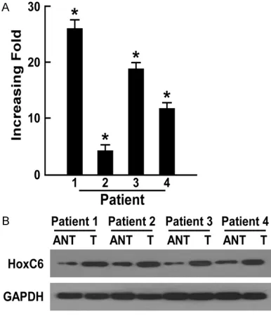
five cervical carcinoma cell lines compared to NCTs (Figure 1). Furthermore, comparative an- alysis showed that HOXC6 mRNA and protein
levels were differentially up-regulated in all four
cervical carcinoma samples compared with
corresponding ANTs derived from the same
patient (Figure 2). With these findings, our
results demonstrated that HOXC6 is up-regulat-ed in cervical cancer.
Association between HOXC6 expression and clinical features in cervical carcinoma
To further investigate the clinical significance of the above finding in cervical carcinoma, the expression of HOXC6 was examined by IHC in 90 paraffin-embedded, archived cervical can
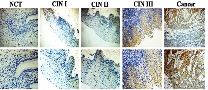
-cer tissues, 20 cases of CINI, 17 cases of CINII, 23 cases of CINIII, and 19 NCTs. An overview of
HOXC6 expression and clinicopathological parameters is given in Table 1. The positive expression rate of HOXC6 in CINI, CINII, CINIII
and cervical cancer were 15.0% (3/20), 29.4% (5/17), 56.5% (13/23) and 75.6% (68/90).
HOXC6 expression gradually increased from
CINI through to cervical cancer, but was absent
in NCTs (P<0.001), and the subcellular location
of HOXC6 was primarily mainly in the cytoplasm of cancer cells (Figure 3 and Table 2).
Statistical analyses were used to examine the correlation between HOXC6 expression and the clinicopathological characteristics of
early-stage SCC patients. As shown in Table 2, analy-sis exhibited that HOXC6 expression was strongly correlated with tumor size (P=0.044),
type of tumor growth (P=0.009), stromal inva-sion (P=0.030), lymph node metastasis (PLNM,
[image:5.612.91.529.72.263.2]ever, there were no significant associations
between HOXC6 high expression and other
clin-ical features including FIGO stage, age, differ -entiation grade, lymphovascular space
inva-sion (LVSI), and parametrial infiltration. Uni- and multi-variable analysis of clinicopath-ologic factors associated with 5-year OS and DFS rates of SCC patients
Kaplan-Meier analysis and the log-rank test
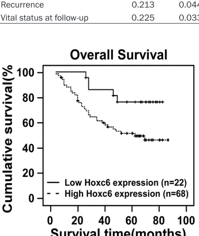
were used to calculate the effect of HOXC6 expression on survival. The log-rank test reve-aled a significantly inverse correlation between
high HOXC6 expression level and patient sur-vival. Patients with low HOXC6 expression had longer survival times, whereas those with high HOXC6 expression had shorter survival times (Figure 4, log-rank, P=0.019). The cumulative 5-year OS rates for the patients with high levels of HOXC6 expression were 51.5%, whereas the rates were 77.3% for the patients with low or no
HOXC6 expression.
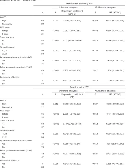
Univariable analysis using log-rank tests sh- owed that HOXC6 expression (P=0.001, P= 0.012), FIGO stage (P<0.001, P<0.001), tumor size (P<0.001, P<0.001), stromal invasion (P= 0.012; P=0.016), LVSI (P<0.001, P<0.001), PLNM (P<0.001, P<0.001) and parametrial
infiltration (P=0.012, P=0.016) as factors sig
-nificantly associated with the 5-year DFS and OS rate for 90 SCC patients. Multivariable anal -ysis showed that tumor size (P=0.013, P= 0.012), LVSI (P=0.025, P=0.013) and PLNM (P=0.017, P=0.027) were factors significantly associated with the 5-year DFS and OS rate for these patients. Thus, according to our findings
that tumor size, LVSI and PLNM served as the
[image:6.612.89.363.98.213.2]independent prognostic factors significantly
Table 2. HOXC6 expression in normal cervical tissue (NCT), CINI,
CINII, CINIII and cervical squamous cell caner (SCC)
N
HOXC6 expression
Chi-squre
value valueP Trend value valueP None or
low (N=80) (N=89)High Case
NCT 19 19 0 55.262 0.000 54.537 0.000
CINI 20 17 3
CINII 17 12 5
CINIII 23 10 13
[image:6.612.92.302.273.406.2]SCC 90 22 68
Table 3. Spearman analysis of correlation
between HOXC6 and clinicopathological charac-teristics
Variables
HOXC6 expression level Spearman
correlation P value
Types of tumor growth 0.324 0.002
Tumor size (cm) 0.213 0.044
Stromal invasion 0.238 0.024
Pelvic lymph node metastasis 0.233 0.020
Positive surgical margins 0.218 0.039
Recurrence 0.213 0.044
Vital status at follow-up 0.225 0.033
Figure 4. Kaplan-Meier curves with univariate analy-ses (log-rank) for cervical cancer patients with low versus high HOXC6 expression. The cumulative 5-year survival rate was 77.3% in the low HOXC6 protein expression group (n=22; thick line), but only 51.5% in the high-expression group (n=68; dotted line).
adjuvant therapy (P=0.012), recurrence (P=0.044) and sur-vival (P=0.033). Moreover, Spe- arman correlation analysis (Ta-
ble 3) confirmed that high
HOXC6 expression level was cor-related with tumor size (R= 0.213, P=0.044), type of
[image:6.612.90.290.379.617.2]associated with the 5-year DFS and OS for
90 SCC patients. Both analyses determined HOXC6 expression as being significant in affecting the 5-year DFS and OS rates (Table 4).
-Table 4. Cox-regression univariate and multivariate analyses of various prognostic parameters in
patients with early-stage SCC
Disease-free survival (DFS)
Univariate analyses Multivariate analyses
N P Regression coefficient (95% CI) P HR (95% CI)
HOXC6
High 68 0.017 2.670 (1.037-6.875) 0.268 0.571 (0.212-1.539)
None or low 22
FIGO stage
I stage 66 <0.001 2.052 (1.569-2.683) 0.052 0.395 (0.155-1.008)
II stage 24
Tumor size (cm)
<4 53 <0.001 5.171 (2.522-10-603) 0.013 0.256 (0.087-0.754)
≥4 37
Stromal invasion
<1/2 40 0.012 0.321 (0.133-0.776) 0.234 0.499 (0.159-1.567)
≥1/2 50
Lymphovascular space invasion (LVSI)
Yes 15 <0.001 0.252 (0.127-0.504) 0.025 2.835 (1.139-7.053)
No 75
Pelvic lymph node metastasis (PLNM)
Yes 43 <0.001 0.205 (0.096-0.436) 0.017 2.734 (1.194-6.261)
No 47
Parametrial infiltration
Yes 7 0.012 0.321 (0.133-0.776) 0.973 1.020 (0.316-3.295)
No 83
Overall survival (OS)
Univariate analyses Multivariate analyses N P Regression coefficient
(SE) P HR (95% CI)
HOXC6
High 68 0.012 2.912 (1.136-7.467) 0.187 0.516 (0.193-1.377)
None or low 22
FIGO stage
I stage 66 <0.001 2.006 (1.549-2.596) 0.052 0.417 (0.173-1.007)
II stage 24
Tumor size (cm)
<4 53 <0.001 5.407 (2.710-10.789) 0.012 0.239 (0.079-0.726)
≥4 37
Stromal invasion
<1/2 40 0.016 0.342 (0.142-0.821) 0.313 0.556 (0.178-1.737)
≥1/2 50
Lymphovascular space invasion (LVSI)
Yes 15 <0.001 0.269 (0.134-0.540) 0.013 3.154 (1.274-7.870)
No 75
Pelvic lymph node metastasis (PLNM)
Yes 43 <0.001 0.217 (0.105-0.451) 0.027 2.434 (1.107-5.352)
No 47
Parametrial infiltration
Yes 7 0.016 0.342 (0.142-0.821) 0.854 1.118 (0.340-3.683)
No 83
[image:7.612.92.528.90.672.2]Discussion
In this study, we showed the overexpression of
HOXC6 in cervical cancer cell lines and tissues at both the mRNA and protein levels. Further- more, immunostaining analysis demonstrated
HOXC6 expression was significantly up-regulat -ed in early-stage cervical cancer, compar-ed to
CIN and NCTs. Kaplan-Meier analysis indicated
that patients with higher HOXC6 expression had shorter overall survival duration than patients with lower HOXC6 expression. Cox-regression analysis demonstrated that HOXC6
expression was a factor significantly associat
-ed with the 5-year DFS and OS rates for SCC patients. Therefore, HOXC6 can be considered prognostic factors and provide a reference for
clinical work.
Patients with PLNM always present higher mor-tality than patients without PLNM [20]. In our study, Cox-regression analysis similarly demon-strated that PLNM is an independent
prognos-tic factor significantly associated with the 5-year DFS and OS for SCC patients. The previ
-ous study reported that 14.7% of patients with early-stage cervical cancer after standard
tre-atment, which includes radical hysterectomy plus lymphadenectomy or co-chemoradiation, would have a recurrence and a suboptimal prognosis [21]. However, our study showed
higher rates of recurrence and mortality, mainly because that the positive rate of PLNM was
higher than the previous literature.
The HOX genes are a group of transcription fac -tors related to developmental processes,
embryonic morphogenesis, and differentiation
[22-24]. HOXC6 has been shown to promote the
proliferation of tumor cells and be poor prog
-nostic factors for some human tumors, such as the squamous cell carcinoma of esophagus,
oral, head and neck [12, 13, 19]. Recent
stud-ies described that elevated levels of HOXC6
mRNA have been demonstrated in CIN and cer-vical cancer [25, 26]. However, there is no study
about profile of HOXC6 expression and its clini
-cal/prognostic significance in cervical cancer. Our findings provided the first evidence that high expression of HOXC6 protein was associ -ated with poor prognosis in early-stage SCC, and it may serve as a prognostic biomarker.
First of all, HOXC6 expression gradually increased from CINI through to cervical cancer, but was absent in NCTs. Secondly, the cumula
-tive 5-year DFS and OS rates for the patients with low or no levels of HOXC6 expression were
higher than the patients with low or no HOXC6
expression. Thirdly, HOXC6 expression was a factor significantly associated with the 5-year DFS and OS rates for SCC patients. However, in
the current study, multivariable analysis showed that tumor size, LVSI and PLNM as the
independent prognostic factors, which not including HOXC6 expression. This is the limita
-tion of our study, the most likely cause is that
this retrospective study only enrolled 90 patients in our hospital and the sample size
was relatively small. Thus, it is very necessary to organize a multicenter study in the future.
HOXC6 is a HOX family member that presum
-ably functions by binding directly to DNA pro -moter elements via its homeodomain, which is a DNA binding domain located within its N ter-minus. Moreover, the previous study showed that HOXC6 binds to similar elements on the neural cell adhesion molecule (N-CAM) promot-er [27]. Furthpromot-ermore, an anothpromot-er study support-ed the new HOXC6 mechanism that involves
direct binding of HOXC6 to specific promoter
elements to activate Bcl-2 expression in HNSCC
cells [19]. Hence, HOXC6 is critical for various
regulated cellular processes and is correlated
with cancer progression, and our future study
needs to explore the related regulatory
mecha-nism of tumorigenesis in SCC.
In conclusion, our study found HOXC6 was over -expressed and closely correlated with poor prognosis in early-stage cervical cancer. In addition, it may be a new therapeutic target
and clinically novel indicator for predicting
survival.
Acknowledgements
This project was supported by the Guangdong
Natural Science Foundation, China (Nos. S20- 13010015599; S2012010008270) and the
Science and Technology Projects of Huangpu
District, Guangdong Province, China (No. 201544-03).
Disclosure of conflict of interest
None.
Gy-necology, The First Affiliated Hospital, Sun Yat-sen University, Guangzhou 510080, P. R. China. Tel: +86-18820016688; Fax: +86-2082379431; E-mail: [email protected] (SZY); [email protected] (SYH)
References
[1] Torre LA, Bray F, Siegel RL, Ferlay J, Lortet-Tieu -lent J and Jemal A. Global cancer statistics, 2012. CA Cancer J Clin 2015; 66: 87-108. [2] Wright TC Jr and Kuhn L. Alternative approach
-es to cervical cancer screening for developing countries. Best Pract Res Clin Obstet Gynaecol 2012; 26: 197-208.
[3] Sankaranarayanan R, Nene BM, Shastri SS, Jayant K, Muwonge R, Budukh AM, Hingmire S, Malvi SG, Thorat R, Kothari A, Chinoy R, Kelkar R, Kane S, Desai S, Keskar VR, Rajeshwarkar R, Panse N and Dinshaw KA. HPV screening for cervical cancer in rural India. N Eng J Med 2009; 360: 1385-94.
[4] Denny L; International Federation of Gynecol -ogy and Obstetrics. Safety of HPV vaccination: a FIGO statement. Int J Gynaecol Obstet 2013; 123: 187-8.
[5] Chen W, Zheng R, Baade PD, Zhang S, Zeng H, Bray F, Jemal A, Yu XQ and He J. Cancer statis-tics in China, 2015. CA Cancer J Clin 2016; 66: 115-32.
[6] Colombo N, Carinelli S, Colombo A, Marini C, Rollo D, Sessa C; ESMO Guidelines Working Group. Cervical cancer: ESMO clinical practice guidelines for diagnosis, treatment and follow-up. Ann Oncol 2012; 23 Suppl 7: vii27-32. [7] Grier DG, Thompson A, Kwasniewska A, McGo
-nigle GJ, Halliday HL and Lappin TR. The patho -physiology of HOX genes and their role in can -cer. J Pathol 2005; 205: 154-71.
[8] Abate-Shen C. Deregulated homeobox gene ex-pression in cancer: cause or consequence? Nat Rev Cancer 2002; 2: 777-85.
[9] Veraksa A, Del Campo M and McGinnis W. De-velopmental patterning genes and their con-served functions: from model organisms to humans. Mol Genet Metab 2000; 69: 85-100. [10] Friedmann Y, Daniel CA, Strickland P and Dan-iel CW. Hox genes in normal and neoplastic mouse mammary gland. Cancer Res 1994; 54: 5981-5985.
[11] Castronovo V, Kusaka M, Chariot A, Gielen J and Sobel M. Homeobox genes: potential can-didates for the transcriptional control of the transformed and invasive phenotype. Biochem Pharmacol 1994; 47: 137-143.
[12] Chen KN, Gu ZD, Ke Y, Li JY, Shi XT and Xu GW. Expression of 11 HOX genes is deregulated in esophageal squamous cell carcinoma. Clin Cancer Res 2005; 11: 1044-1049.
[13] Tucci R, Campos MS, Matizonkas-Antonio LF, Durazzo M, Pinto Junior Ddos S and Nunes FD. HOXB5 expression in oral squamous cell carci-noma. J Appl Oral Sci 2011; 19: 125-129. [14] Hassan NM, Hamada J, Murai T, Seino A, Taka
-hashi Y, Tada M, Zhang X, Kashiwazaki H, Yamazaki Y, Inoue N and Moriuchi T. Aberrant expression of HOX genes in oral dysplasia and squamous cell carcinoma tissues. Oncol Res 2006; 16: 217-224.
[15] Lawrence HJ, Stage KM, Mathews CH, Detmer K, Scibienski R, MacKenzie M, Miqliaccio E, Boncinelli E and Largman C. Expression of HOX homeobox genes in lymphoid cells. Cell Growth Differ 1993; 4: 665-9.
[16] DeVita G, Barba P, Odartchenki N, Givel JC, Fre-schi G, Bucciarelli G, Magli MC, Boncinelli E and Cillo C. Expression of homeobox-contain -ing genes in primary and metastatic colorectal cancer. Eur J Cancer 1993; 29A: 887-93. [17] Cillo C, Barba P, Freschi G, Bucciarelli G, Magli
MC and Boncinelli E. HOX gene expression in normal and neoplastic human kidney. Int J Cancer 1992; 51: 892-7.
[18] Fujiki K, Duerr EM, Kikuchi H, Ng A, Xavier RJ, Mizukami Y, Imamura T, Kulke MH and Chung DC. Hoxc6 is overexpressed in gastrointestinal carcinoids and interacts with JunD to regulate tumor growth. Gastroenterology 2008; 135: 907-16.
[19] Moon SM, Kim SA, Yoon JH and Ahn SG. HOXC6 is deregulated in human head and neck squa-mous cell carcinoma and modulates Bcl-2 ex-pression. J Biol Chem 2012; 287: 35678-88. [20] Noordhuis MG, Fehrmann RS, Wisman GB,
Ni-jhuis ER, van Zanden JJ, Moerland PD, Ver Lo -ren van Themaat E, Volders HH, Kok M, ten Hoor KA, Hollema H, de Vries EG, de Bock GH, van der Zee AG and Schuuring E. Involvement of the TGF-beta and betacatenin pathways in pelvic lymph node metastasis in early-stage cervical cancer. Clin Cancer Res 2011; 17: 1317-30.
[21] Laterza RM, Uccella S, Casarin J, Morosi C, Se-rati M, Koelbl H and Ghezzi F. Recurrence of early stage cervical cancer after laparoscopic versus open radical surgery. Int J Gynecol Can-cer 2016; 26: 547-52.
[22] Gehring WJ and Hiromi Y. Homeotic genes and the homeobox. Annu Rev Genet 1986; 20: 147-173.
[23] Krumlauf R. HOX genes in vertebrate develop -ment. Cell 1994; 78: 191-201.
[25] Wong YF, Chung TK, Wang VW and Smith DI. DNA microchips to identify molecular signa -tures in cervical cancers. Methods Mol Biol 2007; 385: 87-101.
[26] Martin CM, Astbury K, McEvoy L, O’Toole S, Sheils O and O’Leary JJ. Gene expression profil -ing in cervical cancer: identification of novel markers for disease diagnosis and therapy. Methods Mol Biol 2009; 511: 333-59.
![Bis[2 (1H benzimidazol 2 yl)phenolato κ2N3,O]cobalt(II) dimethylformamide disolvate](data:image/gif;base64,R0lGODlhAQABAIAAAP///wAAACH5BAEAAAAALAAAAAABAAEAAAICRAEAOw==)