International Journal of Medical Science and Current Research (IJMSCR) Available online at: www.ijmscr.com
Volume2, Issue 3,Page No: 384-401 May-June 2019
Medicine ID-101739732
Role of computed tomography in the evaluation of intestinal obstruction
Dr. Gnana Swaroop Rao Poladi1, Dr. N. Rachegowda*2, Dr. Anil Kumar Sakalecha3, Dr. Shivaprasad G. Savagave4
1Postgraduate, 2Professor and Head, 3Professor, 4Assistant Professor
Department of Radiodiagnosis, SDUMC, Tamaka, Kolar
*Corresponding Author:
Dr. N. Rachegowda
Professor and Head, Department of Radiodiagnosis, SDUMC, Tamaka, Kolar
Type of Publication: Original Research Paper Conflicts of Interest: Nil
ABSTRACT
Background: Intestinal obstruction (IO) is one of the leading causes of admission in surgical and Emergency units. CT provides information on the viability of affected bowel tissue and helps in treatment planning.
Objectives:To determine the role of CT in the confirmation or exclusion of clinically suspected mechanical intestinal obstruction and to assess the location and cause of obstruction using CT.
Material and methods:This was a prospective observational study conducted over a period of one and half years (January 2017 to
June 2018) and was performed on 33 patients suspected to have intestinal obstruction. Follow up was undertaken for all patients undergoing surgery or those who were managed conservatively. Their surgical and histopathological findings were reported.
Results: There were 21 patients with final diagnosis of intestinal obstruction with 22 instances of bowel obstruction, as one patient had both small and large bowel obstruction. Among SBO, the commonest level of obstruction was ileum and ileocecal junction (n = 8; 80%) of which three patients had intestinal tuberculosis, two patients had postoperative adhesions and benign strictures due to Crohn’s disease each. Among LBO the commonest site of obstruction was rectum in 41.67% of patients (n = 5), all of them are proved to be primary adenocarcinomas. The overall sensitivity, specificity, PPV, NPV and accuracy of CT in diagnosing bowel obstruction was 100%, 83.33%, 64.71%, 91.67% and 94.21% respectively.
Conclusion:CT provides accurate information in determining the cause and level of bowel obstruction. We recommend CT study as
part of evaluation in patients presenting with bowel obstruction.
MeSH Terms: Intestinal obstruction, bowel obstruction, small bowel obstruction, large bowel obstruction, computed tomography, bowel hernia, intestinal hernia, bowel adhesions, gastrointestinal tract, gastrointestinal malignancy, gastrointestinal neoplasms, colon, jejunum, ileum, duodenum, colonic diverticulitis, carcinoma rectum, sigmoid, rectosigmoid junction, cecum, bowel perforation, abdominal CT, Crohn’s disease, inflammatory bowel disease.
Keywords: Antianxiety drugs, Escitalopram, Alprazolam, Venlafaxine, psychomotor performance.
INTRODUCTION
Intestinal obstruction (IO) is one of the leading causes of admission in surgical and emergency units. Early diagnosis of bowel obstruction is critical in preventing complications, particularly perforation and ischemia. Previous studies have demonstrated computed tomography (CT) to be a valuable technique for imaging in intestinal obstruction.
The morbidity and mortality associated with acute small-bowel obstruction is significant accounting for 12–16% of all surgical admissions. Postoperative adhesions being the most common cause accounts for
neoplasms, and Crohn’s disease. Mechanical large bowel obstruction is four to five times less common than small bowel obstructioni
e
385
e
385
e385
e
385
e385
e
385
e385
e
385
e385
e
385
e385
e385
e385
e385
e
385
e385
e
385
e385
e
385
e385
e
385
Its use is therefore limited in patients with markedly diminished intestinal peristalsis.
Given the relative lack of sensitivity and specificity of plain film findings in patients with symptoms of bowel obstruction, in acute settings, CT plays a central role in evaluation.
This study was carried out to evaluate the role of MDCT (multi detector computed tomography) in detecting etiology, diagnosis and management of intestinal obstruction. The MDCT diagnosis was
confirmed by laparotomy
findings/histopathology/clinical outcome.
AIMS AND OBJECTIVES
The objectives of the study are as follows:
1. To determine the role of CT in the confirmation or exclusion of clinically suspected mechanical intestinal obstruction.
2. To assess the location and cause of obstruction using CT.
MATERIALS AND METHODS
Source of data:
This was a prospective observational study that was performed on 33 patients suspected to have intestinal obstruction referred to the Department of Radio Diagnosis at R. L. Jalappa Hospital and Research Centre attached to Sri Devaraj Urs medical college, Kolar. Study was conducted over a period of one and half years (Jan 2017 - June 2018).
Inclusion Criteria:
All patients with clinically suspected intestinal obstructions, who were referred to our department for abdominal CT scan for evaluation of obstruction and whose follow up regarding surgical or conservative management was available.
Exclusion Criteria:
All patients in whom either CECT could not be performed or surgical / conservative follow up was not available.
Method of collection of data:
Informed consent was taken from all patients for their willingness to participate in the study. Baseline data was collected from the patients along with pertinent clinical history and relevant lab investigations.
X-ray erect abdomen was performed as a part of standard protocol for the patients with suspected intestinal obstruction.
CT scan was performed using 16-slice Siemens® Somatom Emotion® machine. Follow up was undertaken for all patients undergoing surgery or those who were managed conservatively. Their surgical and histopathological findings were recorded.
CT Protocol
CT was performed with patients either having oral contrast. The oral contrast used was non-iodinated contrast agent Iohexol 300 (Ultravist®) diluted with plain drinking water in the ratio 1:80 to 1:100. Patients were advised to drink 2 L of water prior to start of intravenous (iv) study. In patients who were nil per oral (npo) no oral contrast was given. Contrast enhanced CT was performed with multiphase study, which included arterial, venous and delayed sequences. The details were as follows:
Slice thickness – 5 mm plain and contrast
Pitch – 1.2
kVp – 130 kVp for plain study followed by 110 kVp for arterial phase and 130 kVp for venous and delayed phases.
mAs – CARE Dose 4D®, which is automated exposure control (AEC) provided by Siemens.
Scan area – From base of lungs to pelvis
Type of CT scan – spiral/ helical CT was done.
Contrast agent – Iohexol 300 (Ultravist®) was injected intravenously at the rate of 3.5 to 4 mL/s. Quantity of contrast used was based on body weight and ranged from 1.25 to 1.5 mL/kg body weight.
Bolus tracking was used to initiate the CT scan following injection of iv contrast.
Arterial phase: The arterial phase was calculated at 3-5 seconds following bolus trigger or about 13-5-20 seconds following contrast administration. Slice thickness of 5 mm was used, which was then reformed to 0.75 mm thin sections. The thin sections were used to create 3D images.
386
386
386
386
386
386
386
386
386
386
386
386
386
386
386
386
386
386
386
386
386
administration. Slice thickness of 5 mm was used, which was reconstructed to 1.5 mm for 3D reformations.
Delayed phase: The delayed phase was calculated about 240 seconds following completion of venous phase or 300 seconds following contrast administration. Slice thickness of 5 mm was used, which was reconstructed to 1.5 mm for 3D reformations.
Image Assessment
The images were transferred to work station (Myrian ® or Osirix ®), where the studies were reported by two radiologists who were blinded to each other’s findings. The radiologists were aware of the clinical question for the study and had access to patient’s files, results of other imaging tests (such as ultrasound and X-rays) and results of any previous studies in the same patient. The radiologists had 10 years and 5 years of experience in reporting abdominal CT studies. The findings were compared with each other and with final diagnosis, which was either through surgery or histopathology. All the patients had not undergone surgery and in cases managed conservatively, the final diagnosis was compared with clinical outcome.
Statistical Analysis
Data will be entered into Microsoft excel data sheet and will be analyzed using SPSS 22 version software. Categorical data will be represented in the form of frequencies and proportions. Chi-square will be used as test of significance. Collected data will be analyzed by sensitivity, specificity, positive predictive value, negative predictive value, accuracy and test of significance. P value <0.05 will be considered as statistically significant.
RESULTS
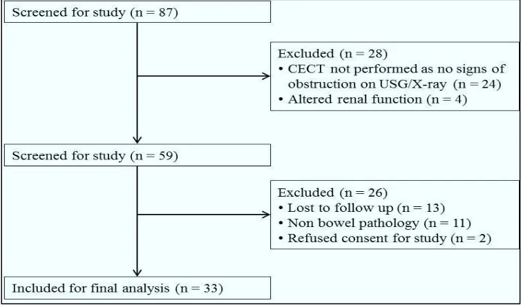
In our study we screened 87 patients who presented with complaints of intestinal obstruction. Among them, 24 patients had showed normal bowel loops and peristalsis on ultrasound and unremarkable erect abdominal radiograph and were managed conservatively and did not undergo CT abdomen. CECT study was performed in finally in 59 patients as four patients had altered renal function and therefore those patients were taken up for surgery. Among them, 13 patients who underwent were lost to follow up. There were 11 patients who were diagnosed with non-bowel pathological causing mass effect and causing pseudo-obstruction and therefore were not considered. Two patients refused to participate in the study and data from 33 patients was included for final analysis (Figure 1).
e
387
e
387
e387
e
387
e387
e
387
e387
e
387
e387
e
387
e
387
e
387
e
387
e387
e
387
e387
e
387
e387
e
387
e387
e
38
7
Table 1: Age and Gender-wise Distribution of Patients
Age group (in years) Male Female Total
11 to 20 0 1 1
21 to 30 2 2 4
31 to 40 0 2 2
41 to 50 4 3 7
51 to 60 2 4 6
61 to 70 4 5 9
71 to 80 1 3 4
Total 13 20 33
There were 33 patients with suspected bowel obstruction who were included in the final analysis. In our study there were more than 60% were females (n = 20) and remaining 13 patients were males (39.4%). The age and gender-wise distribution of patients is mentioned in Table 1. The commonest age groups belonged to patients of age 61 to 70 years (n = 9; 27.3%) followed by 41 to 50 years (n = 7; 21.2%), 51 to 60 years (n = 5; 18.2%), 71 to 80 years and 21 to 30 years (n = 4 each; 12.1%), 31 to 40 years (n = 2; 6.1%) and lastly age group of 11 to 20 years (n = 1; 3%).
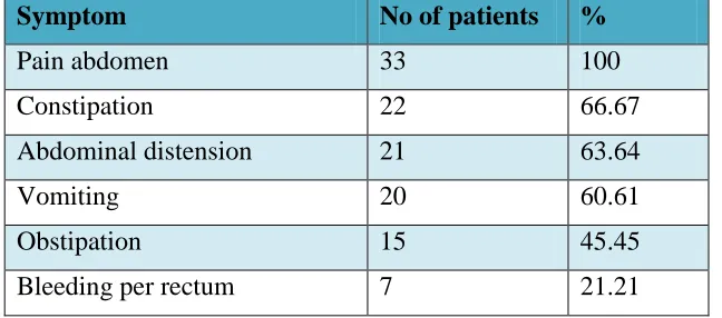
Table 2: Clinical Presentation in Patients with Bowel Obstruction
Symptom No of patients %
Pain abdomen 33 100
Constipation 22 66.67
Abdominal distension 21 63.64
Vomiting 20 60.61
Obstipation 15 45.45
Bleeding per rectum 7 21.21
The commonest presenting complaint was pain abdomen, which was seen in all the patients followed by constipation in 22 patients (66.67%), abdominal distension (n = 21; 63.64%), vomiting (n = 20; 60.61%), obstipation (n = 15; 45.45%) and lastly bleeding per rectum in seven patients (21.21%) (Table 2). Majority of the patients presented with multiple complaints. Almost all the patients with SBO presented with pain abdomen, vomiting, abdominal distension and constipation. Majority of the patients with LBO presented with pain abdomen, constipation, obstipation, abdominal distension, and bleeding per rectum. None of the patients with SBO presented with bleeding per rectum. Similarly obstipation was also primarily a complaint in patients with LBO and only small percentage of patients with SBO had obstipation.
Table 3: Type of Modality & Bowel Obstruction
Type of modality showing bowel obstruction
No of bowel obstruction
%
X-ray 9 42.8
388
388
388
388
388
388
388
388
388
388
388
388
388
388
388
388
388
388
388
388
388
Final diagnosis 22 100
At final diagnosis, there were 21 patients with intestinal obstruction (there were 22 instances of bowel obstruction, as one patient had both small and large bowel obstruction). In all of the patients with clinically suspected bowel erect X-ray abdomen was performed. On erect X-ray abdomen intestinal obstruction was suspected in nine patients (42.8%). The features seen in radiographs were multiple air-fluid levels in six patients (66.67%) followed by fluid-filled bowel loops (n = 4; 44.4%), and lastly gasless abdomen and dilated colon (n = 2 patients each; 22.2%). On CT there were 23 patients with 24 bowel obstruction (109.1%) who were suspected to have bowel obstruction (Table 3). CT over estimated two patients with bowel obstruction, which on surgery proved otherwise. Both these patients were diagnosed with small bowel obstruction. The finding seen in these two patients was dilated small bowel loops (>2.5 cm). The overall sensitivity, specificity, PPV, NPV and accuracy of CT was 100%, 83.33%, 64.71%, 91.67% and 94.21% respectively.
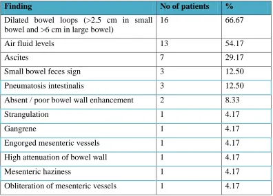
Table 4: CT Findings in Intestinal Obstruction
Finding No of patients %
Dilated bowel loops (>2.5 cm in small bowel and >6 cm in large bowel)
16 66.67
Air fluid levels 13 54.17
Ascites 7 29.17
Small bowel feces sign 3 12.50
Pneumatosis intestinalis 3 12.50
Absent / poor bowel wall enhancement 2 8.33
Strangulation 1 4.17
Gangrene 1 4.17
Engorged mesenteric vessels 1 4.17
High attenuation of bowel wall 1 4.17
Mesenteric haziness 1 4.17
Obliteration of mesenteric vessels 1 4.17
The commonest findings observed on CT were dilated bowel loops (>2.5 cm in small bowel and >6 cm in large bowel) in 16 patients (66.67%), followed by air fluid levels in 13 patients (54.17%), ascites in seven patients (n = 7; 29.17%), pneumatosis intestinalis and ‘small bowel feces’ sign in three patients each (12.5%), and absent or poor bowel wall enhancement in two patients each (8.33%) (Table 4). Other findings of strangulation, gangrene, engorged mesenteric vessels, high attenuation of bowel wall, mesenteric haziness and obliterated mesenteric vessels were seen in one patient each (4.17%).
Table 5: Level and Site of Bowel Obstruction
Site CT Underwent
surgery
Level of obstruction
Large bowel only 11 8
e
389
e
389
e389
e
389
e389
e
389
e389
e
389
e389
e
389
e389
e389
e389
e389
e
389
e389
e
389
e389
e
389
e389
e
389
Both 1 0
Sublevel of SBO
Duodenum 1 0
Jejunum 1 0
Ileum and ileocecal junction 10 8
Sublevel of LBO
AC 1 1
TC 2 0
DC 1 1
SC & RSJ 3 2
Rectum 5 4
In our study we observed a total of 23 patients (with 24 instances) with bowel obstruction of which 11 patients had SBO only, 11 patients had LBO only and one patient had both LBO and SBO, thus making 12 findings of SBO and LBO each. Surgery was performed in eight patients with SBO and LBO each (Table 5). Among patients with SBO only, those with intestinal Koch’s and SMA syndrome were managed conservatively and were not operated. When only LBO was considered, patients with inoperable carcinoma rectal or colon were managed conservatively. One patient with peritoneal carcinomatosis had both SBO and LBO probably caused due to ascites and peritoneal adhesions. Because of peritoneal carcinomatosis, this patient was not operated and was managed conservatively. The lesion was causing obstruction in ileum & transverse colon.
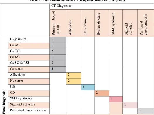
Among SBO only, the commonest level of obstruction was ileum and ileocecal junction (n = 10; 83.3%). Duodenal obstruction and jejunal obstruction were seen in one patient each (8.33%). The patient with duodenal obstruction was diagnosed with SMA syndrome and was managed conservatively. The patient with jejunal obstruction was diagnosed on CT as primary carcinoma which was managed conservatively due to advanced disease and diagnosis was confirmed on biopsy. Among the 10 patients with ileal and ileocecal junction obstruction four patients were suspected to have adhesions of which two cases were confirmed during surgery. Two patients with CT diagnosed adhesions were found to have no evidence of intestinal obstruction on surgery.
The patients improved post-surgery. Three patients had intestinal tuberculosis of which two patients were operated and one patient was managed conservatively. There were two patients who had benign stricture caused due to Crohn’s disease. Both the patients were operated.. The remaining patient was diagnosed with peritoneal carcinomatosis and was managed conservatively (Table 6).
390
390
390
390
390
390
390
390
390
390
390
390
390
390
390
390
390
390
390
390
390
Table 6: Correlation between CT Diagnosis and Final Diagnosis
CT Diagnosis
P
rimar
y
bowe
l
tum
our
Adhe
sions
TB st
ric
ture
B
enign s
trictur
e
S
MA syndr
ome
S
igm
oid
volvul
us
P
eritone
al
ca
rc
inom
atosi
s
Fin
al Diagn
osis
Ca jejunum 1
Ca AC 1
Ca TC 2
Ca DC 1
Ca SC & RSJ 3
Ca rectum 5
Adhesions 2
No cause 2
ITB 3
CD 2
SMA syndrome 1
Sigmoid volvulus 1
Peritoneal carcinomatosis 1
e
391
e
391
e391
e
391
e391
e
391
e391
e
391
e391
e
391
e
391
e
391
e
391
e391
e
391
e391
e
391
e391
e
391
e391
e
391
IMAGES
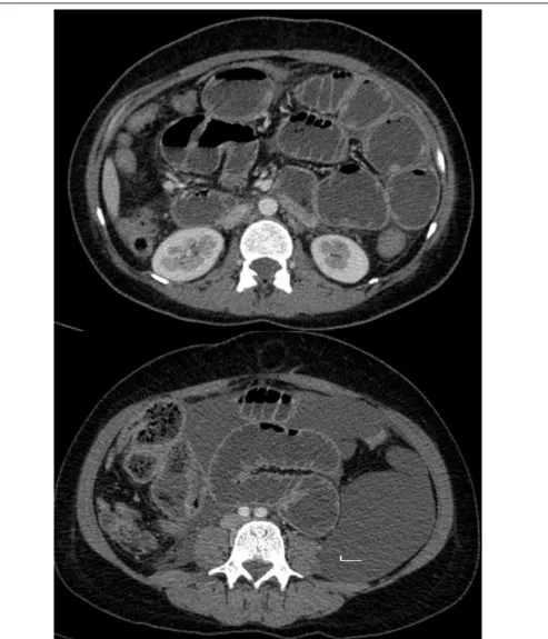
Figure Error! Main Document Only.: Large bowel neoplasm. Contrast enhanced images show a neoplastic lesion at the junction of descending and sigmoid colon with luminal narrowing and proximal bowel
392
392
392
392
392
392
392
392
392
392
392
392
392
392
392
392
392
392
392
392
392
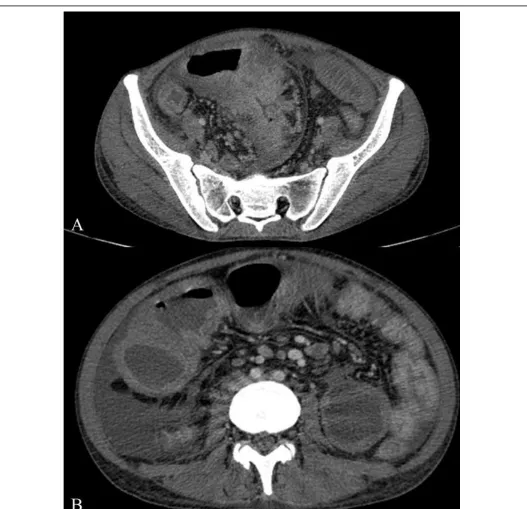
Figure Error! Main Document Only.: Crohn’s disease. Contrast enhanced images show ileocecal wall thickening with pericecal fat stranding. Long segment small bowel thickening with focal strictures also
e
393
e
393
e393
e
393
e393
e
393
e393
e
393
e393
e
393
e
393
e
393
e
393
e393
e
393
e393
e
39
3
e
393
e
393
e393
e
393
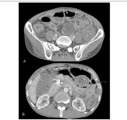
Figure Error! Main Document Only.: Contrast enhanced images show circumferentially enhancing mass in the terminal ileal region with features of proximal obstruction. Multiple lymph nodes also noted.
394
394
394
394
394
394
394
394
394
394
394
394
394
394
394
394
394
394
394
394
394
e
395
e
395
e395
e
395
e395
e
395
e395
e
395
e395
e
395
e
395
e
395
e
395
e395
e
395
e395
e
395
e395
e
395
e395
e
395
396
396
396
396
396
396
396
396
396
396
396
396
396
396
396
396
396
396
396
396
396
e
397
e
397
e397
e
397
e397
e
397
e397
e
397
e397
e
397
e
397
e
397
e
397
e397
e
397
e397
e
397
e397
e
397
e397
e
397
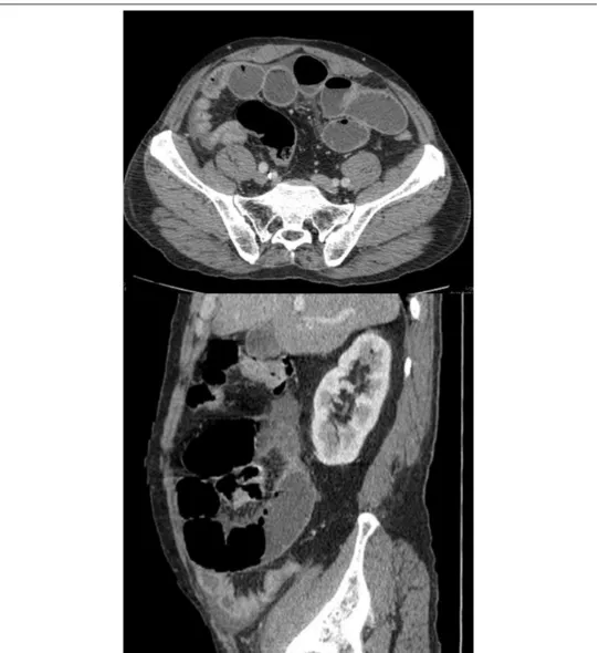
Figure Error! Main Document Only.: Case of superior mesenteric artery syndrome. Contrast enhanced images grossly distended stomach, 1st and 2nd part of Duodenum with transition at 3rd part. There is
marginally reduced aorto –mesenteric distance
DISCUSSION
Intestinal obstruction is one of important differential diagnosis in a patient presenting with acute abdomen. Clinical diagnosis of intestinal obstruction can be challenging and imaging a significant role in diagnosis of intestinal obstruction. Currently CECT abdomen is considered as the most appropriate radiological investigation in evaluation of suspected small and large bowel obstruction. CT is able to demonstrate the level of bowel obstruction, diagnose common causes of bowel obstruction and to differentiate between high- and low-grade obstructions. Furthermore CECT also helps to assess complications of obstruction, such as strangulation
In our study, intestinal obstruction was observed more frequently in females as compared with males (60.6% vs 39.4% respectively). The commonest age groups belonged to patients of age 61 to 70 years (n = 9; 27.3%) followed by 41 to 50 years (n = 7; 21.2%) and 51 to 60 years (n = 5; 18.2%). Least number of patients were in the age group of 11 to 20 years (n = 1; 3%).
The gender distribution in our study is different as compared with findings reported by Saini et al, who in their study of 40 patients in urban set up reported a male predominant populace with males constituting 67% (n = 27). They also reported the commonest age group of patients to be between ages 31 to 45 (n = 13; 33%) followed by 46 to 60 years (n = 12; 30%) and 15 to 30 years (n = 11; 27%). In our study we observed more number of cases of intestinal obstruction with increasing age, whereas Saini et al have reported more cases of intestinal obstruction in middle age groupi. This difference in age-group and gender distribution could be attributed to different cultures and socioeconomic strata of patients observed in both the set ups.
398
398
398
398
398
398
398
398
398
398
398
398
398
398
398
398
398
398
398
398
398
the patients presented with multiple complaints. Almost all the patients with SBO presented with pain abdomen, vomiting, abdominal distension and constipation. Majority of the patients with LBO presented with pain abdomen, constipation, obstipation, abdominal distension, and bleeding per rectum. None of the patients with SBO presented with bleeding per rectum. Similarly obstipation was also primarily a complaint in patients with LBO and only small percentage of patients with SBO had obstipation.
Data from various studies have also shown similar clinical complaints. Saini et al have reported pain abdomen in all the patients with bowel obstruction, abdominal distension in 82.5% of patients, vomiting in 67.5% of patients, followed by constipation/obstipation in 60% of patients, and abdominal tenderness in 65% of patientsError! Bookmark not defined.. Singhania et al in the study on 53 patients with bowel obstruction reported abdominal distension in about 75.47% of patients, constipation in 73.58%, vomiting in 54.72% and abdominal pain in 56.6% of patients
In our study, there were 21 patients with intestinal obstruction (there were 22 instances of bowel obstruction, as one patient had both small and large bowel obstruction). Erect X-ray abdomen showed intestinal obstruction in nine patients (42.8%). The features seen in radiographs were multiple air-fluid levels in six patients (66.67%) followed by fluid-filled bowel loops (n = 4; 44.4%), and lastly gasless abdomen and dilated colon (n = 2 patients each; 22.2%). On CT there were 23 patients (109.1%) (with 24 bowel obstruction) who were diagnosed with bowel obstruction. CT over estimated two patients with bowel obstruction, which on surgery proved otherwise. Both these patients were diagnosed with small bowel obstruction. The finding seen in these two patients was dilated small bowel loops (>2.5 cm). The overall sensitivity, specificity, PPV, NPV and accuracy of CT was 100%, 83.33%, 64.71%, 91.67% and 94.21% respectively. In our study CT evaluation was performed with axial sections and coronal and sagittal reformations for better understanding and delineation of bowel pathology.
Our study results are similar to data seen in other studies. Pongpornsup S et al in their study on 35 patients with SBO reported that CT diagnosed 25 cases with SBO of which one false positive and remaining were true positive. The authors reported that CT had overall sensitivity, specificity, PPV, NPV and accuracy of 96%, 100%, 100%, 90% and 97% respectively for diagnosing SBOi. Filippone et al reported 94% accuracy of CT in diagnosis of SBO. It was shown by the authors that addition of coronal reformations improved accuracy of CT in diagnosis of SBO (88% versus 94% in axial and axial with coronal reformations respectively). They also reported that when compared with final diagnosis axial sections alone were better at delineating SBO when compared with coronal reformations alone (92% vs 82% respectively). The authors also reported improved accuracy in diagnosis of LBO (88% versus 92% in axial and axial with coronal reformations respectively)i. Other studies have however reported lesser accuracy of CT in diagnosis of bowel obstruction. Singhania et al reported that CT identified 81.13% of cases of SBO. The sensitivity and specificity of CT was reported as 97.29% and 63.63% respectively. In their study of 53 patients, bowel obstruction was diagnosed in 43 patients on CT; however the final diagnosis revealed only 37 cases of intestinal obstructionError! Bookmark not defined.. Mallo et al conducted a review where they reported that the sensitivity of CT in diagnosis of SBO ranged from 81 to 100%, specificity 68 to 100%, PPV of 84 to 100% and NPV of 76 to 100%i, which is consistent with our study. It is possible that studies having a lower sensitivity and specificity may be due to inherent selection bias in the studies. CT may show lower performance if the patient population in the study has relatively good number of patients with low grade obstruction. This can be concurred with data reported by Pongpornsup et al, who reported CT could identify all cases of high grade obstruction and could correctly identify only 58% of low grade SBOError! Bookmark not defined.. In our region we tend to get majority of cases with high grade obstruction, where the sensitivity and specificity of CT is high.
e
399
e
399
e399
e
399
e399
e
399
e399
e
399
e399
e
399
e
399
e
399
e
39
9
e
399
e
399
e399
e
399
e399
e
399
e399
e
399
strangulation, gangrene, engorged mesenteric vessels, high attenuation of bowel wall, mesenteric haziness and obliterated mesenteric vessels all of which were seen in one patient each (4.17%).
In our study the ‘small bowel feces’ sign was seen in three patients (12.5%) all of who had high grade bowel obstruction. ‘Small bowel feces’ sign is considered a highly specific sign for bowel obstruction. It is believed that in chronic or high grade obstruction, there is stasis and mixing of small bowel contents, which creates an appearance likened to feces in colon and hence the name ‘small bowel feces’ sign. It is usually present in high grade obstruction. The importance of this sign is that it is present just proximal to the site of obstruction/transition point and therefore helps in identifying the transition point in bowel obstructioni,i. Singhania et al reported presence of ‘small bowel feces’ sign in 5% of casesError! Bookmark not defined.. Lazarus et al reported a high ratio of small bowel feces sign in their study (n = 19 of 34 patients; 55.9%) in patients with SBO only. In their study they had relatively high number of moderate and high grade obstruction and this probably explains the unusually high percentage of ‘small bowel feces’ sign in their studyError! Bookmark not defined..
In our study we observed a total of 23 patients (with 24 instances) with bowel obstruction of which 11 patients had SBO only, 11 patients had LBO only and one patient had both LBO and SBO, thus making 12 findings of SBO and LBO each. Surgery was performed in eight patients with SBO and LBO each. One patient with peritoneal carcinomatosis had both SBO and LBO probably caused due to ascites and peritoneal adhesions. Because of peritoneal carcinomatosis, this patient was not operated and was managed conservatively. The lesion was causing obstruction in ileum & transverse colon. Among SBO only, the commonest level of obstruction was ileum and ileocecal junction with duodenal obstruction and jejunal obstruction seen in one patient each. The patient with duodenal obstruction was diagnosed with SMA syndrome and was managed conservatively. The patient with jejunal obstruction was diagnosed on CT as primary carcinoma which was managed conservatively due to advanced disease and diagnosis was confirmed on biopsy. Among the 10 patients with ileal and ileocecal junction obstruction four patients were suspected to have adhesions of which two cases were confirmed during surgery. Two patients with CT diagnosed adhesions were found to have no evidence of intestinal obstruction on surgery. The patients improved post-surgery. Three patients had intestinal tuberculosis of which two patients were operated and one patient was managed conservatively. There were two patients who had benign stricture caused due to Crohn’s disease. Both the patients were operated. The remaining patient was diagnosed with peritoneal carcinomatosis and was managed conservatively. Among LBO the commonest site of obstruction was rectum in 41.67% of patients followed by sigmoid colon (25%) and transverse colon (16.67%). LBO at ascending colon and descending colon were observed in 8.3%. Four patients with carcinoma rectum were operated and the diagnosis was confirmed histopathologically. One patient with carcinoma rectum was managed conservatively due to advanced disease and diagnosis was confirmed with biopsy. There were two cases with carcinoma sigmoid colon and rectosigmoid junction of which one was operated and finding was confirmed histopathologically. One was managed conservatively due to advanced disease and diagnosis was confirmed on biopsy. There was one case of sigmoid volvulus, which was managed surgically and finding was confirmed. There were carcinomas once each in ascending, transverse & descending colon of which ascending and descending colon tumours were operated and their histopathology report came out to be adenocarcinoma. The carcinoma of transverse colon was managed conservatively due to advanced disease and diagnosis was confirmed as adenocarcinoma with biopsy.
400
400
400
400
400
400
400
400
400
400
400
400
400
400
400
400
400
400
400
400
400
colon in 25% of patients, transverse colon in 17 of patients, and sigmoid colon in 21% of patients. When LBO was considered, neoplasms were cause in majority of patients (n = 17 of 21; 71%), which is consistent with our study. The other causes of LBO were volvulus in three patients and diverticulitis in four patientsError! Bookmark not defined.. Singhania et al in their study on 53 patients reported SBO in 69.8% of patients and LBO in only 11.3% of patients. The commonest cause for SBO in their study was adhesions (22.92%) followed by inflammatory cause (9.3%), volvulus (8.33%), bowel neoplasm, hernia, and intussusceptions (6.25% each). Other uncommon causes were intestinal malrotation, extrinsic cause, and foreign body. No cause could be identified in about 16.28% of casesError! Bookmark not defined.. Pongpornsup et al in their study on 35 patients reported adhesions as commonest cause for SBO in 10 patients followed by metastases in four patients. Other uncommon causes were postradiative enteropathy, internal hernia, inguinal, submucosal hernia, midgut volvulus, SMA syndrome and benign stricture in one patient eachError! Bookmark not defined.. Ali et al in their study in 40 patients demonstrated that both extrinsic and intrinsic causes were equally seen in SBO with commonest cause being adhesions, hernias followed by carcinoid tumour, appendicular cause, mesenteric vein thrombosis, Crohn’s disease, lymphoma, ileal carcinoma. Uncommon causes for SBO reported in their study were gall stone ileus, midgut volvulus and Ladd’s band compressing duodenum. The unusual finding reported by the authors is probably due to the age group of patients (15 to 30 years) and the selective patient population who underwent CECTi. Megibow et al have also reported that adhesions were commonest cause for SBO followed by small bowel neoplasm, metastasis, Crohn’s disease, hernia, hematoma, diverticulitis, intussusception, gall stone ileus and appendicitisi. The variation in the findings may be due to the native patient population and the disease demographics. While abdominal tuberculosis is common in our region and is a known cause of SBO in our population, the same may not be applicable in other patient population. We commonly receive patients with rectal carcinoma and this may the reason of high percentage of patients with rectal and rectosigmoid carcinomas in our studies.
Our study has certain limitations. Our patient population was limited and a more extensive patient population could have shown other factors causing bowel obstruction. Final diagnosis was not available in patients treated conservatively. This could theoretically affect the overall accuracy of CT in evaluation of bowel obstruction. It is possible that majority of patients who present with suspected intestinal obstruction have high grade obstruction and our data may have inadvertently been biased towards high grade obstruction and not general population.
CONCLUSION
Bowel obstruction is a fairly common encounter in clinical and radiological practice. There are various causes of bowel obstruction, which may make accurately diagnosing the cause of bowel obstruction a challenging task. CT provides accurate information in determining the cause and level of bowel obstruction. CT also helps to provide information on the viability of affected bowel tissue and help in treatment planning or identify the need for surgery. We recommend CT study as part of evaluation in patients presenting with bowel obstruction.
BIBLIOGRAPHY
1. Khurana B, Ledbetter S, McTavish J, Wiesner W, Pablo RR. Bowel Obstruction Revealed by Multidetector CT. Am J Roentgenol 2002; 178:1139–44.
2. Nicolaou S, Kai B, Ho S, Su J, Ahamed K. Imaging of acute small-bowel obstruction. AJR Am J Roentgenol 2005;185:1036-44.
3. Furukawa A, Yamasaki M, Takahashi M, Nitta N, Tanaka T, Kanasaki S, Yokoyama, et al. Small Bowel Obstruction: Scanning Technique, Interpretation and Role in the Diagnosis. Semin in Ultrasound CT MR October 2003; 24(5):336-52.
e
401
e
401
e401
e
401
e401
e
401
e401
e
401
e401
e
401
e
401
e
401
e
401
e401
e
401
e401
e
401
e401
e
401
e401
e
401
5. Saini DK, Chaudhary P, Durga CK, Saini K. Role of multislice computed tomography in evaluation and
management of intestinal obstruction. Clin Pract 2013;3:e20.
6. Singhania KV, Mehta R, Kazi Z. Role of multidetector computed tomography in bowel obstruction. Int J Sci Stud 2017;5(5):131-134
7. Pongpornsup S, Tarachat K, Srisajjakul S. Accuracy of 64 sliced multi-detector computed tomography in diagnosis of small bowel obstruction. J Med Assoc Thai 2009;92:1651-61.
8. Filippone A, Cianci R, Storto ML. Bowel obstruction: comparison between multidetector-row CT axial and coronal planes. Abdom Imaging 2007;32:310-6.
9. Mallo RD, Salem L, Flum DR. Computed tomography diagnosis of ischemia and complete obstruction in small bowel obstruction: a systematic review. J Gastrointest Surg 2005; 9(5):690–4.
10.Paulson EK, Thompson WM. Review of small-bowel obstruction: the diagnosis and when to worry. Radiology 2015;275:332-42.
11.Lazarus DE, Slywotsky C, Bennett GL, Megibow AJ, Macari M. Frequency and relevance of the "small-bowel feces" sign on CT in patients with small-"small-bowel obstruction. AJR Am J Roentgenol 2004;183:1361-6.
12.Ali SA, Mansour MGE, Farouk O. Utility of 64-row multidetector computed tomography in diagnosis and management of small bowel obstruction. Egyptian J Radiol Nucl Med 2017;48:839-46.