0095-1137/10/$12.00 doi:10.1128/JCM.00274-10
Copyright © 2010, American Society for Microbiology. All Rights Reserved.
Multilocus Sequence Typing Identifies Evidence for Recombination
and Two Distinct Lineages of
Corynebacterium diphtheriae
䌤
Frances Bolt,
1Pamela Cassiday,
2Maria Lucia Tondella,
2Aruni DeZoysa,
3Androulla Efstratiou,
3Andreas Sing,
4Aleksandra Zasada,
5Kathryn Bernard,
6Nicole Guiso,
7Edgar Badell,
7Marie-Laure Rosso,
7Adam Baldwin,
1and Christopher Dowson
1*
Biological Sciences, Warwick University, Coventry, United Kingdom1; Division of Bacterial Diseases, Centers for Disease Control and Prevention, Atlanta, Georgia2; Health Protection Agency, London, United Kingdom3; the National Consiliary Laboratory for Diphtheria (NCLD), Oberschleissheim, Germany4; the National Institute of Public Health (NIPH), Warsaw, Poland5;
National Microbiology Laboratory, PHAC, Winnipeg, Canada6; and Institut Pasteur, Molecular Prevention and Therapy of Human Diseases Unit. Paris, France7
Received 10 February 2010/Returned for modification 29 March 2010/Accepted 24 August 2010
We describe the development of a multilocus sequence typing (MLST) scheme forCorynebacterium
diphthe-riae, the causative agent of the potentially fatal upper respiratory disease diphtheria. Global changes in
diphtheria epidemiology are highlighted by the recent epidemic in the former Soviet Union (FSU) and also by the emergence of nontoxigenic strains causing atypical disease. Although numerous techniques have been
developed to characterizeC. diphtheriae, their use is hindered by limited portability and, in some instances,
poor reproducibility. One hundred fifty isolates from 18 countries and encompassing a period of 50 years were analyzed by multilocus sequence typing (MLST). Strain discrimination was in accordance with previous ribotyping data, and clonal complexes associated with disease outbreaks were clearly identified by MLST. The data produced are portable, reproducible, and unambiguous. The MLST scheme described provides a valuable
tool for monitoring and characterizing endemic and epidemicC. diphtheriaestrains. Furthermore, multilocus
sequence analysis of the nucleotide data reveals two distinct lineages within the population ofC. diphtheriae
examined, one of which is composed exclusively of biotype belfanti isolates and the other of multiple biotypes.
Diphtheria has historically evoked fear and terror due to its slow suffocating death and previously unknown origin, but so-cioeconomic improvement and the introduction of mass im-munization in the 1940s and 1950s led its near-elimination in the developed world. However, diphtheria remains a global disease and is endemic in many countries. The World Health Organization (WHO) has recorded outbreaks throughout the world, including Afghanistan, Algeria, Iraq, Lao People’s Re-public, Mongolia, Papua New Guinea, Sudan, and Thailand (1). It is also a potentially resurgent infectious disease, exem-plified in the 1990s by a notable epidemic in the newly inde-pendent states of the former Soviet Union (NIS), where vac-cination had been employed since 1958 (33). At least 20 cases were reported beyond these countries, highlighting the poten-tial threat of introduced strains from countries in which it is endemic and epidemic (29). Furthermore, according to sero-logical surveillance studies, the proportion of susceptible indi-viduals in vaccinated populations remains high (7, 10). Ed-munds et al. estimated that there are inadequate protection levels in the United Kingdom for 70 to 75% of those aged 50 to 60 years old (7). Similar observations were recently reported for individuals who had followed the French vaccine recom-mendations (17).
It is therefore apparent that typing tools enabling global
Corynebacterium diphtheriae surveillance are of great
impor-tance. Based upon their biochemical and morphological
prop-erties, fourC. diphtheriaebiotypes have been identified: mitis,
gravis, intermedius, and belfanti (8). Several typing techniques for C. diphtheriae have been developed. Traditionally these techniques were based upon serologic, phage, and biotyping methods. However, since the methods provide limited resolu-tion, molecular typing techniques, including amplified frag-ment length polymorphisms (AFLP) (4), random amplified polymorphic DNA (RAPD) (3, 24), multilocus enzyme elec-trophoresis (MEE) (28), spoligotyping (21), and pulsed-field gel electrophoresis (PFGE) (5), have been developed and show significant intraspecies genetic diversity. Recently a com-parison of the different typing techniques was performed (6). It was shown that the most discriminative was ribotyping, the
current “gold standard” typing method forC. diphtheriae. This
method has identified 86 distinct ribotype patterns and clusters isolates associated with the former Soviet Union (FSU) out-break (5, 11, 28). However, ribotyping is very dependent upon the use of a rigid standardized method, and without this, there are clearly difficulties in reproducibility (25). Additionally, typ-ing methods based upon band matchtyp-ing do not clearly reveal the population structure or underlying evolutionary mecha-nisms of a given species.
Proposed in 1998, multilocus sequence typing (MLST) over-comes the problems encountered with ribotyping by directly indexing nucleotide variation within several core metabolic genes, thereby providing portable, reproducible, and high-res-olution data appropriate for the evhigh-res-olutionary and epidemio-logical investigation of diphtheria (19). We describe the de-velopment of an MLST scheme to examine the genetic
* Corresponding author. Mailing address: Department of Biological Sciences, University of Warwick, Gibbet Hill Road, Coventry CV4 7AL, United Kingdom. Phone: 44 2476 523534. Fax: 44 2476 52356. E-mail: [email protected].
䌤Published ahead of print on 15 September 2010.
4177
on May 16, 2020 by guest
http://jcm.asm.org/
relationship between a temporally and geographically
di-verse collection ofC. diphtheriaeisolates.
MATERIALS AND METHODS
Bacterial strain collection.As shown in Table 1, a total of 150C. diphtheriae
isolates were examined and obtained from the Centers for Disease Control and Prevention (CDC) (Atlanta, GA), the Health Protection Agency (HPA) (Lon-don, United Kingdom), the National Consiliary Laboratory for Diphtheria (Ger-many), and the National Institute of Public Health (Poland). The MLST scheme was validated by comparison to 29 previously ribotyped isolates encompassing 20 different ribotypes (5, 28). The collection is both temporally and geographically diverse, with isolates from 18 countries, covering a time period from 1957 to 2006. Of the 45 human cases with available clinical information, 28.9% were associated with carriage (n⫽13), 42.2% were isolated from diphtheria (n⫽19), 11.1% were isolated from cutaneous lesions (n⫽5), 13.3% were isolated from patients with upper respiratory conditions, including pharyngitis (n⫽1), tonsil-litis (n⫽4), and a sore throat (n⫽1), and 4.3% (n⫽2) were obtained from patients with both osteomyelitis and cutaneous lesions. Both toxigenic (n⫽96) and nontoxigenic (n⫽52) (toxigenic data were unavailable for two isolates) biotype gravis (n⫽43), intermedius (n⫽6), mitis (n⫽85), and belfanti (n⫽
16) strains were examined.
Biotyping and toxigenicity testing.Toxigenicity was determined by the Elek immunoprecipitation method (Elek test) in accordance with the WHO guide-lines (9). All isolates were biotyped using the API Coryne kit (bioMe´rieux, Lyon, France) according to the manufacturer’s instructions.
Locus selection and amplification.Potential housekeeping genes were iden-tified by comparing theC. diphtheriae(1),Corynebacterium glutamicum(C. glu-tamicum) (15), and Corynebacterium efficiens (C. efficiens) (26) genome se-quences using the Artemis Comparison Tool (ACT) and the Double ACT program, available at http://www.sanger.ac.uk/Software/ACT/ and http://www .hpa-bioinfotools.org.uk/pise/double_act.html, respectively. Amplification and nested sequencing primers were designed for the lociatpA,dnaE,dnaK,fusA,
leuA,odhA, andrpoB(Table 2) using Primer3 (30). The sequencing primers for
rpoBwere previously described by Khamis and colleagues (16).
DNA was extracted primarily as described by Mothershed et al. (23). Each 25-l PCR was carried out using 10 ng of chromosomal DNA, 5l Q solution (Qiagen, United Kingdom), 4.0l chromosomal DNA (5 to 20 ng/l), 1.0l forward primer (10 pmol/l), 1.0l reverse primer (10 pmol/l), 2.5l 10⫻PCR buffer (Qiagen) (containing 15 mM MgCl2), 0.5l deoxynucleoside triphosphate (dNTP) solution (Qiagen) (10 mM [each] dNTP), 0.125lTaq polymerase (Qiagen, 5 U/l), 0.5l MgCl2(Qiagen) (25 mM), and PCR-grade water. All primer sets were designed to ensure they had similar melting temperatures, and reaction conditions were as follows: initial denaturation at 94°C for 1 min; 35 cycles of denaturation at 94°C for 1 min; and primer annealing at 58°C for 1 min and extension at 72°C for 2 min, followed by a final extension step of 72°C for 5 min. Amplicons were purified using the MiniElute UF plates (Qiagen, United Kingdom) according to the manufacturer’s instructions and stored at⫺20°C.
Amplicon nucleotide sequences were determined by nested sequencing using the BigDye Terminator ready reaction mix, v3.1 (Perkin-Elmer Applied Biosys-tems, Foster City), following the manufacturer’s protocol. The forward and reverse sequences of a given locus were edited, aligned, and trimmed to the desired length using the SeqManII software program (DNASTAR, Madison, WI).
Allele and sequence type designation.Allelic numbers were assigned to each unique allele for a given locus. For each isolate, the allelic profile was generated by combining the allele numbers for each locus in the orderatpA,dnaE,dnaK,
fusA,leuA,odhA, andrpoB. A novel sequence type (ST) designation was given to all unique allelic profiles, while isolates with identical profiles belonged to the same ST.
Tree congruence.Tree concordance was assessed using the method developed by Holmes and colleagues; however, here tree congruence data were increased to compare 200 rather than 100 randomly generated maximum-likelihood (ML) trees (14).
Phylogenetic analysis.The allelic sequences for each isolate were concate-nated (2,545 bp), and phylogenetic trees, with 1,500 bootstrap replicates, were generated by the neighbor-joining method using the Jukes Cantor algorithm within the MEGA software program, v4 (32). Isolates were grouped based upon the MLST definition of a clonal complex (or eBURST group) being a cluster of isolates sharing at least six of seven alleles, using the eBURST program, available at www.mlst.net. To visualize clustering within the population and to detect recombination between STs, Splits decomposition analysis was performed using the SplitsTree software program, v4. Neighbor-joining phylogenetic analysis was
done (www.mlst.net), and the index of association (IA) was calculated using the
LIAN software program, v3.5 (www.pubmlst.org). SignificantIAvalues were
determined using the Monte-Carlo method with 1,000 resamplings. To ensure sampling bias did not affect the value, one representative of each ST was used. The allelic frequencies, GC content, number of polymorphic sites, and ratio of nonsynonymous substitutions to synonymous substitutions (dN/dSratio) for all
seven loci were calculated using the START software program, v2. Nucleotide identities were calculated using the maximum composite likelihood model within MEGA, v4.
RESULTS
Allelic variation.In order to validate the MLST scheme, a
collection of temporally and geographically diverse isolates were analyzed, including two equine isolates previously de-scribed by Henricson et al. (13). Cultures from both epidemi-ologically linked and unrelated cases were examined to assess the scheme’s performance. Isolates previously designated as
belonging to one of the fourC. diphtheriae biotypes, gravis,
mitis, intermedius, and belfanti, were typed by MLST to better understand their genetic relationships to each other and to determine their epidemiological value.
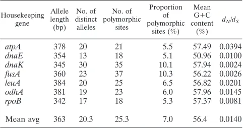
As shown in Table 3, among the 150 isolates investigated, the mean average allele length for each locus was 363 bp and
ranged from 342 bp (rpoB) to 384 bp (leuA). All alleles for a
given locus were of equal lengths and, to aid further analysis, were in the correct reading frame. The proportion of variable
sites at each locus varied from 5.1% (dnaE) to 10.3% (fusA)
(mean average⫽7%).
To determine the degree of selective pressure upon each locus, the ratio of nonsynonymous to synonymous substitutions
(dN/dS) was determined. Since the ratios were significantly less
than 1, it is clear that the genes chosen were not under puri-fying selection and were therefore suitable for MLST analysis (Table 3).
Assignment of allele and sequence types.A total of 73 STs
were assigned to the 150 C. diphtheriaeisolates investigated
and divided into 11 clonal complexes designated by eBURST groups (Table 1). For this study, isolates were assigned as members of an eBURST group when six of seven MLST alleles were shared.
Strain identification.MLST identified two clonal complexes
linked to diphtheria outbreaks. eBURST group 2 (composed of ST-8, ST-12, ST-52, and ST-66 strains) was associated with the FSU epidemic. Six isolates identified as having a
Sankt-Peterburg (n⫽3) or Rossija (n⫽3) ribotype, the two clonally
derived ribotypes linked with the FSU outbreak, clustered within this group, as did three epidemic strains identified by Skogen and colleagues in cultures obtained prior to the out-break (31). Interestingly, the first nontoxigenic gravis strain to cause septicemia and endocarditis in Poland also belonged to eBURST group 2. This strain (493/K/04) was isolated in a region where no diphtheria cases had been reported for 10 years (34).
Twenty-eight of 31 isolates from Haiti (n ⫽ 14) and the
Dominican Republic (n⫽ 17) were collected during a
diph-theria epidemic (2004 to 2006). Eighty-nine percent (n⫽25)
of the outbreak isolates were ST-31, and another (isolate 158)
was ST-4, a single locus variant (SLV) of ST-31 at thefusA
locus (98.06% allelic nucleotide similarity). The SLV is likely to have arisen by recombination, since multiple nucleotide
on May 16, 2020 by guest
http://jcm.asm.org/
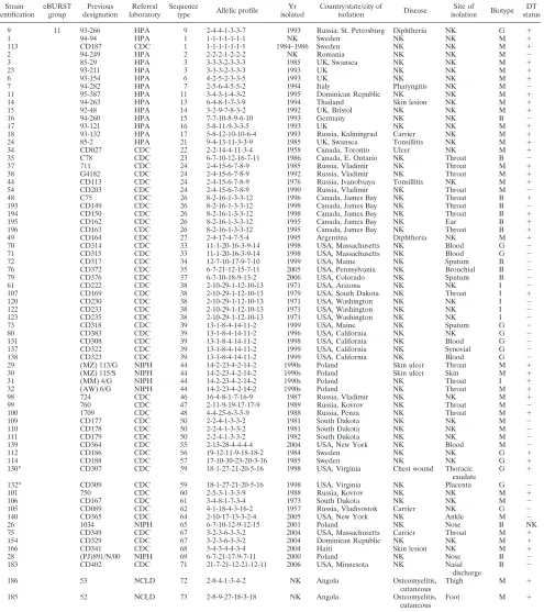
TABLE 1. Details ofC. diphtheriaestrains useda
Strain identification
eBURST group
Previous designation
Referral laboratory
Sequence
type Allelic profile
Yr isolated
Country/state/city of
isolation Disease
Site of
isolation Biotype DT status
27 1 (MS) 7/97 NIPH 32 3-1-18-4-13-3-5 1997 Poland NK NK G NK
64 1 CD293 CDC 32 3-1-18-4-13-3-5 1996 Kazakhstan NK NK M ⫹
66 1 CD295 CDC 32 3-1-18-4-13-3-5 1996 Kazakhstan NK Throat M ⫺
78 1 CD373 CDC 36 3-7-18-4-13-3-5 2005 USA, California Sore throat Throat G ⫺
96 1 722 CDC 43 15-1-18-4-13-3-5 1986 Russia, Kovrov Carrier Throat G ⫹
127 1 CD286 CDC 58 3-1-26-4-13-3-5 1996 Kazakhstan NK NK G ⫹
129 1 CD303 CDC 70 3-1-18-4-20-3-5 1997 USA, Michigan NK Blood G ⫺
8 2 95-135 HPA 8 3-5-6-5-3-3-6 1993 Finland NK NK G ⫹
25 2 493/K/04 NIPH 8 3-5-6-5-3-3-6 2004 Poland NK NK G
-36 2 G4174 CDC 8 3-5-6-5-3-3-6 1993 Russia, Leningrad Diphtheria NK G ⫹
45 2 CD126 CDC 8 3-5-6-5-3-3-6 1985 Russia, Moscow Diphtheria NK G ⫹
46 2 CD130 CDC 8 3-5-6-5-3-3-6 1987 Russia, Sverdlovsk Tonsillitis NK G ⫹
47 2 CD131 CDC 8 3-5-6-5-3-3-6 1987 Russia, Sverdlovsk Carrier NK G ⫹
62 2 CD290 CDC 8 3-5-6-5-3-3-6 1996 Kazakhstan NK NK G ⫹
63 2 CD291 CDC 8 3-5-6-5-3-3-6 1996 Kazakhstan NK NK G ⫹
65 2 CD294 CDC 8 3-5-6-5-3-3-6 1996 Kazakhstan NK Nose M
-126 2 CD283 CDC 8 3-5-6-5-3-3-6 1996 Kazakhstan NK NK G ⫹
13 2 93-146 HPA 12 1-5-6-5-3-3-6 1993 Russia, Kaliningrad Carrier NK G ⫹
124 2 CD267 CDC 52 3-5-6-5-3-10-6 1996 Kazakhstan NK NK G ⫹
69 3 CD306 CDC 42 6-7-10-8-9-12-10 1997 USA, Michigan NK Sputum B
-95 2 749 CDC 66 3-5-6-5-11-3-6 1987 Russia, Kovrov NK Throat G ⫹
116 3 CD198 CDC 51 6-7-10-8-9-19-10 1999 USA, California NK Nose B
-133 3 CD310 CDC 54 6-7-10-22-9-12-10 1998 USA, Ohio NK Sputum B
-10 4 93-32 HPA 10 5-2-7-1-3-5-8 1993 UK NK NK G ⫹
108 4 CD170 CDC 49 2-6-7-1-3-5-8 1973 South Dakota NK NK G ⫹
115 4 CD196 CDC 63 5-6-7-1-3-5-8 1978 USA, Colorado NK Throat G ⫹
19 5 95-115 HPA 18 8-6-7-6-6-3-8 1995 Russia, Vladimir
region
Diphtheria NK G ⫹
20 5 93-181 HPA 19 2-6-7-6-6-3-8 1966 Russia, Moscow Carrier NK G ⫹
12 5 93-69 HPA 25 5-6-7-6-6-3-8 NK Russia, Murmansk Diphtheria NK G ⫹
39 5 CD085 CDC 25 5-6-7-6-6-3-8 1957 Russia, Vladivostok Diphtheria NK G ⫹
40 5 CD087 CDC 25 5-6-7-6-6-3-8 1957 Russia, Vladivostok Carrier NK G ⫹
41 5 CD092 CDC 25 5-6-7-6-6-3-8 1964 Russia, Moscow Carrier NK G ⫹
42 5 CD094 CDC 25 5-6-7-6-6-3-8 1965 Russia, Moscow Carrier NK G ⫹
43 5 CD097 CDC 25 5-6-7-6-6-3-8 1971 Russia, Moscow Carrier NK G ⫹
51 5 PR79 CDC 25 5-6-7-6-6-3-8 1996 USA, South Dakota NK NK G ⫹
125 5 CD268 CDC 25 5-6-7-6-6-3-8 1996 Kazakhstan NK NK G ⫹
97 6 G4212 CDC 45 2-10-24-1-3-15-2 1992 Russia, Murmansk Diphtheria NK M ⫹
128 6 CD301 CDC 53 2-10-24-1-3-3-2 1997 USA, Oregon NK Throat M
-52 7 1507 CDC 28 2-9-3-13-3-3-3 1995 Kyrgyzstan NK NK M ⫹
53 7 1512 CDC 41 2-9-22-13-3-3-3 1995 Kyrgyzstan NK NK M ⫹
21 8 90-39 HPA 20 8-3-3-2-7-3-3 1990 UK, London Tonsillitis NK M ⫹
67 8 CD304 CDC 40 8-3-19-2-7-3-3 1997 Guatemala NK NK M
-103 8 CD081 CDC 40 8-3-19-2-7-3-3 1997 Russia, Moscow NK NK M
-118 8 CD209 CDC 40 8-3-19-2-7-3-3 1997 Russia NK NK M
-4 9 95-385 HPA 4 3-2-4-3-4-4-4 1995 Dominican Republic NK NK M ⫹
158 9 CD333 CDC 4 3-2-4-3-4-4-4 2004 Dominican Republic NK NK M ⫹
59 9 CD216 CDC 31 3-2-4-15-4-4-4 2000 Dominican Republic NK NK M ⫹
74 9 CD340 CDC 31 3-2-4-15-4-4-4 2004 Haiti Diphtheria Throat M ⫹
149 9 CD324 CDC 31 3-2-4-15-4-4-4 2004 Dominican Republic NK NK M ⫹
150 9 CD325 CDC 31 3-2-4-15-4-4-4 2004 Dominican Republic NK NK M ⫹
151 9 CD326 CDC 31 3-2-4-15-4-4-4 2004 Dominican Republic NK NK M ⫹
152 9 CD327 CDC 31 3-2-4-15-4-4-4 2004 Dominican Republic NK NK M ⫹
153 9 CD328 CDC 31 3-2-4-15-4-4-4 2004 Dominican Republic NK NK M ⫹
155 9 CD330 CDC 31 3-2-4-15-4-4-4 2004 Dominican Republic NK NK M ⫹
156 9 CD331 CDC 31 3-2-4-15-4-4-4 2004 Dominican Republic NK NK M ⫹
157 9 CD332 CDC 31 3-2-4-15-4-4-4 2004 Dominican Republic NK NK M ⫹
159 9 CD334 CDC 31 3-2-4-15-4-4-4 2004 Dominican Republic NK NK M ⫹
161 9 CD336 CDC 31 3-2-4-15-4-4-4 2004 Dominican Republic NK NK M ⫹
162 9 CD337 CDC 31 3-2-4-15-4-4-4 2004 Dominican Republic NK NK M ⫹
164 9 CD339 CDC 31 3-2-4-15-4-4-4 2004 Dominican Republic NK NK M ⫹
167 9 CD342 CDC 31 3-2-4-15-4-4-4 2004 Haiti Diphtheria NK M ⫹
169 9 CD344 CDC 31 3-2-4-15-4-4-4 2004 Haiti NK Nose M ⫹
170 9 CD345 CDC 31 3-2-4-15-4-4-4 2004 Haiti Diphtheria NK M ⫹
171 9 CD346 CDC 31 3-2-4-15-4-4-4 2004 Haiti Diphtheria NK M ⫹
172 9 CD347 CDC 31 3-2-4-15-4-4-4 2004 Haiti Diphtheria NK M ⫹
173 9 CD350 CDC 31 3-2-4-15-4-4-4 2004 Haiti NK Throat M ⫹
174 9 CD352 CDC 31 3-2-4-15-4-4-4 2004 Haiti Diphtheria NK M ⫹
176 9 CD354 CDC 31 3-2-4-15-4-4-4 2005 Haiti Diphtheria NK M ⫹
177 9 CD359 CDC 31 3-2-4-15-4-4-4 2005 Haiti Diphtheria NK M ⫹
178 9 CD360 CDC 31 3-2-4-15-4-4-4 2005 Haiti Diphtheria NK M ⫹
179 9 CD371 CDC 31 3-2-4-15-4-4-4 2005 Haiti Diphtheria NK M ⫹
181 9 CD400 CDC 31 3-2-4-15-4-4-4 2006 Haiti Diphtheria NK M ⫹
55 10 CD204 CDC 29 10-8-16-14-10-3-5 1990 Russia, Selivavovo NK Throat M ⫺
57 10 CD211 CDC 30 10-8-16-14-10-3-9 1997 Russia NK NK M ⫺
102 10 CD080 CDC 30 10-8-16-14-10-3-9 1997 Russia, Moscow NK NK M ⫺
104 10 CD082 CDC 30 10-8-16-14-10-3-9 1997 Russia, Moscow NK NK M ⫺
117 10 CD208 CDC 30 10-8-16-14-10-3-9 1997 Russia NK NK M ⫺
5 11 93-186 HPA 5 2-4-4-1-3-3-5 1989 Russia, St. Petersburg Carrier NK M ⫺
22 11 93-218 HPA 5 2-4-4-1-3-3-5 1993 Russia, St. Petersburg Carrier NK M ⫺
56 11 CD207 CDC 5 2-4-4-1-3-3-5 1997 Russia NK NK M ⫺
68 11 CD305 CDC 5 2-4-4-1-3-3-5 1997 USA, Massachusetts NK Sputum M ⫺
Continued on following page
on May 16, 2020 by guest
http://jcm.asm.org/
substitutions were detected within thefusAvariant and both alleles were identified in other isolates within the data set. Two distinct strains that also circulated during the epidemic, 154 and 166, were not closely related to the outbreak strains,
[image:4.585.49.544.82.639.2]shar-ing only two and three of the seven loci, respectively. The epidemic strain was present prior to the suspected outbreak since two of the three isolates obtained in the preepidemic period (1995 to 2000) also belonged to the outbreak clonal
TABLE 1—Continued
Strain identification
eBURST group
Previous designation
Referral laboratory
Sequence
type Allelic profile
Yr isolated
Country/state/city of
isolation Disease
Site of
isolation Biotype DT status
9 11 93-266 HPA 9 2-4-4-1-3-3-7 1993 Russia, St. Petersburg Diphtheria NK G ⫹
1 94-94 HPA 1 1-1-1-1-1-1-1 NK Sweden NK NK M ⫹
113 CD187 CDC 1 1-1-1-1-1-1-1 1984–1986 Sweden NK NK M ⫹
2 94-249 HPA 2 2-2-2-1-2-2-2 NK Romania NK NK M ⫺
3 85-29 HPA 3 3-3-3-2-3-3-3 1985 UK, Swansea NK NK M ⫹
23 93-211 HPA 3 3-3-3-2-3-3-3 1993 UK NK NK M ⫹
6 93-154 HPA 6 4-2-5-2-3-3-5 1993 UK NK NK M ⫹
7 94-282 HPA 7 2-5-6-4-5-5-2 1994 Italy Pharyngitis NK M ⫺
11 95-387 HPA 11 3-4-3-1-4-3-2 1995 Dominican Republic NK NK M ⫹
14 94-263 HPA 13 6-4-8-1-7-3-9 1994 Thailand Skin lesion NK M ⫹
15 92-48 HPA 14 3-2-9-7-8-3-2 1992 UK, Bristol NK NK M ⫹
16 94-260 HPA 15 7-7-10-8-9-6-10 1993 Germany NK NK B ⫺
17 93-121 HPA 16 5-8-11-9-3-3-5 1993 UK NK NK M ⫹
18 93-132 HPA 17 5-8-12-10-10-6-4 1993 Russia, Kaliningrad Carrier NK M ⫹
24 85-2 HPA 21 9-4-13-11-3-3-9 1985 UK, Swansea Tonsillitis NK M ⫹
34 CD027 CDC 22 2-2-14-4-11-3-4 1958 Canada, Toronto Ulcer NK M ⫹
35 C78 CDC 23 6-7-10-12-16-7-11 1986 Canada, E. Ontario NK Throat B ⫺
37 711 CDC 24 2-4-15-6-7-8-9 1985 Russia, Vladimir NK Throat M ⫹
38 G4182 CDC 24 2-4-15-6-7-8-9 1992 Russia, Vladimir NK Throat M ⫹
44 CD113 CDC 24 2-4-15-6-7-8-9 1976 Russia, Ivanobsaya Tonsillitis NK M ⫹
54 CD203 CDC 24 2-4-15-6-7-8-9 1990 Russia, Vladimir NK Throat M ⫺
48 C75 CDC 26 8-2-16-1-3-3-12 1996 Canada, James Bay NK Throat B ⫹
193 CD149 CDC 26 8-2-16-1-3-3-12 1998 Canada, James Bay NK Throat B ⫺
194 CD150 CDC 26 8-2-16-1-3-3-12 1998 Canada, James Bay NK Throat B ⫹
195 CD162 CDC 26 8-2-16-1-3-3-12 1995 Canada, James Bay NK Ear B ⫹
196 CD163 CDC 26 8-2-16-1-3-3-12 1995 Canada, James Bay NK Throat B ⫹
49 CD164 CDC 27 2-4-17-4-7-5-4 1995 Argentina Diphtheria NK M ⫹
70 CD314 CDC 33 11-1-20-16-3-9-14 1998 USA, Massachusetts NK Blood G ⫺
71 CD315 CDC 33 11-1-20-16-3-9-14 1998 USA, Massachusetts NK Blood G ⫺
72 CD317 CDC 34 12-7-10-17-9-7-10 1999 USA, Maine NK Sputum B ⫺
76 CD372 CDC 35 6-7-21-12-15-7-11 2005 USA, Pennsylvania NK Bronchial B ⫺
79 CD376 CDC 37 6-7-10-18-9-13-2 2006 USA, Colorado NK Sputum B ⫺
61 CD222 CDC 38 2-10-29-1-12-10-13 1971 USA, Arizona NK NK I ⫺
107 CD169 CDC 38 2-10-29-1-12-10-13 1979 USA, South Dakota NK Throat I ⫹
120 CD230 CDC 38 2-10-29-1-12-10-13 1971 USA, Washington NK NK I ⫺
122 CD233 CDC 38 2-10-29-1-12-10-13 1971 USA, Washington NK NK I ⫺
123 CD235 CDC 38 2-10-29-1-12-10-13 1971 USA, Washington NK NK I ⫺
73 CD318 CDC 39 13-1-8-4-14-11-2 1999 USA, Maine NK Sputum G ⫺
80 CD383 CDC 39 13-1-8-4-14-11-2 1996 USA, California NK NK G ⫺
131 CD308 CDC 39 13-1-8-4-14-11-2 1998 USA, California NK Blood G ⫺
137 CD322 CDC 39 13-1-8-4-14-11-2 1999 USA, California NK Synovial G ⫺
138 CD323 CDC 39 13-1-8-4-14-11-2 1999 USA, California NK Blood G ⫺
29 (MZ) 113/G NIPH 44 14-2-23-4-2-14-2 1990s Poland Skin ulcer Throat M ⫹
30 (MZ) 115/S NIPH 44 14-2-23-4-2-14-2 1990s Poland Skin ulcer Skin M ⫹
31 (MM) 4/G NIPH 44 14-2-23-4-2-14-2 1990s Poland NK Throat I ⫹
32 (AW) 6/G NIPH 44 14-2-23-4-2-14-2 1990s Poland NK Throat M ⫹
98 724 CDC 46 16-4-8-1-7-16-9 1987 Russia, Vladimir NK NK M ⫹
99 760 CDC 47 2-11-9-19-17-17-9 1989 Russia, Kovrov NK Throat M ⫺
100 1709 CDC 48 4-4-25-6-3-5-9 1988 Russia, Penza NK Throat M ⫹
109 CD177 CDC 50 2-2-4-1-3-3-2 1981 South Dakota NK NK M ⫺
110 CD178 CDC 50 2-2-4-1-3-3-2 1981 South Dakota NK NK M ⫺
111 CD179 CDC 50 2-2-4-1-3-3-2 1982 South Dakota NK NK M ⫺
139 CD364 CDC 55 2-13-28-4-4-4-4 2004 USA, New York NK Blood M ⫺
112 CD186 CDC 56 19-12-11-9-18-18-2 1984 Sweden NK NK G ⫹
114 CD188 CDC 57 17-10-30-23-20-3-16 1985 Sweden NK NK G ⫹
130* CD307 CDC 59 18-1-27-21-20-5-16 1998 USA, Virginia Chest wound Thoracic
exudate
G ⫹
132* CD309 CDC 59 18-1-27-21-20-5-16 1998 USA, Virginia NK Placenta G ⫺
101 750 CDC 60 2-5-3-1-3-3-9 1988 Russia, Kovrov NK NK M ⫹
106 CD167 CDC 61 3-4-8-1-7-3-4 1973 South Dakota NK NK M ⫺
105 CD089 CDC 62 4-1-18-4-3-16-2 1957 Russia, Vladivostok Carrier NK G ⫺
140 CD365 CDC 64 2-10-17-13-3-2-4 2005 USA, New York NK Ankle M ⫺
26 1034 NIPH 65 6-7-10-12-9-12-15 2001 Poland NK Nose B NK
75 CD349 CDC 67 3-2-3-6-3-3-2 2004 USA, Massachusetts Carrier Throat M ⫹
154 CD329 CDC 67 3-2-3-6-3-3-2 2004 Dominican Republic NK NK M ⫹
166 CD341 CDC 68 3-4-3-4-4-3-4 2004 Haiti Skin lesion NK M ⫹
28 (PJ)891/N/00 NIPH 69 6-7-21-17-9-7-11 2000 Poland NK Nose B ⫺
183 CD402 CDC 71 21-7-21-12-21-12-11 2006 USA, Minnesota NK Nasal
discharge
B ⫺
186 53 NCLD 72 2-8-4-1-3-4-2 NK Angola Osteomyelitis,
cutaneous
Thigh M ⫹
185 52 NCLD 73 2-8-9-27-18-3-18 NK Angola Osteomyelitis,
cutaneous
Foot M ⫹
a”NK” is used where clinical data are not known. Toxigenicity information is provided by “⫹” for toxin positive and “⫺” for toxin negative under the diphtheria
toxin status (DT) column. Biotypes are indicated as follows: B, biotype belfanti; I, intermedius; G, gravis; and M, mitis. All isolates were cultured from humans with the exception of those highlighted by an asterisk (ⴱ), which were obtained from horses. Strains were obtained from the Health Protection Agency (HPA), Centers for Disease Control (CDC), the National Consiliary Laboratory for Diphtheria (NCLD), and the National Institute of Public Health (NIPH).
on May 16, 2020 by guest
http://jcm.asm.org/
complex. The remaining isolate (isolate 11) was distinct from the epidemic cluster but was a double locus variant (DLV) of isolate 166 obtained in 2004.
MLST analysis indicates that some strains examined in this study are geographically dispersed while others are associated with specific geographical regions. For example, eBURST group 1 is composed of isolates from Poland, Russia, Kazakh-stan, and the United States, while only isolates from the Ca-ribbean region belong to eBURST group 9.
C. diphtheriaehas been isolated from animals (2, 12, 18, 13,
27). Zoonotic infections withC. diphtheriae, although currently
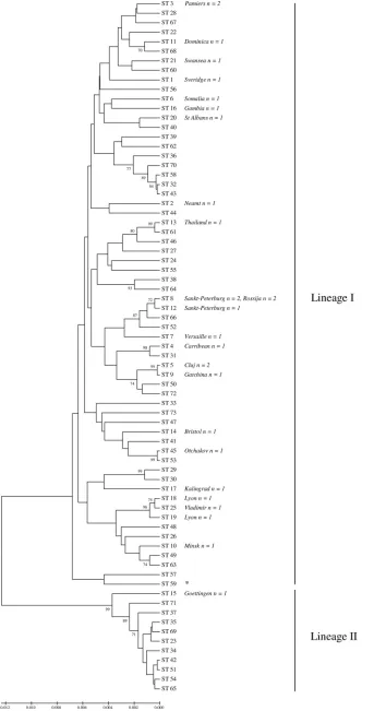
rare, may act as a reservoir for human infection. It is therefore important to characterize the isolates to understand their re-lationship to human strains. Two biotype gravis equine isolates, previously described by Henricson (13), were identical by MLST, and although comprising a unique ST, they clearly
cluster within the typical humanC. diphtheriaepopulation, as
shown by Fig. 1.
Relationship between MLST and ribotyping. In total, 86
validated ribotypes have been assigned to theC. diphtheriae
ribotype database (11). As shown by Fig. 1, the examination of 29 previously ribotyped isolates by MLST was in concordance with the ribotyping data. This study of 20 of the total 86 ribotype patterns by MLST represents a preliminary compar-ison but is sufficient to help validate the MLST scheme in
terms of correctly assigning strains that were identical and part of an outbreak and those believed to be clearly distinct strains. In two instances, MLST provided greater strain discrimina-tion than ribotyping. First, the Lyon ribotype isolates, 19 and
20, were SLVs of each other, differing at theatpAlocus, and
were identified as ST-18 and ST-19. This clonal expansion is likely to have arisen by recombination rather than point mu-tation, since four base pair changes (4/378) were identified and both alleles were frequently detected in other STs. De Zoysa was also able to differentiate between the two isolates using AFLP, where a single band difference was observed (4). Sec-ond, one of three Sankt-Peterburg ribotype isolates (isolate 13)
was an SLV at theatpAlocus, where two base pairs
substitu-tions were identified.
Previous studies identified two predominant ribotypes asso-ciated with the FSU outbreak: Sankt-Peterburg and Rossija. Three isolates of each ribotype were analyzed by MLST. With the exception of isolate 13, discussed previously, all were in-distinguishable by MLST (ST-8). AFLP, RAPD, and PFGE typing studies were also unable to differentiate between the two ribotypes, and only a single band difference was detected by ribotyping (3, 4, 5). Therefore, the preliminary MLST data further support the suggestion (4, 5) that Sankt-Peterburg and Rossija ribotyped isolates are part of a shared clonal complex, along with ST-12 and ST-52, here forming eBURST group 2. Likewise, isolates belonging to ribotypes Vladimir (ST-25) and Lyon (ST-18 and ST-19) are clonally derived (eBURST group 5), as are isolates belonging to ribotypes Cluj (ST-5) and Gatchina (ST-9), closely related to each other (eBURST group 11).
Relationship between sequence type, biotype, and toxin
sta-tus.There was some linkage between ST, biotype, and toxin
status. However, of the 73 STs containing more than one iso-late, 21% were associated with multiple biotypes and 32% with variable toxin status. There was no clear association between disease status and ST, and carriage isolates could not be dif-ferentiated from disease-causing strains.
[image:5.585.41.544.80.243.2]Importantly, while some of the clusters identified by MLST contained isolates of only one biotype, isolates with either the gravis, intermedius, or mitis biotype were not found within
TABLE 2. C. diphtheriaehousekeeping genes, gene functions, and amplification and sequencing primers used in the MLST schemea
Gene Gene function
Amplification primer, 5⬘–3⬘ Amplicon size (bp)
Sequencing primer, 5⬘–3⬘ Allele size (bp)
Fwd Rev Fwd Rev
atpA ATP synthase alpha chain GCGATTGCGAAC TACACC CTCGAGGAAT ACCTRACC 1,029 AGAAGGCGACGA AGTMAAGC CRGAATCAGAA GCTGGWGCA 378
dnaE DNA polymerase III alpha subunit TGCGTCATCTGA TTGAAA CGGTCCAATA AGACACCA 858 GTGCGACAAGCT GGTGTG GGCTTWCGGCC ATTYTTG 354
dnaK Chaperone protein DnaK ACTTGGGTGGCG GTACTT TGGTGAACGT CTCGGAAC 696 AGATGGCTATGC AGCGTCT GATGAGCTTGG TCATCACG 345
fusA Elongation factor G TACCGCGAGAAG CTCGTT GAAGGTTGG GTCCTCTTC 683 CGTAAGCTGACC GTTAACTC CCATGGACTCR AGGATGA 360
leuA 2-Isopropylmalate synthase CGTGCACTTCTA CAACTC ACCGTGATCG GTCTTCAT 865 CCYATCATCATCA AYCTGCC CAGCTGGTTGC AGTAYTC 384
odha 2-Oxoglutarate dehydrogenase CGGCAAGGAAAS CATGAC GTTGTCGCCR AACATCTG 505 TBCAAGATCGCA TYGARRC TWGGCTCGATG TGKCCTTC 382
rpoB RNA polymerase beta chain AAGCGCAAGATC CAGGAC TCGAACTCGT CGTCATCC 845 CGWATGAACATY GGBCAGGT TCCATYTCRCC RAARCGCTG 342 a
Fwd, forward; Rev, reverse.
TABLE 3. Characteristics of the seven loci used in theC. diphtheriaeMLST scheme
Housekeeping gene Allele length (bp) No. of distinct alleles No. of polymorphic sites Proportion of polymorphic sites (%) Mean G⫹C content
(%)
dN/dS
atpA 378 20 21 5.5 57.49 0.0394
dnaE 354 13 18 5.1 50.96 0.0100
dnaK 345 30 35 10.1 57.94 0.0024
fusA 360 23 37 10.3 56.22 0.0026
leuA 384 20 25 6.5 56.82 0.0201
odhA 381 19 23 6.0 57.96 0.0145
rpoB 342 17 18 5.3 57.37 0.0081
Mean avg 363 20.3 25.3 7.0 56.4 0.0140
on May 16, 2020 by guest
http://jcm.asm.org/
[image:5.585.43.284.598.725.2]FIG. 1. Genetic relationships between the 73 sequence types identified and their relationship to ribotyping results. Where available, ribotyping data and the number of each ribotyped isolate are shown in italics to the right of the ST to which the ribotype corresponded. ST-59 (identified by an asterisk) refers to equine isolates 130 and 132. Lineage I and lineage II are highlighted by the line to the right of the phylogenetic tree.
4182
on May 16, 2020 by guest
genetically distinct subgroups. However, as illustrated by Fig. 1 and 2, two distinct lineages (I and II) were identified within our
C. diphtheriaecollection. Lineage I contained the largest pro-portion of isolates and exclusively all of the isolates biotyped as mitis, gravis, and intermedius. Five “belfanti” isolates were also found within lineage I; however, these “belfanti” isolates were all of ST-26, from James Bay, Canada, and all bar one produced diphtheria toxin (DT), which is atypical for the bio-type (20). By comparison, lineage II was composed exclusively of all 11 remaining biotype belfanti isolates, representing 11 distinct STs. These lineage II belfanti isolates are of typical phenotype, being nontoxigenic. Furthermore, based upon con-catenated MLST nucleotide data, lineage I strains were, on average, 2.5% divergent from those in lineage II, each forming distinct subgroups when represented by neighbor-joining trees (Fig. 1).
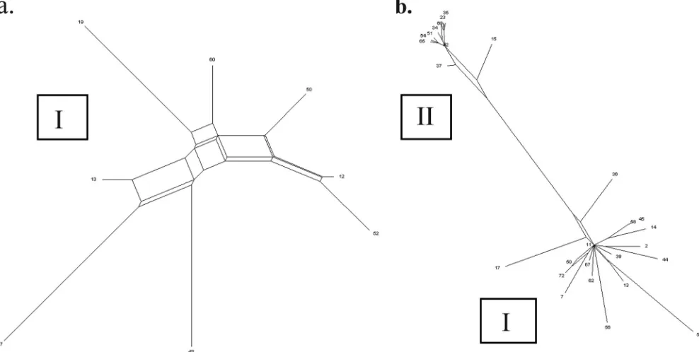
Recombination. The balance between recombination and
mutation has a significant impact upon the population biology of bacteria and their ability to evolve under strong selective pressures. As previously discussed, many SLVs within a clonal complex are likely to have arisen by recombination since mul-tiple substitutions were observed and the allele was identified in other strains within the data set. The MLST data were further analyzed to determine the potential for genetic
ex-change within isolates ofC. diphtheriae.
The degree of recombination within a bacterial population
can be determined using the index of association (IA), which
measures the level of linkage between alleles at different loci.
AnIAnot significantly greater than zero after 1,000 computer
randomizations suggests the organism is in linkage equilibrium and is therefore freely recombining, while a population with an
IA significantly greater than zero is considered to be clonal.
Overall, theIAvalue was 0.1176 (P⫽ ⬍0.01). To minimize any
distortions created when analyzing two relatively distinct
pop-ulations, theIAvalue was also individually calculated for both
lineages I (0.0667;P⫽ ⬍0.01) and II (0.0395;P⫽0.14).
To further examine the impact of recombination upon theC.
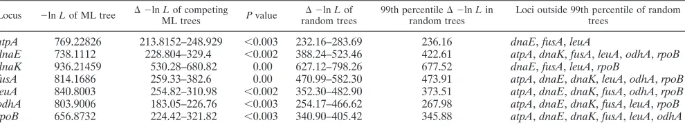
diphtheriaecollection, the congruence observed between gene tree topologies was determined using the method described by Holmes (14). In clonal populations, the genetic relationships at all loci will be the same, while for recombinogenic species or subpopulations, the phylogenetic trees will appear incongru-ent. As shown by Table 4, pairwise comparisons between the gene tree topologies revealed significant discordance for all
loci. However, only the association between theatpAML tree
and thednaK,odhA, andrpoBtrees and that between thednaK
tree andatpAandodhAwere deemed to have no more
simi-larity than that with trees of random topology. It is therefore apparent that while recombination plays a role in the evolution ofC. diphtheriae, it has not obscured all phylogenetic signals.
DISCUSSION
Diphtheria is still endemic in many countries and, as exem-plified by the FSU outbreak, is a potentially resurgent disease. Furthermore, given the low levels of protection within adult populations (in particular seniors), accurate and reproducible
typing methods are required to monitor and characterizeC.
[image:7.585.45.539.71.320.2]diphtheriae. Although numerous typing techniques for C. diphtheriaehave been described, their use is often hindered by limited reproducibility and subjective analysis. MLST is able to circumvent these limitations by directly analyzing nucleotide information within selectively neutral house-keeping genes. The data produced are objective and, due to
FIG. 2. Splits decomposition analysis illustrating the population structure withinC.diphtheriae. (a) Splits within 8 STs from lineage I. (b) Relationship between representative STs from lineages I and II.
on May 16, 2020 by guest
http://jcm.asm.org/
their portability, amenable to international collaborations. TheC. diphtheriaeMLST database can be accessed at http: //pubmlst.org/cdiphtheriae.
This is the first use of MLST to characterize isolates ofC.
diphtheriae. MLST effectively typed 150 diverseC. diphtheriae
isolates and confirmed findings of previous studies indicating that there is significant intraspecies genetic diversity. The data presented demonstrate that recombination has played a role in
the evolution ofC. diphtheriae. This was made evident by splits
decomposition analysis and in the significant discordance ob-served between all MLST gene trees. However, since the con-gruence was deemed to show no greater similarity than that to trees of random topology within only two MLST loci, genetic exchange does not obscure all phylogenetic signals. Analysis of
the complete genome sequence ofC. diphtheriaereveals recent
acquisition of pathogenicity factors. The observed recombina-tion in this study highlights an obvious opportunity for these
and other determinants to move across the population ofC.
diphtheriae.
To validate the accuracy and discriminatory power of the MLST scheme, the data were compared to an available subset of strains typed by the current gold standard, ribotyping. The MLST data were generally in concordance with the ribotyping findings. However, MLST provided greater strain resolution in two instances and ribotyping in one. MLST identified an SLV within three Sankt-Peterburg ribotype isolates and was able to distinguish between two ribotype Lyon isolates, which De Zoysa et al. had previously distinguished (5). As with AFLP, PFGE, and RAPD studies (3, 4, 5), MLST was unable to differentiate between the two predominant ribotypes associ-ated with the FSU outbreak: Sankt-Peterburg and Rossija. Spoligotyping studies of this epidemic clonal group showed a clear divergence between these two ribotypes and suggested that the Rossija ribotype may have originated from one par-ticular subpopulation of ribotype Sankt-Peterburg (21, 22). Likewise, it is clear by MLST that the Vladimir and Lyon ribotypes are clonally derived (eBURST group 5), as are the Cluj and Gatchina ribotypes (eBURST group 11).
MLST also identified a clonal complex (eBURST group 9) associated with an epidemiologically linked diphtheria outbreak in Haiti and the Dominican Republic, which both share the island of Hispaniola. A suspected diphtheria outbreak is believed to have originated in the Fond des Blancs region of Haiti in 2004 (CDC, personal communication). From 2001 to 2003, 13 diph-theria cases in Haiti and 120 in the Dominican Republic were reported (www.who.int/immunization_monitoring/data/en/). This increased significantly, to 253 cases in Haiti and 177 in the Do-minican Republic, from 2004 to 2006 (see above URL). Of 28
isolates obtained during the outbreak, 93% belonged to a clonal complex comprising ST-31 and ST-4 isolates (eBURST group 9). Since two further isolates, collected in 2000 and 1995, were ST-31 and ST-4, respectively, it is evident that the outbreak strains were circulating in the preepidemic period and may belong to the
regions of endemicity for aC. diphtheriaereservoir.
While members of some clonal complexes were globally distributed, other groups were associated with specific loca-tions. Notably, members of eBURST group 8 were obtained from geographically disparate countries (Thailand, Russia, and Guatemala), whereas exclusively Russian isolates belonged to eBURST group 10. However, these findings may be the result of sampling limitations and require a wider sample analysis.
It was not possible to identify a definitive association be-tween ST and toxigenicity for biotypes mitis, intermedius, and
gravis. Likewise, there was not always a clear association
be-tween biotype and ST, indicating that biotypes are not neces-sarily stable epidemiological markers, which is wholly
consis-tent withC. diphtheriaebeing identified in this work as having
only a weakly clonal structure. However, isolates identified by biotype and nontoxigenic status as typical belfanti strains clus-tered together within lineage II.
MLST provides a valuable tool for monitoring and
charac-terizing endemic and epidemicC. diphtheriaestrains. The data
produced are portable, reproducible, and unambiguous. Strain discrimination was in accordance with ribotyping data, and clonal complexes associated with disease outbreaks were iden-tified.
ACKNOWLEDGMENTS
We thank Keith Jolley for his assistance with the gene tree congru-ence data and for setting up the MLST database.
We thank the BBSRC, Micropathology LTD, and Society for Gen-eral Microbiology for financial support at Warwick University. We also thank the Centers for Disease Control and the Institut Pasteur Foun-dation, which currently curates the C. diphtheriae MLST database (http://pubmlst.org/cdiphtheriae/).
REFERENCES
1.Cerden˜o-Ta´rraga, A. M., A. Efstratiou, L. G. Dover, M. T. G. Holden, M. Pallen, S. D. Bentley, G. S. Besra, C. Churcher, K. D. James, A. De Zoysa, T. Chillingworth, A. Cronin, L. Dowd, T. Feltwell, N. Hamlin, S. Holroyd, K. Jagels, S. Moule, M. A. Quail, E. Rabbinowitsch, K. M. Rutherford, N. R. Thomson, L. Unwin, S. Whitehead, B. G. Barrell, and J. Parkhill.2003. The complete genome sequence and analysis of Corynebacterium diphtheriae
NCTC13129. Nucleic Acids Res.31:6516–6523.
2.Corboz, L., R. Thoma, U. Braun, and R. Zbinden.1996. Isolation of Coryne-bacterium diphtheriae subsp. belfantifrom a cow with chronic active derma-titis. Schweiz. Arch. Tierheilkd.138:596–599.
[image:8.585.46.541.81.173.2]3.De Zoysa, A. S., and A. Efstratiou.1999. PCR typing ofCorynebacterium diphtheriaeby random amplification of polymorphic DNA. J. Med. Micro-biol.48:335–340.
TABLE 4. Congruence tests for each gene tree compared to other gene tree topologies and random tree data
Locus ⫺lnLof ML tree ⌬ ⫺lnLof competing
ML trees Pvalue ⌬ ⫺ lnLof random trees
99th percentile⌬ ⫺lnLin random trees
Loci outside 99th percentile of random trees
atpA 769.22826 213.8152–248.929 ⬍0.003 232.16–283.69 236.16 dnaE,fusA,leuA
dnaE 738.1112 228.804–329.4 ⬍0.002 388.24–523.46 422.61 atpA,dnaK,fusA,leuA,odhA,rpoB dnaK 936.21459 530.28–680.82 0.00 627.12–798.26 677.52 dnaE,fusA,leuA,rpoB
fusA 814.1686 259.33–382.6 0.00 470.99–582.30 473.91 atpA,dnaE,dnaK,leuA,odhA,rpoB leuA 840.8003 254.82–310.98 ⬍0.002 352.30–482.90 373.51 atpA,dnaE,dnaK,fusA,odhA,rpoB odhA 803.9006 183.05–226.76 ⬍0.003 254.17–466.62 267.98 atpA,dnaE,dnaK,fusA,leuA,rpoB rpoB 656.8732 224.42–321.82 ⬍0.003 340.90–405.42 345.88 atpA,dnaE,dnaK,fusA,leuA,odhA
on May 16, 2020 by guest
http://jcm.asm.org/
4.De Zoysa, A. S., and A. Efstratiou.2000. Use of amplified fragment length polymorphisms for typingCorynebacterium diphtheriae. J. Clin. Microbiol.
38:3843–3845.
5.De Zoysa, A., A. Efstratiou, R. C. George, M. Jahkola, J. Vuopio-Varkila, S. Deshevoi, G. Tseneva, and Y. Rikushin.1995. Molecular epidemiology of
Corynebacterium diphtheriae from northwestern Russia and surrounding countries studied by using ribotyping and pulsed-field gel electrophoresis. J. Clin. Microbiol.33:1080–1083.
6.De Zoysa, A., P. Hawkey, A. Charlett, and A. Efstratiou.2008. Comparison of four molecular typing methods for characterization ofCorynebacterium diphtheriaeand determination of transcontinental spread ofC. diphtheriae
based on BstEII rRNA gene profiles. J. Clin. Microbiol.46:3626–3635. 7.Edmunds, W. J., R. G. Pebody, H. Aggerback, S. Baron, G. Berbers, M. A.
Conyn-van Spaendonck, H. O. Hallander, R. Olander, P. A. Maple, H. E. Melker, P. Olin, F. Fievret-Groyne, C. Rota, S. Salmaso, A. Tischer, C. von-Hunolstein, and E. Miller.2000. The sero-epidemiology of diphtheria in Western Europe. ESEN Project. European Sero-Epidemiology Network. Epidemiol. Infect.125:113–125.
8.Efstratiou, A., K. H. Engler, I. K. Mazurova, T. Glushkevich, J. Vuopio-Varkila, and T. Popovic.2000. Current approaches to the laboratory diag-nosis of diphtheria. J. Infect. Dis.181:S138–S145.
9.Engler, K. H., T. Glushkevich, I. K. Mazurova, R. C. George, and A. Efstra-tiou.1997. A modified Elek test for detection of toxigenic corynebacteria in the diagnostic laboratory. J. Clin. Microbiol.35:495–498.
10.Gidding, H. F., J. L. Backhouse, M. A. Burgess, and G. L. Gilbert.2005. Immunity to diphtheria and tetanus in Australia: a national serosurvey. Med. J. Aust.183:301–304.
11.Grimont, P. A. D., F. Grimont, A. Efstratiou, A. De Zoysa, I. Mazurova, C. Ruckly, M. Lejay-Collin, S. Martin-Delautre, B. Regnault and members of the European Laboratory Working Group on Diphtheria.2004. Interna-tional nomenclature forCorynebacterium diphtheriaeribotypes. Res. Micro-biol.155:162–166.
12.Hall, A. J., P. K. Cassiday, K. A. Bernard, F. Bolt, A. G. Steigerwalt, D. Bixler, L. C. Pawloski, A. M. Whitney, M. Iwaki, A. Baldwin, C. G. Dowson, T. Komiya, M. Takahashi, H. P. Hinrikson, and M. L. Tondella.2010. Novel
Corynebacterium diphtheriaein domestic cats. Emerg. Infect. Dis.16:688– 691.
13.Henricson, B., M. Segarra, J. Garvin, J. Burns, S. Jenkins, C. Kim, T. Popovic, A. Golaz, and B. Akey.2000. ToxigenicCorynebacterium diphtheriae
associated with an equine wound infection. J. Vet. Diagn. Invest.12:253–257. 14.Holmes, E. C., R. Urwin, and M. C. Maiden.1999. The influence of recom-bination on the population structure and evolution of the human pathogen
Neisseria meningitidis. Mol. Biol. Evol.16:741–749.
15.Kalinowski, J., B. Bathe, D. Bartels, N. Bischoff, M. Bott, A. Burkovski, N. Dusch, L. Eggeling, B. J. Eikmanns, L. Gaigalat, A. Goesmann, M. Hart-mann, K. Huthmacher, R. Kra¨mer, B. Linke, A. C. McHardy, F. Meyer, B. Mo¨ckel, W. Pfefferle, A. Pu¨hler, D. A. Rey, C. Ru¨ckert, O. Rupp, H. Sahm, V. F. Wendisch, I. Wiegra¨be, and A. Tauch.2003. The complete Corynebac-terium glutamicumATCC 13032 genome sequence and its impact on the production of L-aspartate-derived amino acids and vitamins. J. Biotechnol.
104:5–25.
16.Khamis, A., D. Raoult, and B. La Scola.2004.rpoBgene sequencing for identification ofCorynebacteriumspecies. J. Clin. Microbiol.42:3925–3931. 17.Launay, O., C. Toneatti, C. Berne`de, E. Njamkepo, K. Petitprez, A. Leblond, S. Larnaudie, C. Goujon, M. N. Ungeheuer, F. Ajana, C. Raccurt, J. Beytout, C. Chidiac, D. Bouhour, D. Guillemot, and N. Guiso.2009. Antibodies to tetanus, diphtheria and pertussis among healthy adults vaccinated according to the French vaccination recommendations. Hum. Vaccin.5:341–346.
18.Leggett, B. A., A. De Zoysa, Y. E. Abbott, N. Leonard, B. Markey, and A. Efstratiou. 2010. Toxigenic Corynebacterium diphtheriae isolated from a wound in a horse. Vet. Rec.166:656–657.
19.Maiden, M. C., J. A. Bygraves, E. Feil, G. Morelli, J. E. Russell, R. Urwin, Q. Zhang, J. Zhou, K. Zurth, D. A. Caugant, I. M. Feavers, M. Achtman, and B. G. Spratt.1998. Multilocus sequence typing: a portable approach to the identification of clones within populations of pathogenic microorganisms. Proc. Natl. Acad. Sci. U. S. A.95:3140–3145.
20.Marston, C. K., F. Jamieson, F. Cahoon, G. Lesiak, A. Golaz, M. Reeves, and T. Popovic.2001. Persistence of a distinctCorynebacterium diphtheriaeclonal group within two communities in the United States and Canada where diphtheria is endemic. J. Clin. Microbiol.39:1586–1590.
21.Mokrousov, I., O. Narvskaya, E. Limeschenko, and A. Vyazovaya.2005. Efficient discrimination within aCorynebacterium diphtheriaeepidemic clonal group by a novel macroarray-based method. J. Clin. Microbiol.43:1662– 1668.
22.Mokrousov, I., A. Vyazovaya, V. Kolodkina, E. Limeschenko, L. Titov, and O. Narvskaya.2009. Novel macroarray-based method ofCorynebacterium diph-theriaegenotyping: evaluation in a field study in Belarus. Eur. J. Clin. Mi-crobiol. Infect. Dis.28:701–703.
23.Mothershed, E. A., P. K. Cassiday, K. Pierson, L. W. Mayer, and T. Popovic.
2002. Development of a real-time PCR assay for rapid detection of the diphtheria toxin gene. J. Clin. Microbiol.40:4713–4719.
24.Nakao, H., and T. Popovic.1998. Use of random amplified polymorphic DNA for rapid molecular subtyping ofCorynebacterium diphtheriae. Diagn. Microbiol. Infect. Dis.30:167–172.
25.Neal, S., and A. Efstratiou.2009. Diphtheria Surveillance Network: focus on microbiological diagnostics and molecular typing in Europe, abstr. S-11.2. Abstr. Eur. Sci. Conf. Appl. Infect. Dis. Epidemiol.
26.Nishio, Y., Y. Nakamura, Y. Kawarabayasi, Y. Usuda, E. Kimura, S. Sugi-moto, K. Matsui, A. Yamagishi, H. Kikuchi, K. Ikeo, and T. Gojobori.2003. Comparative complete genome sequence analysis of the amino acid replace-ments responsible for the thermostability ofCorynebacterium efficiens. Ge-nome Res.13:1572–1579.
27.Parish, H. J., and C. C. Okell.1926. A note on the isolation of virulent diphtheria bacilli from wounds of horses. J. Exp. Pathol.7:173–174. 28.Popovic, T., S. Y. Kombarova, M. W. Reeves, H. Nakao, I. K. Mazurova, M.
Wharton, I. K. Wachsmuth, and J. D. Wenger.1996. Molecular epidemiol-ogy of diphtheria in Russia, 1985–1994. J. Infect. Dis.174:1064–1072. 29.Popovic, T., I. K. Mazurova, A. Efstratiou, J. Vuopio-Varkila, M. W. Reeves,
A. De Zoysa, T. Glushkevich, and P. Grimont.2000. Molecular epidemiology of diphtheria. J. Infect. Dis.181:S168–S177.
30.Rozen, S., and H. J. Skaletsky.2000. Primer3 on the WWW for general users and for biologist programmers, p. 365–386.InA. Krawetz and S. Misener (ed.), Bioinformatics methods and protocols: methods in molecular biology. Humana Press, Totowa, NJ.
31.Skogen, V., V. V. Cherkasova, N. Maksimova, C. K. Marston, H. Sjursen, M. V. Reeves, O. Olsvik, and T. Popovic.2002. Molecular characterization of
Corynebacterium diphtheriaeisolates, Russia, 1957–1987. Emerg. Infect. Dis.
8:516–518.
32.Tamura, K., J. Dudley, M. Nei, and S. Kumar.2007. MEGA4: Molecular Evolutionary Genetics Analysis (MEGA) software version 4.0. Mol. Biol. Evol.24:1596–1599.
33.Vitek, C. R., and M. Wharton.1998. Diphtheria in the Former Soviet Union: reemergence of a pandemic disease. Emerg. Infect. Dis.4:539–550. 34.Zasada, A. A., M. Zaleska, R. B. Podlasin, and I. Seferyn´ska.2005. The first
case of septicemia due to nontoxigenicCorynebacterium diphtheriaein Po-land: case report. Ann. Clin. Microbiol. Antimicrob.4:8.