Diarrhea Virus by Use of Isothermal Amplification Methods and
High-Speed Real-Time Reverse Transcriptase PCR
Andrea Aebischer, Kerstin Wernike, Bernd Hoffmann, Martin Beer Institute of Diagnostic Virology, Friedrich-Loeffler-Institut, Greifwald-Insel Riems, Germany
Over the past few years, there has been an increasing demand for rapid and simple diagnostic tools that can be applied outside centralized laboratories by using transportable devices. In veterinary medicine, such mobile test systems would circumvent bar-riers associated with the transportation of samples and significantly reduce the time to diagnose important infectious animal diseases. Among a wide range of available technologies, high-speed real-time reverse transcriptase quantitative PCR (RT-qPCR) and the two isothermal amplification techniques loop-mediated isothermal amplification (LAMP) and recombinase polymerase amplification (RPA) represent three promising candidates for integration into mobile pen-side tests. The aim of this study was to investigate the performance of these amplification strategies and to evaluate their suitability for field application. In order to enable a valid comparison, novel pathogen-specific assays have been developed for the detection of Schmallenberg virus and bo-vine viral diarrhea virus. The newly developed assays were evaluated in comparison with established standard RT-qPCR using samples from experimentally or field-infected animals. Even though all assays allowed detection of the target virus in less than 30 min, major differences were revealed concerning sensitivity, specificity, robustness, testing time, and complexity of assay design. These findings indicated that the success of an assay will depend on the integrated amplification technology. Therefore, the ap-plication-specific pros and cons of each method that were identified during this study provide very valuable insights for future development and optimization of pen-side tests.
S
imilar to human medicine, the demands for diagnostic tests that can be applied directly at the point of care are increasing in veterinary science also. These mobile “pen-side” tests would cir-cumvent the delays in diagnosis associated with the transportation of the sample to a centralized laboratory and a resource-intensive processing. Furthermore, a rapid confirmation of clinical diagno-sis directly on-site would enable timely intervention and imple-mentation of control measures (e.g., during an outbreak of a transboundary animal disease). This has already been demon-strated for diagnosis of foot-and-mouth disease using rapid and simple lateral-flow devices (1, 2). However, over the past few years, a huge variety of innovative rapid technologies for amplifi-cation and detection of viral nucleic acid have been developed (3–5). These molecular approaches provide higher test sensitivity and specificity than the immunoassays mentioned before and are therefore attractive alternatives for integration into a new gener-ation of mobile pen-side testing systems. Among the available technologies, high-speed reverse transcriptase quantitative PCR (RT-qPCR) and the two isothermal amplification techniques re-combinase polymerase amplification (RPA) and loop-mediated isothermal amplification (LAMP) represent three promising can-didates for applications in veterinary medicine. Real-time quanti-tative PCR (qPCR) is currently the most widely used method to detect genomes of viral pathogens, since it is highly sensitive and specific, allows quantitative analysis, and minimizes the risk of contamination. Nevertheless, commonly used RT-qPCR proto-cols require more than 1 h. Therefore, many efforts have been made to develop strategies that reduce reaction time to less than 20 min. However, the majority of these approaches required special-ized PCR machines (6–8). Opposed to that, the application of high-speed RT-qPCR using conventional PCR machinesrepre-sents a more feasible approach for common use as has recently been described (9).
The RPA method is based on the formation of a recombinase filament, a complex between oligonucleotide primers and a re-combinase enzyme. Upon recognition of the target-specific se-quence by the recombinase filament, strand exchange is initiated and primers are subsequently extended by a strand-displacing polymerase (10). Real-time detection can be performed by using TwistAmp exo probes. These probes contain an abasic nucleotide analogue (tetrahydrofuran [THF]), which is flanked by an inter-nal fluorophore and a corresponding quencher group. Upon binding to the target sequence, the abasic site is recognized and cleaved by the DNA repair enzyme exonuclease III. This leads to separation of both the fluorophore and the quencher and subse-quent generation of a fluorescent signal. RPA is a newly emerging technology, but present literature hints toward a promising tool for pen-side application (11–14).
In contrast, LAMP is the most widely researched and employed isothermal amplification method (15). It uses a strand-displacing DNA polymerase along with two internal primers (FIP and BIP)
Received17 January 2014 Returned for modification13 February 2014
Accepted13 March 2014
Published ahead of print19 March 2014
Editor:Y.-W. Tang
Address correspondence to Bernd Hoffmann, [email protected]. Supplemental material for this article may be found athttp://dx.doi.org/10.1128 /JCM.00167-14.
Copyright © 2014, American Society for Microbiology. All Rights Reserved.
doi:10.1128/JCM.00167-14
on May 16, 2020 by guest
http://jcm.asm.org/
and two outer primers (F3 and B3), which recognize 6 different regions on the target gene (16). Two additional primers (Loop-F, Loop-B) that anneal at the loop structures of the LAMP amplicons enhance reaction speed and specificity (17). An animation that is helpful for understanding the amplification principle can be found online (http://loopamp.eiken.co.jp/e/lamp/index.html). An abundance of pathogen-specific assays have already been de-scribed as having performance equal to or better than that of the equivalent PCR. This also includes assays for the detection of transboundary animal diseases, such as, e.g., foot-and-mouth-disease virus (18), classical swine fever virus (19), and avian influ-enza (20).
The aim of the present study was to evaluate high-speed RT-qPCR, RPA, and LAMP and to define their application-specific pros and cons with regard to integration in molecular pen-side tests. In order to enable a fair and valid comparison, novel opti-mized pathogen-specific assays were developed for the detection of bovine viral diarrhea virus (BVDV) and Schmallenberg virus (SBV). BVDV is classified as a member of the genusPestivirus
within the familyFlaviviridae(21). It possesses a single-stranded positive-sense RNA genome that encodes one single large poly-protein. The 5=untranslated region (UTR) is used to assign species and genotype and harbors the majority of pestivirus-specific RT-qPCR assays (22–25). A diagnostic tool for rapid detection of per-sistent BVDV-infections in the field would be highly attractive, since identification and subsequent elimination of persistently in-fected cattle are essential for a successful BVD eradication strategy (26). SBV is anOrthobunyavirusfrom the familyBunyaviridaeand
belongs to the Simbu serogroup viruses (27). It has a segmented single-stranded RNA genome of negative polarity that comprises a small (S) segment, medium (M) segment, and large (L) segment. SBV was detected for the first time in Europe in autumn 2011, and over the last 2 years, it has spread rapidly over large parts of north-western Europe. Adult animals develop no or mild clinical disease, whereas transplacental infection can lead to severe congenital malformations (28,29).
The diagnostic accuracy of the newly developed SBV- and BVDV-specific tests was assessed in comparison to that of estab-lished standard RT-qPCRs. Special emphasis was placed on the suitability of the tests for rapid and reliable detection of viral in-fections in the field.
MATERIALS AND METHODS
Standard RT-qPCR.Previously established standard RT-qPCR assays were used to assess the diagnostic accuracy of the newly developed tests (23,25,30). The primers and probes are indicated inTable 1along with the applied concentrations. Unless stated otherwise, all primers were syn-thesized by Metabion (Martinsried, Germany). Reactions were carried out in a 12.5-l volume using the SuperScript III One-Step RT-PCR sys-tem with PlatinumTaq(Invitrogen, Carlsbad, CA) according to the man-ufacturer=s instructions. RT-qPCR was performed using an Eco real-time PCR system (amplifa Labortechnik GmbH, Wasserburg, Germany) and the following thermal profile: reverse transcription for 15 min at 50°C and then polymerase activation for 2 min at 95°C, followed by 45 cycles of 95°C for 15 s, 56°C for 30 s, and 68°C for 30 s.
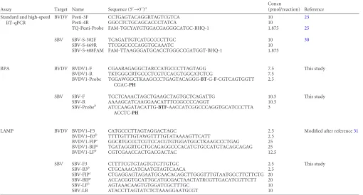
[image:2.585.40.550.79.354.2]High-speed RT-qPCR.The primers and probes and their concentra-tions used in the different assays are indicated inTable 1. Reactions were TABLE 1Primers and probes used in this study
Assay Target Name Sequence (5=¡3=)a Concn
(pmol/reaction) Reference Standard and high-speed
RT-qPCR
BVDV Pesti-3F CCTGAGTACAGGRTAGTCGTCA 10 23
Pesti-4R GGCCTCTGCAGCACCCTATCA 10
TQ-Pesti-Probe FAM-TGCYAYGTGGACGAGGGCATGC-BHQ-1 1.875 25
SBV SBV-S-382F TCAGATTGTCATGCCCCTTGC 10 30
SBV-S-469R TTCGGCCCCAGGTGCAAATC 10 SBV-S-408FAM FAM-TTAAGGGATGCACCTGGGCCGATGGT-BHQ-1 1.875
RPA BVDV BVDV1-F CGAARAGAGGCTARCCATGCCCTTAGTAGG 7.5 This study BVDV1-R TKTGGGCRTGCCCTCGTCCACGTGGCATCTCG 7.5
BVDV1-Probe TGGAWGGCTKAAGCCCTGAGTACAGGG-BT-G-F-CGTCAGTGGTT CGAC-PH
2.5
SBV SBV-F TCCTCAAACTAGCTGAAGCTAGTGCTCAGATTG 10.5 This study SBV-R AAAAGCATCAAGGAACATTTCGGCCCCAGGT 10.5
SBV-Probeb
ATCCAAGATACATTG-BTF-AACCATCGGCCCAGGTGCATCCCTTA ACCTC-PH
3
LAMP BVDV BVDV1–F3 CATGCCCTTAGTAGGACTAGC 2.5 Modified after reference31
BVDV1–B3b TTTTGTTTGTAWGTTTTGTATAAAAGTTCATT 2.5
BVDV1-FIPc
GGCRTGCCCTCGTCCACGTGTGGATGGCTKAAGCCCTGAG 25 BVDV1-BIPc
TGATAGGRTGCTGCAGAGGCCCACATGTGCCATGTACAGCAGAG 25 BVDV1-LFb CGTCGAACCACTGACGACTAC 12.5
SBV SBV-F3 CTTTTCGTGTAGTGTGTTGTGC 2.5 This study SBV-B3b CTGCAAACATCAATGTAGTCAACA 2.5
SBV-FIPc CTGAGGAGTAGAATGCAACACAGCTTGGGTTTGTAATGCCTTCTTCTG 20
SBV-BIPc
ACCACGGTGCATTGCATGCGACTAACTATRCGTTGACATCGTTCTT 20 SBV-LFb
AGTAAACAAGTGTGGATCGCTTTGC 10 SBV-LB ATACCTTAGTATCTCTAAAGGAATGCGT 10
a
RPA assay sequence abbreviations:B, thymidine nucleotide carrying BHQ-1 quencher;T, abasic tetrahydrofuran site;F, thymidine nucleotide carrying FAM fluorophore;PH, phosphate.
b
Designed on the antisense strand.
cHPLC purified.
on May 16, 2020 by guest
http://jcm.asm.org/
conducted in a total reaction volume of 12.5l using the SuperScript III One-Step RT-PCR system with PlatinumTaq(Invitrogen) according to the manufacturer=s instructions with 1l of 5 mM magnesium sulfate added per reaction. In order to omit the reverse transcription step of the qPCR, 2.5l template RNA was added to the master mix at room tem-perature. Amplification was performed on an Eco real-time PCR system, using the Eco software version 4.0 (amplifa Labortechnik GmbH).
For the BVDV-specific assay, the following thermal profile was used: polymerase activation for 1 min at 95°C, followed by 45 cycles of denatur-ation at 98°C for 3 s and annealing and extension at 60°C for 1 s. For the SBV-specific assay, the denaturation time was shortened to 1 s, and the annealing temperature increased to 64°C.
Recombinase polymerase amplification.Sequences of primers and probes used for RPA, as well as details of the assay design are shown in Tables 1and2, respectively. Both TwistAmp exo RPA probes were syn-thesized by TIB Molbiol (Berlin, Germany) with an inverse arrangement of fluorophore (6-carboxyfluorescein [FAM]) and quencher (black hole quencher 1 [BHQ-1]). RPA reactions were performed in a 25-l volume using the enzyme pellets of the TwistAmp exo kit (TwistDx, Cambridge, United Kingdom), 1.7⫻rehydration buffer, 1.5l of 280 mM magnesium acetate (TwistDx), 2 mM dithiothreitol (DTT) (Invitrogen); and 5 U Transcriptor reverse transcriptase (Roche Diagnostics, Mannheim, Ger-many). Mixtures of primers and probes according to the concentrations indicated inTable 1were prepared and added to an empty 0.2-ml reaction tube. A master mixture containing the rehydration buffer, DTT, water, and Transcriptor RT was prepared separately and added to the dried en-zyme pellets. Twenty microliters of the resuspended pellet was then added to the primer-probe mixtures. Finally, magnesium acetate was pipetted into the tube lid, and 1l of template RNA was added to the reaction mixture. The lids were closed, and the magnesium acetate was centrifuged into the tubes. The tubes were then immediately placed into an ESEQuant tube scanner (Qiagen, Hilden, Germany). Fluorescence measurements using the FAM channel were performed for 20 min at 42°C. Optimal reaction conditions were defined after testing different incubation tem-peratures (39 to 42°C), as well as different concentrations of template (0.5 to 2l), magnesium acetate (1 to 2.5l), and DTT (2 to 4 mM). For interpretation of the collected fluorescence signals, a signal slope analysis combined with a 2nd derivative analysis was performed (Tube Scanner Studio software; Qiagen).
Loop-mediated isothermal amplification.The BVDV LAMP assay described inTables 1and2was designed based on a previously published primer set (31). The original primers were modified using a sequence alignment of the 5=UTR of BVDV-1 strains available in GenBank. In addition, a Loop-F primer was included in the set. Placement of a Loop-B primer was not possible due to low sequence conservation in the respec-tive genomic region of the 5=UTR.
The L segment was chosen as target for the SBV LAMP. Sequences available in GenBank were aligned using ClustalW in order to find con-served regions. The final primer set (Tables 1and2) was constructed by using the LAMP primer design software Primer Explorer V4 (http: //primerexplorer.jp/elamp4.0.0/index.html). For both assays, FIP and
BIP primers were purified by high-performance liquid chromatography (HPLC).
The RT-LAMP reactions were carried out in a 12.5-l reaction volume containing the primer concentrations indicated inTable 1; a 1⫻ concen-tration of ThermoPol buffer (New England BioLabs [NEB], Ipswich, MA), 8 mM magnesium sulfate (Invitrogen), 0.8 M betaine (Sigma-Al-drich, St. Louis, MO), 1.4 mM each deoxynucleoside triphosphate (dNTP) (Qiagen, Hilden, Germany), 0.25l ResoLight dye (Roche Diag-nostics), 3 U of Bst DNA polymerase (large fragment; NEB), and 3 U of cloned avian myeloblastosis virus (AMV) reverse transcriptase (Invitro-gen). Finally, 2.5l of template RNA was added to the reaction mixture. Optimization was performed by testing different concentrations of mag-nesium sulfate (4 to 10 mM) and betaine (0.6 to 1 M) as well as different reaction temperatures ranging from 60 to 65°C. For the final assays, am-plification was performed on the Eco real-time PCR system (amplifa Labortechnik GmBH), using 60 cycles of 1 min at 63°C followed by a standard melting curve analysis. Real-time data were analyzed in conjunc-tion with melt curve data to exclude nonspecific fluorescence interference (Eco software version 4.0; amplifa Labortechnik GmbH).
Viruses, reference RNA, and clinical samples.Simbu serogroup vi-ruses (Sabo, Sango, Shamonda, Shuni, Aino, Simbu, Peaton, Douglas, and Sathuperi viruses) were kindly provided by Peter Kirkland (Elizabeth Macarthur Agricultural Institute, Australia) and Robert Tesh (University of Texas Medical Branch). Full-length viral RNAs from BVDV strains 1a, 1b, 1d, 1e, 1f, 1 h, 1x, 2a, and 2c, as well as from classical swine fever virus (CSFV), border disease virus (BDV), and atypical pestiviruses, were taken from the EPIZONE pestivirus reference RNA panel (32). SBV reference RNA was produced from cell-culture-grown SBV. The RNA copy number of the starting dilution was determined using an external SBV standard.
Previouslyin vitro-transcribed and quantified RNA from a BVDV-DI9 plasmid construct (33,34) was used to determine the analytical sensitivity of the BVDV assays. BVDV-positive blood and serum samples were sup-plied by various veterinary diagnostic laboratories from different parts of Germany as well as by the BVDV National Reference Laboratory at the Institute of Diagnostic Virology of the Friedrich-Loeffler-Institut (FLI). SBV-positive blood and serum samples were obtained during animal tri-als conducted at the FLI. All experimental protocols were reviewed by a state ethics commission and have been approved by the competent au-thority (State Office for Agriculture, Food Safety and Fisheries of Meck-lenburg-Vorpommern, Rostock, Germany; reference no. LALLF M-V TSD/7221.3-1.1-004/12). Additional blood and tissue samples were taken from the collection of SBV-positive field samples at the Institute of Diag-nostic Virology of the FLI.
SBV and BVDV reference RNAs were tested in three independent runs to determine the analytical sensitivity of the assays. Clinical samples were tested in duplicate by standard and high-speed RT-qPCR, and the mean value of the replicates was calculated. For RPA and LAMP assays, only samples yielding negative results in the first run were tested a second time.
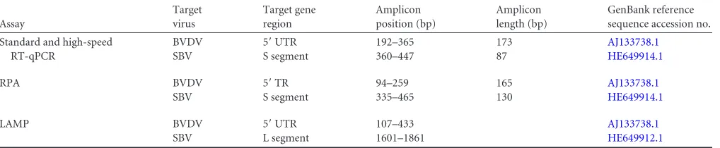
[image:3.585.41.550.78.183.2]RNA extraction.RNA was extracted from 140l of sera or infected cell culture supernatant, 75l of whole blood, or 140l of homogenized tissue by using the QIAamp viral RNA minikit (Qiagen) or the RNeasy TABLE 2Details of the BVDV- and SBV-specific assays used in this study
Assay
Target virus
Target gene region
Amplicon position (bp)
Amplicon length (bp)
GenBank reference sequence accession no.
Standard and high-speed RT-qPCR
BVDV 5=UTR 192–365 173 AJ133738.1
SBV S segment 360–447 87 HE649914.1
RPA BVDV 5=TR 94–259 165 AJ133738.1
SBV S segment 335–465 130 HE649914.1
LAMP BVDV 5=UTR 107–433 AJ133738.1
SBV L segment 1601–1861 HE649912.1
on May 16, 2020 by guest
http://jcm.asm.org/
mini kit (Qiagen) according to the manufacturer=s instructions. All sam-ples were eluted in 100l.
Statistical analysis.Linear regression analyses, the Kruskal-Wallis test, and Dunn=s test were performed using the SigmaPlot software v11 (Systat Software GmbH, Erkrath, Germany). PCR efficiency (E) was cal-culated using the following equation:E⫽10(⫺1/slope)⫺1.
RESULTS
Assay design and optimization. (i) High-speed RT-qPCR.For BVDV, a number of previously described primers have been eval-uated (22–24). Primers Pesti-3F and Pesti-4R (23) in conjunction with the TQ-Pesti-probe (25) yielded the best results using the high-speed profile. The SBV-specific assay could be established by using the previously published RT-qPCR (30). Both of the proto-cols were optimized by variations of the denaturation time and of the annealing and extension temperature. Using the Eco cycler, the final run times were 26 min for the BVDV-specific assay and 22 min for the SBV-specific assay, respectively.
(ii) LAMP.A BVDV-RT-LAMP primer set located in the same genomic region on the 5=UTR as the selected RT-qPCR assay has been described before (31). However, amplification of BVDV RNA could only be achieved after manual modification of the published primer set (Tables 1and2). For the SBV LAMP, initial primer sets designed for the S segment repeatedly produced non-specific amplification products. For this reason, additional assays were also designed for target regions in the M and L segments. Among those, only one primer set located in the L segment al-lowed specific amplification of SBV RNA and was therefore cho-sen for the final assay. The specificity and rapidity of the assay could be further optimized by variation of the outer primer B3. The concentration of the individual primers and their ratio to each other were found to have a crucial influence on the specificity of the LAMP assays. Optimal performance was achieved using primer ratios of 10:1:5 (inner to outer to loop) for the BVDV-specific assay and 8:1:4 for the SBV-BVDV-specific assay.
(iii) RPA.For each RPA, several forward and reverse primers were designed and evaluated in combination with the respective TwistAmp exo probe. During initial experiments, the original 50-l volume of the RPA reaction was successfully reduced to 25
l. The optimal concentration of primers and probes was found to be assay specific (Table 1). After evaluation of several RT enzymes, the Transcriptor RT was selected since it outperformed the
re-maining candidates with regard to amplification speed (data not shown).
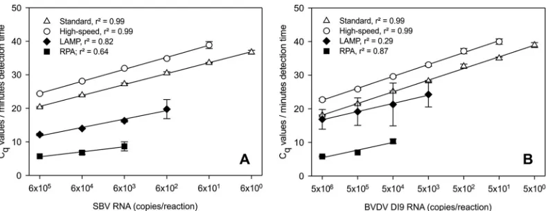
Analysis of assay parameters.Serial 10-fold dilutions of in vitro-transcribed BVDV-1-specific RNA were used to determine the analytical sensitivity of the newly developed BVDV assays. Five RNA copies per reaction could be amplified using the standard RT-qPCR, whereas the detection limits were 50 copies for the high-speed assay, 5⫻103copies for LAMP, and only 5⫻104 copies for RPA (Fig. 1B). Accordingly, the analytical sensitivity of the SBV-specific assays was defined using serial 10-fold dilutions of SBV reference RNA. The standard RT-qPCR protocol (30) was able to amplify the dilution series down to 6 genome copies per reaction. In comparison, the sensitivities of the high-speed RT-qPCR, LAMP, and RPA were 1-, 2-, and 3-log10steps lower, re-spectively (Fig. 1A).
Quantitative parameters of all assays were further assessed by linear regression analysis. Calculations were performed using the quantification cycle (Cq) values for the standard and high-speed
RT-qPCR assays and the detection time (in minutes) for the re-spective LAMP and RPA assays. Consequently, the standard curves presented inFig. 1AandBdo not allow a direct comparison of the reaction times. An overview of statistical analyses is given in Table S1 in the supplemental material. In summary,r2 values reached⬎0.9 for standard and high-speed RT-qPCR, whereas for LAMP and RPA, ther2values were⬍0.9 (see Table S1).
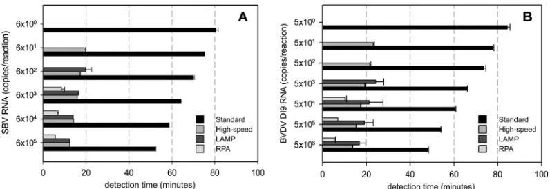
Using the detection time for comparison of the individual as-says, high-speed RT-qPCR, LAMP, and RPA evidently required less time to detect equal amounts of target RNA than the respec-tive standard RT-qPCR (Fig. 2A and B). Statistical analysis (Kruskal-Wallis test followed by Dunn‘s test) confirmed that these differences with regard to reaction speed were significant for both the BVDV- and SBV-specific assays (P⬍0.05).
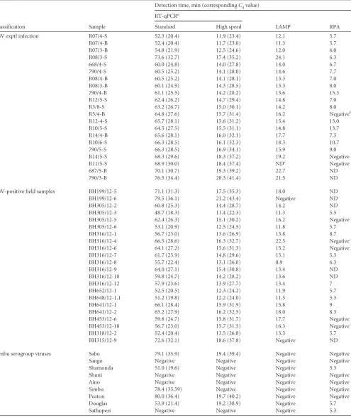
Diagnostic sensitivity and specificity. (i) SBV.The applicabil-ity of the SBV assays was tested using samples from SBV-infected animals (Table 3). Analysis revealed similar levels of performance of LAMP and high-speed RT-qPCR, with slightly reduced sensi-tivity of LAMP for samples with low viral loads. Using RPA, 8 false-negative results were obtained. This included samples with
Cqvalues of⬎27 as well as 2 tissue samples. No amplification of
nontarget RNA could be observed using previously characterized SBV-negative samples (data not shown). The cross-reactivity of FIG 1Analytical sensitivity and standard curves for (A) SBV-specific assays and (B) BVDV-specific assays. Serial 10-fold dilutions of reference RNA samples were tested in three independent runs. Linear regression analysis was performed usingCqvalues (white symbols) for standard and high-speed RT-qPCR and the time (minutes) to detection (black symbols) for LAMP and RPA.
on May 16, 2020 by guest
http://jcm.asm.org/
[image:4.585.96.490.65.217.2]the assays was evaluated using 9 viruses of the Simbu serogroup. Standard and high-speed RT-qPCR, as well as RPA, cross-de-tected several of these viruses. In contrast, the LAMP assay proved to be highly specific for SBV (Table 3).
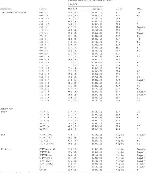
(ii) BVDV.The suitability of the BVDV assays to detect viremic cattle in the field was investigated using serum and blood samples collected in different parts of Germany. All virus-positive samples were readily detected by high-speed RT-qPCR and LAMP. In con-trast, RPA yielded 5 false-negative results (Table 4). Each assay iden-tified positively all of the investigated BVDV-1 reference strains (Ta-ble 4). Using standard and high-speed RT-qPCR assays, all of the additional pestiviruses included in the EPIZONE reference panel could be amplified, whereas the RPA detected only BVDV-2 strains and the atypical pestivirus Giraffe (Table 4). The BVDV LAMP assay was specific for BVDV-1, with the exception of one BVDV-2a strain (Table 4). The latter could be distinguished from BVDV-1 strains by melting curve analysis (data not shown).
Finally, no amplification of nontargeted RNA was observed during testing of BVDV-negative samples with all systems (data not shown).
DISCUSSION
Over the past few years, a variety of rapid nucleic acid amplifica-tion technologies have been developed and used for integraamplifica-tion into molecular pen-side tests. This includes PCR-based ap-proaches and isothermal amplification techniques. However, each of these methods has application-specific pros and cons that make a valid comparison challenging. In order to identify the most suit-able strategy for the future development of pen-side test systems, we aimed to directly compare the applicabilities of high-speed RT-qPCR, RPA, and LAMP. For this purpose, we developed novel pathogen-specific assays for the detection of BVDV and SBV and evaluated these assays in comparison with standard RT-qPCRs.
The high-speed RT-qPCR assays both showed a higher analyt-ical sensitivity than the respective LAMP or RPA assays, with only a 1-log10-step reduction compared to the reference standard RT-qPCR (Fig. 1AandB). They further displayed a larger linear dy-namic range and a higher reproducibility than the isothermal am-plification techniques (see Table S1 in the supplemental material). The fast assay development was an additional very valuable advan-tage of the high-speed RT-qPCRs. As shown before, primers and probes of established RT-qPCR assays can be applied and
opti-mized for the high-speed protocol (9). However, we also found that the size of the amplification product critically influenced the assay performance. In the case of BVDV, the primer pair Pesti-3F and Pesti-4R, which produced the shortest amplicon among all the primer candidates (173 bp), yielded the best results in the high-speed profile. Use of the SBV-specific assay, which amplifies a product of only 87 bp, allowed reduction of the denaturation time during two-step cycling to 1 s and an increase in the anneal-ing and extension temperature to 64°C, which further decreased the total running time of the protocol. Consequently, the highest reaction speed can be achieved by using RT-qPCR assays with amplicons of less than 100 bp. Considering future applications in the field, the high-speed RT-qPCR protocols were established us-ing the portable Eco real-time system (amplifa Labortechnik GmbH). In addition to a small size, the Eco cycler enables a sample ramping rate of 5.5°C/s, which is significantly faster than ramp rates of conventional Peltier block-based cyclers. Therefore, the reaction speed of the high-speed protocol depends on the avail-able thermocycler, as was previously shown (9). This finding, to-gether with the costly and nonstabilized PCR reagents, might re-strict application of high-speed RT-qPCR in resource-limited settings.
Opposed to that, the RPA technology has several important advantages with regard to field use. (i) RPA reagents are available in a lyophilized format, with only the separately added RT enzyme requiring refrigeration. (ii) The very-small-footprint ESEQuant tube scanner can be easily transported and installed on site. (iii) The low reaction temperature of 42°C is an advantage with regard to miniaturization and integration into battery-driven devices (3). This has already been proven by the development of a microfluidic lab-on-a-foil system and a digital RPA SlipChip (35,36). How-ever, in the present study, the newly developed SBV and BVDV RPA tests showed a low analytical sensitivity (Fig. 1AandB). As indicated inTables 3and4, virus-positive samples with RT-qPCR
Cqvalues of⬎27 were not reliably detected using RPA. In case of
BVDV, the RPA detected only 27 out of 32 field samples collected from persistently infected cattle. This was surprising, since the 5 false-negative samples yieldedCqvalues of⬍26 in the
correspond-ing reference RT-qPCR (Table 4). The failure of the test can there-fore not be explained by low viral loads in the samples. More likely, the selected primers and probe do not optimally recognize all of the currently circulating BVDVs. Thus, the assay is not suit-FIG 2Assay times of the different SBV-specific (A) and BVDV-specific (B) detection systems. The time (minutes) until detection of a positive signal is plotted against the concentration of target RNA in the sample. For standard and high-speed RT-qPCR assays, the time to detection was calculated from the obtainedCq values.
on May 16, 2020 by guest
http://jcm.asm.org/
[image:5.585.93.491.65.202.2]TABLE 3Evaluation of specific high-speed RT-qPCR, LAMP, and RPA assays in comparison to standard RT-qPCR using samples from SBV-infected animals and supernatants of cells SBV-infected with different Simbu serogroup viruses
Classification Sample
Detection time, min (correspondingCqvalue)
RT-qPCRa
LAMP RPA
Standard High speed
SBV exptl infection R07/4-S 52.3 (20.4) 11.9 (23.4) 12.1 5.7
R07/4-B 52.4 (20.4) 11.7 (23.0) 11.3 5.7
R07/3-B 54.8 (21.9) 12.5 (24.6) 12.0 6.0
R08/3-S 73.6 (32.7) 17.4 (35.2) 24.1 6.3
668/4-S 60.0 (24.8) 14.0 (27.8) 14.0 6.7
790/4-S 60.5 (25.2) 14.1 (28.0) 14.6 7.7
R08/4-B 60.5 (25.2) 14.1 (28.1) 13.3 7.0
R08/3-B 60.1 (24.9) 14.3 (28.5) 13.3 8.0
790/4-B 61.1 (25.5) 14.2 (28.2) 13.6 15.3
R12/5-S 62.4 (26.2) 14.7 (29.4) 14.8 7.0
R3/8-S 63.2 (26.7) 15.0 (30.1) 14.2 8.0
R5/4-B 64.8 (27.6) 15.7 (31.4) 16.2 Negativeb
R12-4-S 65.7 (28.1) 15.6 (31.2) 15.4 13.0
R10/5-S 64.5 (27.5) 15.5 (31.1) 14.8 13.7
R14/4-B 65.6 (28.1) 16.0 (32.1) 17.7 7.3
R10/6-S 66.3 (28.5) 16.1 (32.3) 18.3 10.7
790/5-S 66.3 (28.5) 16.9 (34.1) 15.9 9.0
R14/5-S 68.3 (29.6) 18.3 (37.2) 19.2 Negative
R11/5-S 68.9 (30.0) 18.4 (37.4) NDc Negative
687/5-B 70.1 (30.7) 19.3 (39.2) 22.7 ND
790/3-B 76.5 (34.4) 20.3 (41.4) 21.5 ND
SBV-positive field samples BH199/12-5 71.1 (31.3) 17.5 (35.3) 18.0 ND
BH199/12-6 79.5 (36.1) 21.2 (43.4) Negative ND
BH305/12-2 60.8 (25.3) 14.4 (28.7) 14.2 ND
BH305/12-3 48.7 (18.3) 11.4 (22.3) 11.3 5.5
BH305/12-5 62.4 (26.3) 15.1 (30.2) 16.2 Negative
BH305/12-6 53.1 (20.9) 12.5 (24.5) 11.8 5.7
BH316/12-1 56.7 (23.0) 13.6 (26.9) 13.8 8.7
BH316/12-4 66.5 (28.6) 16.3 (32.7) 22.5 Negative
BH316/12-6 64.1 (27.2) 15.6 (31.3) 15.2 Negative
BH316/12-7 61.7 (25.9) 14.8 (29.6) 15.1 5.3
BH316/12-8 55.7 (22.4) 13.1 (26.0) 8.9 6.3
BH316/12-9 64.0 (27.1) 15.4 (30.8) 13.4 ND
BH316/12-10 59.8 (24.7) 14.2 (28.2) 13.6 ND
BH316/12-12 57.9 (23.6) 13.9 (27.7) 13.4 7
BH652/12-1 52.5 (20.5) 12.3 (24.2) 11.9 5.7
BH648/12-1.1 51.2 (19.8) 12.2 (24.0) 11.5 5.3
BH641/12-1 66.1 (28.4) 15.9 (31.9) 15.8 9
BH641/12-2 65.2 (27.9) 16.2 (32.5) 18.0 8.3
BH453/12-6 59.8 (24.7) 15.8 (31.7) 17.7 Negative
BH453/12-10 56.7 (23.0) 15.7 (31.5) 16.3 Negative
BH318/12-2 52.4 (20.4) 13.5 (26.8) 13.3 5.7
BH315/12-9 72.6 (32.1) 18.6 (37.8) Negative ND
Simbu serogroup viruses Sabo 79.1 (35.9) 19.4 (39.4) Negative Negative
Sango Negative Negative Negative Negative
Shamonda 51.0 (19.6) Negative Negative 5.3
Shuni Negative Negative Negative Negative
Aino Negative Negative Negative Negative
Simbu 78.4 (35.59) Negative Negative Negative
Peaton 80.0 (36.4) 19.7 (40.2) Negative Negative
Douglas 53.9 (21.4) 19.2 (38.9) Negative 5.7
Sathuperi Negative Negative Negative 5.3
aShown are mean values from 2 replicates. b
Negative results were those negative in 2 consecutive runs.
cND, not determined.
on May 16, 2020 by guest
http://jcm.asm.org/
TABLE 4Evaluation of BVDV-specific high-speed RT-qPCR, LAMP, and RPAs in comparison to standard RT-qPCR using BVDV-positive field samples and pestivirus reference RNA
Classification Sample
Detection time, min (correspondingCqvalue)
RT-qPCRa
LAMP RPA
Standard High speed
BVDV-positive field samples 699/12-8 55.0 (22.0) 15.2 (25.7) 32.1 10
699/12-9 58.5 (24.0) 15.8 (26.7) 24.8 9.3
699/12-10 54.7 (21.8) 16.1 (27.2) 27.5 7.7
699/12-11 60.0 (24.8) 16.3 (27.6) 27.6 9
699/12-12 54.8 (21.8) 14.8 (24.8) 22.8 7
696/12-1 62.3 (26.2) 16.6 (28.1) 37.0 Negativeb
696/12-2 60.2 (24.9) 16.1 (27.1) 25.5 9.3
696/12-3 57.0 (23.1) 15.4 (26.0) 28.3 Negative
696/12-4 55.4 (22.2) 15.3 (25.8) 25.9 8.3
720/12-1 58.0 (23.7) 16.3 (27.7) 27.7 10
720/12-2 54.0 (21.4) 15.1 (25.5) 23.5 8.7
720/12-3 57.8 (23.6) 17.3 (29.4) 25.8 12
699/12-1 51.4 (19.9) 14.8 (24.9) 21.1 8
699/12-4 55.5 (22.3) 15.7 (26.4) 19.6 9.7
699/12-5 52.7 (20.6) 15.0 (25.2) 27.7 11
699/12-15 52.7 (20.7) 14.6 (24.5) 18.2 8.3
696/12-14 59.6 (24.6) 16.9 (28.7) 23.8 7
696/12-15 55.9 (22.5) 15.8 (26.7) 23.5 6.3
720/12-9 53.1 (20.9) 14.2 (23.8) 18.5 8.3
720/12-10 59.6 (24.6) 16.9 (28.6) 26.0 11
720/12-12 53.1 (20.9) 14.6 (24.5) 22.6 8.3
720/12-13 57.0 (23.1) 15.9 (26.9) 25.4 9
720/12-16 57.8 (23.6) 15.7 (26.5) 20.1 8.3
720/12-17 61.6 (25.8) 16.8 (28.5) 22.2 Negative
720/12-18 56.7 (23.0) 15.3 (25.8) 30.3 7.7
720/12-20 57.3 (23.3) 15.8 (26.7) 29.9 9.3
720/12-21 51.4 (19.9) 14.5 (24.3) 17.7 8.7
720/12-23 60.3 (25.0) 16.8 (28.4) 27.8 Negative
720/12-24 59.8 (24.8) 16.8 (28.5) 29.5 Negative
720/12-25 53.9 (21.3) 15.0 (25.2) 26.7 8.7
720/12-19 53.1 (20.8) 15.1 (25.4) 19.6 8.3
Reference RNA
BVDV-1 BVDV-1a 51.4 (19.9) 14.1 (23.7) 22.8 7.3
BVDV-1b 51.1 (19.7) 14.5 (24.3) 26.9 6
BVDV-1d 57.5 (23.4) 15.9 (26.9) 21.9 6.7
BVDV-1e 53.3 (21.0) 15.3 (25.7) 24.4 7.7
BVDV-1f 58.9 (24.2) 18.8 (32.0) 31.8 9.7
BVDV-1h 60.2 (25.0) 17.9 (30.4) 27.3 7.7
BVDV-1x 60.8 (25.3) 17.6 (29.9) 49.8 9
BVDV-2 BVDV-2a US 61.8 (25.9) 19.1 (32.5) Negative Negative
BVDV-2a G 59.3 (24.4) 18.1 (30.8) 38.87 7.3
BVDV 2c 62.0 (26.0) 19.0 (32.4) Negative 9.7
BVDV 2c NRW 56.5 (22.8) 16.6 (28.1) Negative 8.3
Pestivirus CSFV Alfort 187 53.0 (20.8) 16.4 (27.8) Negative Negative
CSFV Pader 57.6 (23.5) 18.0 (30.6) Negative Negative
CSFV Koslov 56.8 (23.0) 17.1 (29.1) Negative Negative
CSFV Uelzen 55.1 (22.0) 17.2 (29.1) Negative Negative
BDV Gifhorn 53.2 (20.9) 15.1 (25.4) Negative Negative
BDV Moredun 54.0 (21.4) 16.8 (28.5) Negative Negative
Hobi 60.9 (25.4) 20.5 (35.1) Negative Negative
Giraffe 55.6 (22.3) 16.2 (27.5) Negative 9
aShown are mean values from 2 replicates. b
Negative results are those negative in 2 consecutive runs.
on May 16, 2020 by guest
http://jcm.asm.org/
able as a screening test for the detection of a broad range of differ-ent BVDV strains.
The SBV RPA assay correctly identified samples from experi-mentally infected animals that were sampled at the peak of viremia. However, field samples yieldingCqvalues of⬎25 were
not reliably detected (Table 3). Therefore, the RPA technology in its current format is not suitable for field detection of transiently SBV-infected animals with only low to moderate viral loads. Fur-thermore, the test did not detect SBV RNA in two tissue samples withCqvalues of⬍25 in standard RT-qPCR. This indicates that
the reaction might be inhibited by the complex genomic back-ground present in those samples. However, since tissue samples are not applicable for pen-side testing, this finding is of minor importance. More significant drawbacks of the RPA are the re-quirement of relatively long primers (30 nucleotides [nt]) in com-bination with a probe with a length of at least 50 nt. The probe further requires internal modifications that are restricted to T residues with fewer than 6 intervening nucleotides (TwistAmp Combined Manual, TwistDx, Cambridge, United Kingdom). This makes the assay design challenging, especially in the case of highly variable viruses. Furthermore, all candidate primers and probes have to be evaluated empirically, which renders assay develop-ment not only time-consuming and labor intensive but also quite expensive. Nevertheless, with regard to reaction speed, the RPA was superior to LAMP and high-speed RT-qPCR, since it yielded positive results in less than 10 min. For this reason, the technique represents a promising tool for rapid local decision-making dur-ing a confirmed outbreak of a highly contagious disease, such as, e.g., foot-and-mouth disease.
In contrast to the experiences with RPA, testing of various clin-ical samples using the SBV and BVDV LAMP assays showed good agreement with RT-qPCR (Tables 3and4). This implies the suit-ability of these assays for use in the field, even though the analytical sensitivity was lower than for standard RT-qPCR (Fig. 1AandB). The reaction speed of the SBV-specific LAMP was comparable to that of the tested high-speed RT-qPCRs; i.e., a positive result was obtained in less than 20 min. The BVDV-LAMP required longer reaction times, which can be explained by the lack of a Loop-B primer. Hence, a significant enhancement of reaction speed can only be expected by using a combination of two Loop primers (17). In comparison to RPA and to high-speed RT-qPCR, the LAMP assays displayed a very high specificity. This finding can be explained by the principle of the LAMP reaction using a set of 6 primers that recognize 8 distinct regions on the target sequence. Amplification occurs only when all 8 regions within the target gene are correctly recognized by the primers (16, 17). Conse-quently, the BVDV LAMP assay specifically amplified BVDV-1 strains, whereas the RT-qPCR and RPAs cross-detected several other pestivirus strains, as indicated inTable 4. In a similar man-ner, the SBV LAMP assay proved to be specific for SBV and did not detect any of the related Simbu serogroup viruses (Table 3). Thus, the LAMP assays represent attractive tools for confirmatory diag-nosis and rapid differentiation of target viruses. However, it has to be considered that due to its high specificity, the LAMP assay might not be suitable for reliable detection of highly variable vi-ruses and for initial screening investigations, which require a max-imum of test sensitivity. Real-time monitoring using an interca-lating dye was chosen as the detection strategy for LAMP, in order to prevent contamination and to enable a direct comparison to RT-qPCR and RPA. This approach has previously been described
and successfully applied (18,37,38). However, the LAMP reaction can also be performed using a simple heat block or a water bath. Furthermore, visual monitoring by the naked eye of the reaction is possible through color change by addition of a fluorescent dye (39). Thus, LAMP is not dependent on sophisticated equipment, which makes the technique especially attractive for application in resource-limited settings and for integration into pen-side tests. An additional feature of the LAMP method is its previously de-scribed tolerance to various biological substances that inhibit PCR (40). Successful amplification has been described with little or no sample preparation (41–43). This indicates that the extraction step can be omitted in LAMP, which saves time, labor, and costs. However, we found the complex primer design to be the major drawback of LAMP. Even though primer design software is avail-able online, the whole process is time-consuming, and the success of the LAMP reaction relies on the selected primer set. Thus, care-ful primer design and evaluation of several primer sets for differ-ent target regions are required. We further had the experience that the primers created by the software do not guarantee optimal per-formance. During development of the SBV LAMP, several primer sets were designed for different target regions on the S segment and M segment. However, with each of these sets, nonspecific amplification products were detected, probably due to primer dimer formation. Among additional primers designed for the SBV L segment, only 1 out of 4 sets specifically amplified SBV RNA.
Contrary problems occurred using a previously published BVDV-specific LAMP (31): amplification of target RNA was not possible using the described assay. A successful amplification of BVDV RNA was achieved only after several manual modifications of the primers. These experiences illustrate the importance of careful primer design. However, they also demonstrate that assay development can be complicated and labor intensive. As discussed before, similar experiences were made using the RPA technology. Thus, both isothermal techniques are not suitable for a rapid es-tablishment of novel pathogen-specific assays. In our view, the complexity of the test design might even represent one of the major obstacles for routine application of LAMP and RPA.
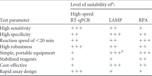
Conclusions.In our study, newly developed high-speed RT-qPCR, RPA, and LAMP assays enabled rapid detection of BVDV and SBV in less than 30 min. However, the tests revealed major differences with regard to sensitivity and specificity, robustness, assay time, complexity of test design, and field applicability (Table 5). Based on these findings, we conclude that none of the investi-TABLE 5Level of suitability of high-speed RT-qPCR, LAMP, and RPA with regard to important properties of a pen-side test
Test parameter
Level of suitability ofa:
High-speed
RT-qPCR LAMP RPA
High sensitivity ⫹⫹⫹ ⫹⫹ ⫹
High specificity ⫹⫹ ⫹⫹⫹ ⫹⫹
Reaction speed of⬍20 min ⫹⫹ ⫹⫹ ⫹⫹⫹
High robustness ⫹⫹⫹ ⫹⫹ ⫹⫹
Simple, portable equipment ⫹ ⫹⫹⫹b ⫹⫹⫹
Stabilized reagents ⫹ ⫹ ⫹⫹
Cost-effective ⫹ ⫹⫹⫹ ⫹⫹
Rapid assay design ⫹⫹⫹ ⫹ ⫹
a⫹⫹⫹, very high;⫹⫹, high;⫹, medium to low. b
Based on the possibility to perform LAMP with a simple heat block or water bath.
on May 16, 2020 by guest
http://jcm.asm.org/
[image:8.585.299.545.87.210.2]gated amplification techniques represents a generic platform that can be used across a variety of diagnostic questions and for a broad range of pathogens. Since the success of a pen-side test relies on the integrated amplification strategy, the application-specific properties of the available technologies have to be assessed care-fully prior to assay development. Thus, the findings of the present study deliver a valuable contribution to the future development of rapid and reliable molecular pen-side test systems.
ACKNOWLEDGMENTS
We thank Qiagen for providing the ESEQuant tube scanner, Ahmed Abd El Wahed (Department of Virology, University Medical Center, Goettin-gen, Germany) for advice on the RPA technology, Mario Ziller (FLI Insel Riems) for help with statistical analysis, and Christian Korthase for excel-lent technical assistance.
This work was funded by Zentrales Innovationsprogramm Mittel-stand (ZIM) of the German Federal Ministry of Economics and Tech-nology.
REFERENCES
1.Ferris NP, Nordengrahn A, Hutchings GH, Reid SM, King DP, Ebert K, Paton DJ, Kristersson T, Brocchi E, Grazioli S, Merza M.2009. Devel-opment and laboratory validation of a lateral flow device for the detection of foot-and-mouth disease virus in clinical samples. J. Virol. Methods 155:10 –17.http://dx.doi.org/10.1016/j.jviromet.2008.09.009.
2.Ferris NP, Nordengrahn A, Hutchings GH, Paton DJ, Kristersson T, Brocchi E, Grazioli S, Merza M. 2010. Development and laboratory validation of a lateral flow device for the detection of serotype SAT 2 foot-and-mouth disease viruses in clinical samples. J. Virol. Methods163: 474 – 476.http://dx.doi.org/10.1016/j.jviromet.2009.09.022.
3.Asiello PJ, Baeumner AJ. 2011. Miniaturized isothermal nucleic acid amplification, a review. Lab Chip 11:1420 –1430. http://dx.doi.org/10 .1039/c0lc00666a.
4.Craw P, Balachandran W.2012. Isothermal nucleic acid amplification technologies for point-of-care diagnostics: a critical review. Lab Chip12: 2469 –2486.http://dx.doi.org/10.1039/c2lc40100b.
5.Niemz A, Ferguson TM, Boyle DS. 2011. Point-of-care nucleic acid testing for infectious diseases. Trends Biotechnol.29:240 –250.http://dx .doi.org/10.1016/j.tibtech.2011.01.007.
6.Wheeler EK, Hara CA, Frank J, Deotte J, Hall SB, Benett W, Spadaccini C, Beer NR.2011. Under-three minute PCR: probing the limits of fast amplifi-cation. Analyst136:3707–3712.http://dx.doi.org/10.1039/c1an15365j. 7.Fujimoto T, Konagaya M, Enomoto M, Tsuboi K, Hashimoto K,
Taniguchi K, Kodama T, Okabe N.2010. Novel high-speed real-time PCR method (Hyper-PCR): results from its application to adenovirus diagnosis. Jpn. J. Infect. Dis.63:31–35.
8.Sakurai A, Nomura N, Nanba R, Sinkai T, Iwaki T, Obayashi T, Hashimoto K, Hasegawa M, Sakoda Y, Naito A, Morizane Y, Hosaka M, Tsuboi K, Kida H, Kai A, Shibasaki F.2011. Rapid typing of influenza viruses using super high-speed quantitative real-time PCR. J. Virol. Meth-ods178:75– 81.http://dx.doi.org/10.1016/j.jviromet.2011.08.015. 9.Wernike K, Beer M, Hoffmann B.2013. Rapid detection of
foot-and-mouth-disease virus, influenza A virus and classical swine fever virus by high-speed real-time RT-PCR. J. Virol. Methods193:50 –54.http://dx.doi .org/10.1016/j.jviromet.2013.05.005.
10. Piepenburg O, Williams CH, Stemple DL, Armes NA. 2006. DNA detection using recombination proteins. PLoS Biol.4:e204.http://dx.doi .org/10.1371/journal.pbio.0040204.
11. Euler M, Wang Y, Heidenreich D, Patel P, Strohmeier O, Hakenberg S, Niedrig M, Hufert FT, Weidmann M.2013. Development of a panel of recombinase polymerase amplification assays for detection of biothreat agents. J. Clin. Microbiol.51:1110 –1117.http://dx.doi.org/10.1128/JCM .02704-12.
12. Euler M, Wang Y, Nentwich O, Piepenburg O, Hufert FT, Weidmann M.2012. Recombinase polymerase amplification assay for rapid detection of Rift Valley fever virus. J. Clin. Virol.54:308 –312.http://dx.doi.org/10 .1016/j.jcv.2012.05.006.
13. Euler M, Wang Y, Otto P, Tomaso H, Escudero R, Anda P, Hufert FT, Weidmann M.2012. Recombinase polymerase amplification assay for
rapid detection of Francisella tularensis. J. Clin. Microbiol.50:2234 –2238. http://dx.doi.org/10.1128/JCM.06504-11.
14. Abd El Wahed A, El-Deeb A, El-Tholoth M, Abd El Kader H, Ahmed A, Hassan S, Hoffmann B, Haas B, Shalaby MA, Hufert FT, Weidmann M.2013. A portable reverse transcription recombinase polymerase am-plification assay for rapid detection of foot-and-mouth disease virus. PLoS One8:e71642.http://dx.doi.org/10.1371/journal.pone.0071642. 15. Parida M, Sannarangaiah S, Dash PK, Rao PV, Morita K.2008. Loop
mediated isothermal amplification (LAMP): a new generation of innova-tive gene amplification technique; perspecinnova-tives in clinical diagnosis of in-fectious diseases. Rev. Med. Virol.18:407– 421.http://dx.doi.org/10.1002 /rmv.593.
16. Notomi T, Okayama H, Masubuchi H, Yonekawa T, Watanabe K, Amino N, Hase T.2000. Loop-mediated isothermal amplification of DNA. Nucleic Acids Res.28:E63.http://dx.doi.org/10.1093/nar/28.12.e63.
17. Nagamine K, Hase T, Notomi T.2002. Accelerated reaction by loop-mediated isothermal amplification using loop primers. Mol. Cell. Probes 16:223–229.http://dx.doi.org/10.1006/mcpr.2002.0415.
18. Dukes JP, King DP, Alexandersen S.2006. Novel reverse transcription loop-mediated isothermal amplification for rapid detection of foot-and-mouth disease virus. Arch. Virol. 151:1093–1106.http://dx.doi.org/10 .1007/s00705-005-0708-5.
19. Yin S, Shang Y, Zhou G, Tian H, Liu Y, Cai X, Liu X.2010. Develop-ment and evaluation of rapid detection of classical swine fever virus by reverse transcription loop-mediated isothermal amplification (RT-LAMP). J. Biotechnol.146:147–150.http://dx.doi.org/10.1016/j.jbiotec .2009.11.006.
20. Imai M, Ninomiya A, Minekawa H, Notomi T, Ishizaki T, Tashiro M, Odagiri T.2006. Development of H5-RT-LAMP (loop-mediated isother-mal amplification) system for rapid diagnosis of H5 avian influenza virus infection. Vaccine 24:6679 – 6682. http://dx.doi.org/10.1016/j.vaccine .2006.05.046.
21. Neill JD.2013. Molecular biology of bovine viral diarrhea virus. Biologi-cals41:2–7.http://dx.doi.org/10.1016/j.biologicals.2012.07.002. 22. Hoffmann B, Depner K, Schirrmeier H, Beer M. 2006. A universal
heterologous internal control system for duplex real-time RT-PCR assays used in a detection system for pestiviruses. J. Virol. Methods136:200 –209. http://dx.doi.org/10.1016/j.jviromet.2006.05.020.
23. Hyndman L, Vilcek S, Conner J, Nettleton PF.1998. A novel nested reverse transcription PCR detects bovine viral diarrhoea virus in fluids from aborted bovine fetuses. J. Virol. Methods71:69 –76.http://dx.doi .org/10.1016/S0166-0934(97)00206-1.
24. McGoldrick A, Lowings JP, Ibata G, Sands JJ, Belak S, Paton DJ.1998. A novel approach to the detection of classical swine fever virus by RT-PCR with a fluorogenic probe (TaqMan). J. Virol. Methods72:125–135.http: //dx.doi.org/10.1016/S0166-0934(97)00208-5.
25. Gaede W, Reiting R, Schirrmeier H, Depner KR, Beer M.2005. Detec-tion and species-specific differentiaDetec-tion of pestiviruses using real-time RT-PCR. Berl. Munch. Tierarztl. Wochenschr.118:113–120. (In Ger-man.)
26. Presi P, Struchen R, Knight-Jones T, Scholl S, Heim D.2011. Bovine viral diarrhea (BVD) eradication in Switzerland— experiences of the first two years. Prev. Vet. Med. 99:112–121. http://dx.doi.org/10.1016/j .prevetmed.2011.01.012.
27. Hoffmann B, Scheuch M, Höper D, Jungblut R, Holsteg M, Schirrmeier H, Eschbaumer M, Goller KV, Wernike K, Fischer M, Breithaupt A, Mettenleiter TC, Beer M.2012. Novel orthobunyavirus in cattle, Europe, 2011. Emerg. Infect. Dis.18:469 – 472.http://dx.doi.org/10.3201/eid1803 .111905.
28. Beer M, Conraths FJ, van der Poel WH.2012. ‘Schmallenberg virus’—a novel orthobunyavirus emerging in Europe. Epidemiol. Infect.141:1– 8. http://dx.doi.org/10.1017/S0950268812002245.
29. Wernike K, Hoffmann B, Beer M.2013. Schmallenberg virus. Dev. Biol. (Basel)135:175–182.http://dx.doi.org/10.1159/000312546.
30. Bilk S, Schulze C, Fischer M, Beer M, Hlinak A, Hoffmann B.2012. Organ distribution of Schmallenberg virus RNA in malformed newborns. Vet. Microbiol.159:236 –238.http://dx.doi.org/10.1016/j.vetmic.2012.03 .035.
31. Fan Q, Xie Z, Xie L, Liu J, Pang Y, Deng X, Peng Y, Wang X.2012. A reverse transcription loop-mediated isothermal amplification method for rapid detection of bovine viral diarrhea virus. J. Virol. Methods186:43– 48.http://dx.doi.org/10.1016/j.jviromet.2012.08.007.
on May 16, 2020 by guest
http://jcm.asm.org/
32. Elmore S.2007. Apoptosis: a review of programmed cell death. Toxicol. Pathol.35:495–516.http://dx.doi.org/10.1080/01926230701320337. 33. Behrens SE, Grassmann CW, Thiel HJ, Meyers G, Tautz N. 1998.
Characterization of an autonomous subgenomic pestivirus RNA replicon. J. Virol.72:2364 –2372.
34. Meyers G, Tautz N, Becher P, Thiel HJ, Kummerer BM.1996. Recovery of cytopathogenic and noncytopathogenic bovine viral diarrhea viruses from cDNA constructs. J. Virol.70:8606 – 8613.
35. Lutz S, Weber P, Focke M, Faltin B, Hoffmann J, Muller C, Mark D, Roth G, Munday P, Armes N, Piepenburg O, Zengerle R, von Stetten F. 2010. Microfluidic lab-on-a-foil for nucleic acid analysis based on isother-mal recombinase polymerase amplification (RPA). Lab Chip10:887– 893. http://dx.doi.org/10.1039/b921140c.
36. Shen F, Davydova EK, Du W, Kreutz JE, Piepenburg O, Ismagilov RF. 2011. Digital isothermal quantification of nucleic acids via simultaneous chemical initiation of recombinase polymerase amplification reactions on SlipChip. Anal. Chem.83:3533–3540.http://dx.doi.org/10.1021/ac200247e. 37. Tian CJ, Lin ZX, He XM, Luo Q, Luo CB, Yu HQ, Chen R, Wu XW,
Zhu DZ, Ren ZJ, Bi YZ, Ji J. 2012. Development of a fluorescent-intercalating-dye-based reverse transcription loop-mediated isothermal amplification assay for rapid detection of seasonal Japanese B encephalitis outbreaks in pigs. Arch. Virol.157:1481–1488.http://dx.doi.org/10.1007 /s00705-012-1330-y.
38. Peyrefitte CN, Boubis L, Coudrier D, Bouloy M, Grandadam M, Tolou
HJ, Plumet S.2008. Real-time reverse-transcription loop-mediated iso-thermal amplification for rapid detection of Rift Valley Fever virus. J. Clin. Microbiol.46:3653–3659.http://dx.doi.org/10.1128/JCM.01188-08. 39. Tomita N, Mori Y, Kanda H, Notomi T.2008. Loop-mediated
isother-mal amplification (LAMP) of gene sequences and simple visual detection of products. Nat. Protoc.3:877– 882.http://dx.doi.org/10.1038/nprot.2008.57. 40. Kaneko H, Kawana T, Fukushima E, Suzutani T.2007. Tolerance of
loop-mediated isothermal amplification to a culture medium and biolog-ical substances. J. Biochem. Biophys. Methods70:499 –501.http://dx.doi .org/10.1016/j.jbbm.2006.08.008.
41. Enomoto Y, Yoshikawa T, Ihira M, Akimoto S, Miyake F, Usui C, Suga S, Suzuki K, Kawana T, Nishiyama Y, Asano Y.2005. Rapid diagnosis of herpes simplex virus infection by a loop-mediated isothermal amplifica-tion method. J. Clin. Microbiol.43:951–955.http://dx.doi.org/10.1128 /JCM.43.2.951-955.2005.
42. Curtis KA, Rudolph DL, Owen SM.2008. Rapid detection of HIV-1 by reverse-transcription, loop-mediated isothermal amplification (RT-LAMP). J. Virol. Methods 151:264 –270. http://dx.doi.org/10.1016/j .jviromet.2008.04.011.
43. Yamada Y, Itoh M, Yoshida M.2006. Sensitive and rapid diagnosis of human parvovirus B19 infection by loop-mediated isothermal amplifica-tion. Br. J. Dermatol.155:50 –55.http://dx.doi.org/10.1111/j.1365-2133 .2006.07379.x.