© 2017, IRJET | Impact Factor value: 5.181 | ISO 9001:2008 Certified Journal | Page 1546
Brain Tumor Segmentation Based on SFCM using Neural Network
B.Akshaya
1, J.Nandhini
2, J.Pauline Shilpa Ashwanthy
3, Mrs.M.Therasa
41
B.Akshaya, IV-CSE, Panimalar Institute of Technology
2
J.Nandhini, IV-CSE, Panimalar Institute of Technology
3
J.Pauline Shilpa Ashwanthy,IV-CSE,Panimalar Institute of Technology
4
Mrs.M.Therasa, Assisstant Professor, Department of Computer Science and Engineering,
Panimalar Institute of Technology, Chennai, Tamilnadu, India
---***---Abstract :
Programmed surrenders identification in MR pictures is vital in numerous demonstrative and restorative applications. In light of high amount information in MR pictures and obscured limits, tumor division and characterization is hard. This work has presented one programmed cerebrum tumor discovery technique to expand the precision and yield and lessening the finding time. The objective is ordering the tissues to three classes of ordinary, start and harmful. . In MR pictures, the measure of information is a lot for manual elucidation and examination. Amid recent years, mind tumor division in attractive reverberation imaging (MRI) has turned into a new research range in the field of medicinal imaging framework. Precise identification of size and area of cerebrum tumor assumes an imperative part in the analysis of tumor. The conclusion technique comprises of four phases, pre-handling of MR pictures, highlight extraction, and characterization. After histogram balance of picture, the components are separated in view of Dual-Tree Complex wavelet change (DTCWT). In the last stage, Back Propagation Neural Network (BPN) are utilized to characterize the Normal and strange cerebrum. An effective calculation is proposed for tumor identification in light of the Spatial Fuzzy C-Means Clustering.Keywords: Brain tumor segmentation, Magnetic Resonance Imaging (MRI),Dual-Tree Complex wavelet change (DTCWT),Back Propagation Neural Network (BPN),Spatial Fuzzy C-Means Clustering.
1. INTRODUCTION
Gliomas are the mind tumors with the most elevated mortality rate and pervasiveness [1]. These neoplasms can be reviewed into Second rate Gliomas (LGG) and High Grade Gliomas (HGG), with the previous being less forceful and infiltrative than the last [1], [2]. Indeed, even under treatment, patients don't survive by and large over 14 months after analysis [3]. Current medicines incorporate surgery, chemotherapy, radiotherapy, or a mix of them [4]. X-ray is particularly valuable to survey gliomas in clinical practice, since it is conceivable to gain MRI arrangements giving correlative data [1]. The precise division ofgliomas and its intra-tumoralstructures is critical for treatment arranging, as well as for follow-up assessments. Be that as it may, manual division is tedious and subjected to between and intra-rater mistakes
© 2017, IRJET | Impact Factor value: 5.181 | ISO 9001:2008 Certified Journal | Page 1547
model. This sort of methodologies normally consider voxels as autonomous and indistinguishably appropriated [12], in spite of the fact that setting data might be presented through the highlights. Along these lines, some secluded voxels or little bunches might be erroneously grouped with the wrong class, now and then in physiological and anatomically impossible areas. To defeat this issue, a few creators incorporate data of more recently, Random Forests (RF) [14]–[21] were successfullyapplied in brain tumor segmentation. The RF becamevery used due to its natural capability in handling multi-classproblems and large feature vectors. A variety of features wereproposed in the literature: encoding context [15], [16], [21],first-order and fractals-based texture [14], [15], [18], [21],[22], gradients [14], [15], brain symmetry [14], [15], [19], and physical properties [19]. Using supervised classifiers, some authors developed other ways of applying them. Tustison et al[19] developed a two-stage segmentation framework based on RFs, using the output of the first classifier to improve a second stage of segmentation. Geremia et al. [20] proposed a Spatial. Adaptive RF for hierarchical segmentation, going from coarser to finer scales. Meier et al. [23] used a semi-supervised RF totrain a subject-specific classifier for post-operative brain tumor segmentation.2.RELATED WORK
[1]Twofold Classification of Brain Tumors Using a Discrete Wavelet Transform and Energy Criteria by Carlos Arizmendi, Alfredo Vellido, Enrique Romero. The exact conclusion of human mind tumors is a touchy restorative assignment, for which radiology specialists frequently should depend on backhanded flag estimations. There is in this way a requirement for creating PC based choice bolster apparatuses to help specialists in their demonstrative undertaking. The investigations in this short paper address such issue as parallel grouping, for which the pre-handling of the Magnetic Resonance Spectroscopy (MRS) flag is a most applicable information examination organize. A blend of the Discrete Wavelet Transform (DWT) for flag disintegration and a vitality model for flag remaking is utilized to pre-handle the MRS information preceding the component determination and characterization with Bayesian Neural Networks.
[2]Input Feature Selection for Classification Problems by NojunKwak and Chong-Ho Choi. Highlight determination assumes an essential part in arranging frameworks, for example, neural systems (NNs). We utilize an arrangement of qualities which are important, unessential or excess and from the perspective of dealing with a dataset which can be enormous, lessening the number of traits by selecting just the significant ones is alluring. In doing as such, higher exhibitions with lower computational exertion is normal. In this paper, we propose two element choice calculations. The impediment of shared data include selector (MIFS) is broke down and a technique to conquer this confinement is examined. One of the proposed calculations makes more considered utilization of common data between info properties and yield classes than the MIFS. What is exhibited is that the proposed strategy can give the execution of the perfect avaricious determination calculation when data is appropriated consistently. The computational stack for this calculation is about the same as that of MIFS. Moreover, another element choice calculation utilizing the Taguchi technique is proposed. This is progressed as an answer for the question regarding how to distinguish great components with as few analyses as could reasonably be expected. The proposed calculations are connected to a few arrangement issues and contrasted and MIFS. These two calculations can be consolidated to supplement each other's impediments. The consolidated calculation performed well in a few analyses and ought to demonstrate to be a valuable technique in selecting highlights for grouping issues.
[3]Order of cerebrum tumors utilizing PCA-ANN by Vinod Kumar, JainySachdeva, Indra Gupta NiranjanKhandelwal, Chirag Kamal Ahuja. The present review is directed to help radiologists in stamping tumor limits and in basic leadership prepare for multiclass grouping of mind tumors. Essential mind tumors what's more, optional mind tumors alongside typical locales are fragmented by Gradient Vector Flow (GVF)- a limit based procedure. GVF is a client intelligent model for extricating tumor limits. These sectioned areas of intrigue (ROIs) are than ordered by utilizing Principal Component Analysis-Artificial Neural.Organize (PCA-ANN) approach. The review is performed on differentiated dataset of 856 ROIs from 428 post differentiate T1- weighted MR pictures of 55 patients. 218 surface and force components are separated from ROIs. PCA is utilized for lessening of dimensionality of the component space. Six classes which incorporate essential tumors, for example, Astrocytoma (AS), GlioblastomaMultiforme (GBM), tyke tumor-Medulloblastoma (MED) and Meningioma (MEN), auxiliary tumor-Metastatic (MET) along with ordinary areas (NR) are separated utilizing ANN. Test comes about demonstrate that the PCA–ANN approach has upgraded the general exactness of ANN from 72.97 % to 95.37%. The proposed technique has conveyed a high exactness for every class: AS-90.74%, GBM-88.46%, MED-85.00%, MEN-90.70%, MET-96.67% and NR-93.78%. It is watched that PCA-ANN gives better outcomes than the current techniques.
© 2017, IRJET | Impact Factor value: 5.181 | ISO 9001:2008 Certified Journal | Page 1548
k-closest neighbor (k-NN), parzenwindow and counterfeit neural system (ANN) are utilized. Our work is the change and augmentation of the past reviews on the analysis of mind infections, while we get better grouping rate with the less number of elements and we too utilize bigger and rather unique database.[5]Order of cervical cell nuclei using morphological segmentation and textural feature extraction by Ross F. Walker' Paul Jackway’Brian Lovell' I. D. Longstaff. This paper presents preparatory outcomes for the grouping of Pap Smear cell cores, utilizing Gray Level CO-event Matrix (GLCM) textural highlights. We layout a strategy for atomic division utilizing quick morphological dim scale changes. For each portioned core, highlights inferred from an altered type of the GLCM are extricated more than a few point and separation measures. Straight Discriminant Analysis is performed on these elements to diminish the dimensionality of the element space, and a classifier with hyperquadricchoice surface is actualized to group a little arrangement of ordinary and strange cell cores. Utilizing 2 highlights, we accomplish a misclassification rate of 3.3% on an informational collection of 61 cells.
3. PROPOSED SYSTEM
The experiments were carried out on real patient data ob-tained from the 2013 brain tumorsegmentation challenge(BRATS2013), as part of the MICCAI conference [15]. The
BRATS2013 dataset is comprised of 3 sub-datasets. The train-ing dataset, which contains 30 patient subjects all with pixel- accurate ground truth (20 high grade and 10 low grade tumors);the test dataset which contains 10 (all high grade tumors) and the leaderboard dataset which contains 25 patient subjects (21high grade and 4 low grade tumors). There is no ground truth provided for the test and leaderboard datasets. All brains in the dataset have the same orientation. For each brain there exists 4 modalities, namely T1, T1C, T2 and Flair which are co-registered. The training brains come with groundtruth for which segmentation labels are provided, namely non-tumor, necro-sis, edema, non-enhancing tumor and enhancing tumor. In total, the model iterates over about 2.2 million examples of tumorous patches (this consists of all the 4 sub-tumor classes)and goes through 3.2 million of the healthy patches. As men-tioned before during the first phase training, the distribution of examples introduced to the model from all 5 classes is uniform.
SYSTEM ARCHITECTURE
Fig1.1 Proposed System Architecture
BRAIN TUMOR SEGMENTATION BASED ON SFCM USING NEURAL NETWORKS
Phase 1: Preprocessing and Dual-tree complex wavelet transforms(DT-CWT)
© 2017, IRJET | Impact Factor value: 5.181 | ISO 9001:2008 Certified Journal | Page 1549
contrast stretching or de-blurring by a nearest neighbor procedure) provided by "Imaging packages" use no a priori model of the process that created the image. With image enhancement noise can be effectively be removed by sacrificing some resolution, but this is not acceptable in many applications. In a Fluorescence Microscope resolution in the z-direction is bad as it is. More advanced image processing techniques must be applied to recover the object. De-Convolution is an example of image restoration method. It is capable of: Increasing resolution, especially in the axial direction removing noise increasing contrast.The dual-tree complex wavelet transform (CWT) is a relatively recent enhancement to the discrete wavelet transform (DWT), with important additional properties: It is nearly shift invariant and directionally selective in two and higher dimensions. It achieves this with a redundancy factor of only 2d for d-dimensional signals, which is substantially lower than the undecimated DWT. The multidimensional (M-D) dual-tree CWT is no separable but is based on a computationally efficient, separable filter bank (FB). The theory behind the dual-tree transforms shows how complex wavelets with good properties can be designed, and illustrates a range of applications in signal and image processing.
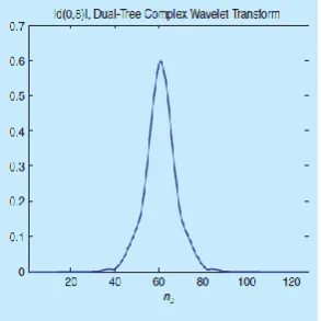
Figure 1.2: The value of the wavelet coefficient in “Real-Valued Discrete Wavelet Transform and Filter Banks
Phase 3
:
Clustering Model
Clustering can be considered the most important un supervised learning problem, so, it deals with finding a structure in a collection of unlabeled data. Acluster is therefore a collection of objects which are “similar” between them and are “dissimilar” to the objects belonging to other clusters.
Phase 4: Feature Extraction And Neural
Network
Originally proposed by R.M. Haralick, the co-occurrence matrix representation of texture features explores the grey level spatial dependence of texture [2]. A mathematical definition of the co-occurrence matrix is as follows [4]:
Given a position operator P(i,j), - let A be an n x n matrix
- whose element A[i][j] is the number of times that points with grey level (intensity the position -Let C be the n x n matrix that isproduced by dividing A with the total number of point pairs that satisfy P. C[i][j] is a measure of the joint probability that a pair of points satisfying P will have values g[i], g[j].
[image:4.595.225.372.249.396.2]© 2017, IRJET | Impact Factor value: 5.181 | ISO 9001:2008 Certified Journal | Page 1550
Phase 5: KNN Classifier
In pattern recognition, the k-nearest neighbor algorithm (k-NN) is a method for classifying objects based on closest training examples in the feature space. k-NN is a type of instance-based learning, or lazy learning where the function is only approximated locally and all computation is deferred until classification. The k-nearest neighbor algorithm is amongst the simplest of all machine learning algorithms: an object is classified by a majority vote of its neighbors, with the object being assigned to the class most common amongst its k nearest neighbors (k is a positive integer, typically small). If k = 1, then the object is simply assigned to the class of its nearest neighbor.The same method can be used for regression, by simply assigning the property value for the object to be the average of the values of itsk nearest neighbors. It can be useful to weight the contributions of the neighbors, so that the nearer neighbors contribute more to the average than the more distant ones. (A common weighting scheme is to give each neighbor a weight of 1/d, where d is the distance to the neighbor. This scheme is a generalization of linear interpolation.)
Phase 6: Back Propagation Networks (BPN):
Back Propagation (BPN) and General Regression Neural Networks (GRNN) have similar architectures, but there is a fundamental difference: Probabilistic networks perform classification where the target variable is categorical, whereas general regression neural networks perform regression where the target variable is continuous. If you select a BPN/GRNN network, DTREG will automatically select the correct type of network based on the type of target variable.
Fig1.3: Network Diagram
4.EXPERIMENTAL RESULTS
4.1 MRI IMAGE
This image represents the MRI image of the scanned human brain.
© 2017, IRJET | Impact Factor value: 5.181 | ISO 9001:2008 Certified Journal | Page 1551
4.2RESTORED IMAGE
This image represents the restored image of the human brain with tumor
4.3FUZZY CLUSTERING
This image uses fuzzy clustering method to display the results.
5.CONCLUSION
In outline, we propose a novel CNN-based technique for division of mind tumors in MRI pictures. We begin by a pre-handling stage comprising of inclination field amendment, power and fix standardization. From that point forward, amid preparing, the quantity of preparing patches is falsely enlarged by turning the preparation fixes, and utilizing tests of HGG to increase the quantity of uncommon LGG classes. The CNN is manufactured over convolutional layers with little 33 pieces to permit more profound designs. In planning our strategy, we address the heterogeneity brought about by multi-site multi-scanner acquisitions of MRI pictures utilizing force standardization as proposed by Ny'ul et al. We demonstrate this is vital in accomplishing a decent division.Cerebrum tumors are profoundly factor in their spatial restriction furthermore, basic organization, so we have explored the utilization of information growth to adapt to such fluctuation. We examined expanding our preparation informational collection by turning the patches and also by examining from classes of HGG that were underrepresented in LGG. We found that information enlargement was likewise very compelling, in spite of the fact that not altogether investigated in Profound Learning techniques for cerebrum tumor division. Moreover, we explored the capability of profound models through little bits by contrasting our profound CNN and shallow models with bigger channels. We found that shallow designs introduced a lower execution, notwithstanding when utilizing a bigger number of highlight maps. At long last, we confirmed that the initiation work LReLU was more imperative than ReLU in adequately preparing our CNN.
REFERENCES
© 2017, IRJET | Impact Factor value: 5.181 | ISO 9001:2008 Certified Journal | Page 1552
[2] D. N. Louis et al., “The 2007 who classification of tumours of the central nervous system,” Actaneuropathologica, vol. 114, no. 2, pp. 97–109,2007.[3] E. G. Van Meir et al., “Exciting new advances in neuro-oncology:The avenue to a cure for malignant glioma,” CA: a cancer journal for clinicians, vol. 60, no. 3, pp. 166–193, 2010.
[4] G. Tabatabai et al., “Molecular diagnostics of gliomas: the clinical perspective,” Actaneuropathologica, vol. 120, no. 5, pp. 585–592, 2010.
[5] B. Menze et al., “The multimodal brain tumor image segmentation benchmark (brats),” IEEE Transactions on Medical Imaging, vol. 34,no. 10, pp. 1993–2024, 2015.
[6] N. J. Tustison et al., “N4itk: improved n3 bias correction,” IEEE Transactions on Medical Imaging, vol. 29, no. 6, pp. 1310–1320, 2010.
[7] L. G. Ny´ul, J. K. Udupa, and X. Zhang, “New variants of a method of mri scale standardization,” IEEE Transactions on Medical Imaging, vol. 19, no. 2, pp. 143–150, 2000.
[8] M. Prastawa et al., “A brain tumor segmentation framework based on outlier detection,” Medical image analysis, vol. 8, no. 3, pp. 275–283,2004.
[9] B. H. Menze et al., “A generative model for brain tumor segmentation in multi-modal images,” in Medical Image Computing and Computer- Assisted Intervention–MICCAI 2010. Springer, 2010, pp. 151–159.