Development 139, 4582-4590 (2012) doi:10.1242/dev.083246 © 2012. Published by The Company of Biologists Ltd
INTRODUCTION
DNA is wrapped around a histone octamer to form the basic unit of chromatin structure. During embryogenesis, dynamic changes of chromatin structure and chromatin modification occur after fertilization; subsequently, the epigenetic information is inherited through many rounds of the cell cycle. Thus, chromatin is essential for the determination of cell identity. Two strategies are used to modulate a chromatin environment: the covalent modification of histone tails and energy-dependent chromatin remodeling. The acetylation, methylation or phosphorylation of histone tails can have profound effects on chromatin structure and transcription (Jenuwein and Allis, 2001). Chromatin remodeling reactions are catalyzed by large protein complexes that use the energy of ATP hydrolysis to alter the structure or positioning of nucleosomes (Becker and Hörz, 2002; Clapier and Cairns, 2009). In addition to these events, histone variants play important roles in modulating chromatin structure (Henikoff and Ahmad, 2005; Kamakaka and Biggins, 2005; Sarma and Reinberg, 2005).
One histone variant, H3.3, has been studied extensively. Although histone H3.3 differs by only four amino acids from canonical histone H3.1, they differ in their mechanisms of chromatin deposition. Histone H3.1 is assembled into chromatin during DNA replication, whereas deposition of histone H3.3 occurs throughout the cell cycle (Ahmad and Henikoff, 2002).
Furthermore, the chaperone complex for H3.1 contains CAF-1, p150, p60 and p48, whereas the complex for H3.3 contains HIRA (Tagami et al., 2004). Biochemical analysis revealed that H3.3 is associated with active histone modifications such as H3 Lys 4 methylation (McKittrick et al., 2004). Moreover, a nucleosome containing H3.3 exhibits instability (Jin and Felsenfeld, 2007). Therefore, histone H3.3 replacement can change pre-existing epigenetic states.
Chromatin boundaries are transcriptional regulatory elements that modulate interaction between enhancer and promoter, and protect gene expression from positive or negative effects of the adjacent chromatin (Bell et al., 2001; Maeda and Karch, 2007). Establishment and maintenance of chromatin boundaries play a crucial role in development. For example, the Fab-7boundary governs space-specific expression of Abd-B by ensuring the autonomous activities of the iab-6and iab-7 cis-regulatory regions (Mihaly et al., 1997). Previously, we have shown that GAGA factor interacts with FACT, induces chromatin remodeling and directs H3.3 replacement through an interaction with HIRA at chromatin boundaries d1 and Fab-7 to counteract the spreading of silent chromatin (Shimojima et al., 2003; Nakayama et al., 2007). Furthermore, H3.3 replacement peaks at multiple boundaries in the Bithoraxcomplex, including Mcp, Fab-7and Fab-8(Mito et al., 2007). Thus, histone H3.3 replacement can influence chromatin boundary functions.
Although FACT displaces a histone H2A-H2B pair from a nucleosome (Belotserkovskaya et al., 2003), it alone cannot accomplish the H3.3 replacement that accompanies extensive chromatin remodeling, as revealed by DNase hypersensitivity (Nakayama et al., 2007). Furthermore, chromatin remodeling factors Chd1 and ATRX contribute to H3.3 deposition on the male pronucleus after fertilization and on pericentric or telomeric chromatin, respectively (Konev et al., 2007; Drané et al., 2010; Department of Developmental Genetics, National Institute of Genetics, Mishima,
Shizuoka-ken 411-8540, Japan.
*These authors contributed equally to this work
‡Present address: Kazusa DNA Research Institute, 2-6-7 Kazusa-kamatari, Kisarazu, Chiba 292-0818, Japan
§Author for correspondence ([email protected])
Accepted 18 September 2012 SUMMARY
Establishment and maintenance of epigenetic memories are essential for development. Replacement of canonical histone H3 by its variant H3.3 has been implicated in cellular memory. Drosophila sequence-specific DNA-binding protein GAGA factor and a chromatin factor FACT direct H3.3 replacement in conjunction with H3.3-specific chaperone HIRA at chromatin boundaries to counteract the spreading of silent chromatin. However, little is known about which ATP-driven chromatin remodeling factor is responsible for the H3.3 replacement at chromatin boundaries. Here, we report that GAGA factor associates with the Polybromo-associated Brm (PBAP) remodeling complex, which consists of many Trithorax group proteins, and recruits this complex to chromatin boundaries d1(which is downstream of w), the Fab-7DNase-hypersensitive site (HS) 1 of Abd-Band the bxdregion of Ubx. Trl -encoding GAGA factor, brmand polybromo/bap180mutations compromise the H3.3 replacement and boundary functions in a synergistic manner. Furthermore, Polybromo is necessary for generation of the DNase HS at d1, and HIRA functions to restore the alteration. Taken together, we propose that FACT and PBAP complexes are recruited to chromatin boundaries in a GAGA factor-dependent manner, and are needed for H3.3 replacement to execute boundary functions. Our results provide new insight into the function of the trithoraxgroup during development.
KEY WORDS: PBAP complex, Histone H3.3, Chromatin boundary, FACT, trithoraxgroup, Drosophila
The PBAP remodeling complex is required for histone H3.3
replacement at chromatin boundaries and for boundary
functions
Takahiro Nakayama*, Tsukasa Shimojima*,‡and Susumu Hirose§
D
E
V
E
LO
P
M
E
N
Goldberg et al., 2010). Therefore, we assume that an ATP-dependent chromatin remodeling factor is necessary for the replacement at chromatin boundaries. Among the ATP-driven remodelers, a Swi/Snf-type complex is thought to be most suitable for the replacement because it has a unique activity to transfer a histone octamer from one piece of DNA to another (Lorch et al., 1999; Workman, 2006). There are two subclasses of the Swi/Snf-type complex that consist of many Trithorax group proteins in Drosophila(Mohrmann et al., 2004). One is the Brm-associated proteins (BAP) complex, which contains Osa in addition to core subunits that are shared by the two complexes: Brm, Mor/Bap155, Bap111, Bap60, Bap55, Actin, and Snr1. The other is PBAP complex, which harbors Polybromo/Bap180 and Bap170 in place of Osa. These two complexes correspond to mammalian Brg1- or Brm-associated factors (BAF), and Polybromo-associated BAF (PBAF) complexes, respectively. However, involvement of these complexes in H3.3 replacement is not clear in either Drosophilaor mammals.
In this study, we isolated GAGA factor-associated proteins to identify an ATP-dependent chromatin remodeling factor responsible for the H3.3 replacement at chromatin boundaries. We found association of the GAGA factor with PBAP complex and GAGA factor-dependent recruitment of the PBAP complex to chromatin boundary regions. Furthermore, our data indicate that PBAP complex is required for the H3.3 replacement at chromatin boundaries and for boundary functions.
MATERIALS AND METHODS Antibodies
For generation of antibodies against Brm, Polybromo or Bap60, we amplified cDNA encoding a polypeptide corresponding to residues 1 to 500 of Polybromo, residues 1301 to 1630 of Brm, or full length Bap60, followed by subcloning it into the pET21 expression vector. Recombinant proteins were expressed in Escherichia coliBL21 (DE3) pLysS at 25°C for 3 hours and were purified with Ni-NTA (Qiagen), followed by immunization of rabbits. Mouse anti-DrosophilaOsa monoclonal antibody (Concentrate) was purchased from Developmental Studies Hybridoma Bank (DSHB, Iowa University, IA, USA). The purified anti-HIRA antibodies PG (Bonnefoy et al., 2007) were a gift from B. Loppin (Universite Lyon, Lyon, France). The anti-SSRP1, SPT16 and GAGA factor antibodies have been described previously (Shimojima et al., 2003; Nakayama et al., 2007).
Isolation of FLAG-GAGA factor associated proteins
All procedures were performed at 4°C. Drosophilaembryos (125 g, 0-24 hours after egg laying) were collected from the fly line expressing FLAG-GAGA factor and dechorionated. Nuclear extract (NE, 100 mg protein) was prepared from the embryos, and FLAG-GAGA factor and its associated proteins were purified using 10 ml settled volume of FLAG M2 beads (Sigma-Aldrich) as described previously (Shimojima et al., 2003). To identify FLAG-GAGA factor-associated proteins, the eluted proteins were resolved by SDS-PAGE and transferred to a PVDF membrane (Roche Diagnostics) by a Trans-Blot Semi-Dry Apparatus (Bio-Rad, CA, USA). The membrane was subsequently stained with Coomassie Brilliant Blue, and protein in each band was subjected to MALDI-TOF mass analysis after digestion with lysylendopeptidase. For Superose 6 gel filtration, the pooled eluate was concentrated up to 500 μl with Amicon Ultra-15 (MWCO1000, Millipore). The concentrated proteins were loaded on a column of Superose 6 10/32 (GE Healthcare) and eluted with buffer F [20 mM HEPES Na+(pH 7.9), 50 mM NaCl, 0.2 mM EGTA, 10% (v/v) glycerol, 0.5 mM DTT, 0.01% (v/v) NP-40 and 0.5 mM PMSF]. Each 500 μl fraction was collected and proteins in each fraction were analyzed by western blotting.
Immunoprecipitation
NE containing 500 μg proteins from 0- to 24-hour-old Drosophilaembryos of the ywline, the transgenic line expressing HIRA-FLAG or third instar
larvae homozygous for bap180Δ86was pre-cleared by a 60-minute rotation with 10 μl of pre-immune rabbit IgG pre-adsorbed to 25 μl settled volume of Protein G agarose (Millipore) followed by centrifugation at 10,000 rpm (8000 g) for 5 minutes. The pre-cleared NE was incubated with 10 μl of rabbit antibodies against GAGA factor, Polybromo, Brm, SSRP1, SPT16, HIRA or anti-mouse monoclonal antibody against Osa or FLAG M2 pre-adsorbed to 25-μl settled volume of Protein G agarose in 500 μl of a binding buffer [20 mM HEPES Na+(pH 7.9), 50 mM (Fig. 1A,B) or 150 mM NaCl (Fig. 1C,D), 3 mM MgCl2, 10% (v/v) glycerol, 0.2 mM EDTA, 1/1000 volume of Protease inhibitor cocktail, 500 μg/ml BSA, 0.5 mM DTT, and 0.1% (v/v) NP-40] for 2 hours. The beads were washed five times with a wash buffer [the binding buffer + 0.4% (v/v) NP-40] by rotating tubes slowly for 5 minutes. Twenty-five μl of 2⫻SDS sample buffer was added to the beads and eluted proteins were heat-denatured at 95°C for 5 minutes and then were centrifuged at 3000 rpm (700 g) for 5 minutes. Proteins in the supernatant were loaded on a 6.5% SDS polyacrylamide gel. Whenever positive results were obtained by co-immunoprecipitation, we confirmed that the association was not mediated through nucleic acids by repeating the experiments in the presence of ethidium bromide.
Western blotting
Proteins were resolved on a 6.5% or 8% SDS polyacrylamide gel and were transferred to a PVDF membrane (Millipore) in a transfer buffer [48 mM Tris base, 390 mM glycine, 20% (v/v) methanol, 0.1% SDS] for 180 minutes at 35 V. The membrane was blocked with 5% skim milk in TBST [20 mM Tris-HCl (pH 8.0), 150 mM NaCl and 0.1% (v/v) Tween 20] for 30 minutes at room temperature. The membrane was incubated with each primary antibody (1:2000) in 1% skim milk in TBST for 1-2 hours at room temperature or at 4°C overnight. The membrane was washed three times with TBST for 5 minutes at room temperature. The membrane was incubated with horseradish peroxidase (HRP)-conjugated secondary antibody (1:5000) in 1% skim milk in TBST for 1 hour at room temperature. Signals were detected with Supersignal+ (Pierce) and a LAS-4000 Mini Imaging System (FUJIFILM, Tokyo, Japan).
Chromatin immunoprecipitation (ChIP)
ChIP assays using larval nuclei were performed as follows. Approximately 3500 third instar larvae homozygous for Trl13Cwere collected and stored
at −80°C. The frozen larvae were thawed and homogenized in 5 ml of the buffer A [10 mM HEPES Na+(pH 7.9), 10 mM KCl, 3 mM MgCl
2, 0.2 mM EGTA, 1 M sucrose, 0.5 mM DTT, 0.1% (v/v) NP-40, 0.5 mM PMSF and 1/1000 volume of Protease inhibitor cocktail (Sigma-Aldrich, P8340)]. The homogenate was filtered through two layers of 63-μm nylon mesh and was centrifuged at 1400 rpm (180 g) for 90 seconds to remove debris. The supernatant was centrifuged at 6000 rpm (3200 g) for 10 minutes. The pelleted nuclei were suspended with 5 ml of buffer G [20 mM HEPES Na+(pH 7.9), 10 mM KCl, 0.2 mM EGTA, 10% (v/v) glycerol, 0.5 mM DTT, 0.1% (v/v) NP-40, 0.5 mM PMSF and 1/1000 volume of Protease inhibitor cocktail]. The suspended nuclei were transferred into a 15-ml tube and equal volume of buffer H [20 mM HEPES Na+(pH 7.9), 10 mM KCl, 0.2 mM EGTA, 10% (v/v) glycerol, 0.1% (v/v) NP-40, 0.5 mM PMSF, 1/1000 volume of Protease inhibitor cocktail and 2% formaldehyde] was added. Crosslinking was carried out by rotating the tube for 15 minutes at 4°C. To stop the crosslink, one-ninth the volume of 1.25 M glycine was added and the tube was rotated for another 5 minutes. The tube was centrifuged at 3500 rpm (1600 g) for 10 minutes. Sonicated chromatin was prepared from the pelleted nuclei and subjected to ChIP as described previously (Nakayama et al., 2007). After phenol/chloroform extraction followed by ethanol precipitation, the DNA was dissolved in 100 μl of TE. Quantitative (q) PCR using 2-10 μl of the DNA was performed with a Light Cycler 2.0 (Roche Diagnostics, Mannheim, Germany). The experiments were repeated at least three times. For ChIP assays of Trl+ (wild type) or polybromomutant samples, ~800 third instar larvae of wild type or bap180Δ86(Carrera et al., 2008) homozygote were collected and processed as above. ChIP assays using embryos were carried out as described previously (Nakayama et al., 2007), except that amounts of DNA
were quantified with qPCR as above.
D
E
V
E
LO
P
M
E
N
Reverse transcription (RT)-qPCR
Total RNAs were prepared from third instar larvae using Sepasol (Nakalai Tesque). cDNAs were synthesized using a Transcriptor First Strand cDNA Synthesis Kit (Roche Diagnostics) in a 20 μl-reaction mixture according to the manufacturer’s protocol. Subsequently, 20 μl of sterilized water were added to the reaction mixtures. The real-time qPCR were conducted using the Light Cycler 2.0 (Roche Diagnostics, Mannheim, Germany). Reactions were carried out using each 2 μl of the cDNA as a template in a 20-μl reaction mixture. The amount of starting cDNA was normalized to that of β-tubulin. The experiments were repeated at least three times. Primer sequences used for qPCR are shown in supplementary material Table S1.
Position effect variegation
For position effect variegation (PEV) assays, wm4or wm4; TrlR85/TM6B
females were crossed with wild-type or a mutant males, and male progenies with a desired genotype were used for quantification of eye pigment levels. Twenty adult male flies eclosed within 24 hours were collected and processed as described previously (Nakayama et al., 2007). More than eight independent samples were measured.
Abdominal segment (A) 6 to A5 transformation
yw, yw; TrlR85/TM6Bor yw;Δspt16/TM3females were crossed with
wild-type or a mutant males, and male progenies with a desired genowild-type were used for detection of ectopic bristles on A6.
SpeI accessibility assay
The assays were carried out as described (Jack and Eggert, 1990). Nuclei were isolated from third instar larvae and ~5 μg DNA was treated with 10 units of SpeI (Takara, Kyoto) at 37°C for 30 minutes. Ten independent incubations were made for each line.
Transgenic lines and other fly lines
Full-length Polybromo cDNA was obtained from National Institute of Genetics. The cDNA was subcloned into pET21 and a FLAG-encoding
sequence was introduced to the N-terminal region. The full-length cDNA of FLAG-Polybromo was digested with MluI and NotI, and inserted into a germline transform vector pCasperHS83_EGFP (Nakayama et al., 2007). Transgenic flies were generated by DrosophilaEmbryo Injection Services (Best Gene). We used a line termed yw; P[bap180+], in which the
transgene was inserted into the second chromosome. We also established fly lines such as P[hsp83-H3.3-FLAG,EGFP];bap180Δ86/TM3,
P[hsp83-H3-FLAG,EGFP];bap180Δ86/TM3, P[hsp83-H3.3-FLAG,EGFP];
brm2/TM3, P[hsp83-H3-FLAG,EGFP];brm2/TM3,
P[hsp83-H3.3-FLAG,EGFP];Trl13C/bap180Δ86, P[hsp83-H3-FLAG,EGFP];Trl13C/
bap180Δ86, P[hsp83-H3.3-FLAG,EGFP];Trl13C/brm2,
P[hsp83-H3-FLAG,EGFP];Trl13C/brm2, wm4;TrlR85/TM6B, P[bap180+];bap180Δ86,
P[hsp83-H3-FLAG,EGFP];osa2/TM3,P[hsp83-H3.3-FLAG,EGFP];osa2/
TM3, ssm/FM7;P[hsp83-H3-FLAG,EGFP],
ssm/FM7;P[hsp83-H3.3-FLAG,EGFP], ssm/FM7GFP and ssm/FM7GFP;bap180Δ86/TM6B
through appropriate crosses. bap180Δ86/TM6Band TrlR85/TM6Bwere gifts
from Drs J. Treisman (New York University School of Medicine, NY, USA) and C. Wu (National Institutes of Health, MD, USA), respectively.
brm2/TM6Band osa2/TM6Bwere obtained from the Bloomington Stock
Center (Indiana University, IN, USA). Other fly lines have been described previously (Shimojima et al., 2003; Nakayama et al., 2007).
RESULTS
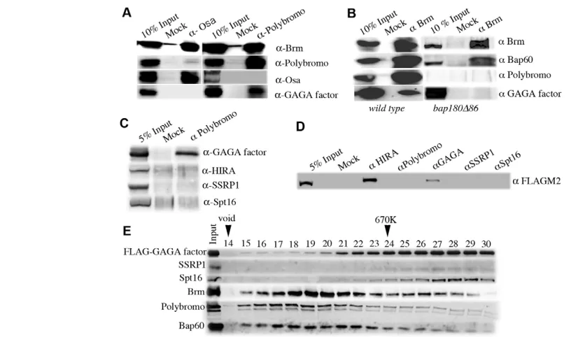
[image:3.612.62.470.58.299.2]Isolation of GAGA factor-associated proteins GAGA factor induces chromatin remodeling at chromatin boundaries (Nakayama et al., 2007) and, hence, we presumed that an ATP-dependent chromatin remodeler would be recruited to the boundaries through association with GAGA factor. When we isolated FLAG-GAGA factor-associated proteins in the previous study, the anti-FLAG immunoaffinity purification was conducted in the presence of 420 mM NaCl. Under this condition, only FACT Fig. 1. PBAP complex is associated with GAGA factor. (A) Co-immunoprecipitation analyses using nuclear extracts (NEs) from ywembryos with anti-Osa antibody (left panel) or with anti-Polybromo antibodies (right panel). Eluted proteins were immunoblotted with antibodies against Brm, Polybromo, Osa or GAGA factor. (B) Co-immunoprecipitation analyses using NEs from ywembryos (left panel) or bap180Δ86homozygous larvae (right panel) with anti-Brm antibodies. Eluted proteins were immunoblotted with antibodies against Brm, Bap60, Polybromo or GAGA factor. (C) Co-immunoprecipitation analyses using NE from ywembryos with anti-Polybromo antibodies. Eluted proteins were immunoblotted with antibodies against GAGA factor, HIRA, SSRP1 or Spt16. (D) Co-immunoprecipitation analyses of HIRA-FLAG. Proteins in NEs from a fly line expressing HIRA-FLAG were immunoprecipitated with antibodies against HIRA, Polybromo, GAGA factor, SSRP1 or Spt16, and analyzed by immunoblotting using anti-FLAG antibody M2. (E) Gel filtration profile. Immunoaffinity-purified FLAG-GAGA factor-associated proteins were fractionated by Superose 6 gel filtration and proteins in each fraction were analyzed by immunoblots using antibodies against FLAG, FACT subunits, Brm, Polybromo or Bap60.
D
E
V
E
LO
P
M
E
N
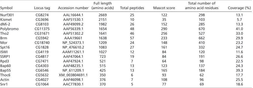
was co-eluted with FLAG-GAGA factor (Shimojima et al., 2003). In the present study, we decreased the concentration of NaCl to 50 mM throughout sample application, washing and elution to isolate proteins that are weakly associated with GAGA factor. MALDI-TOF mass spectrometric analyses revealed that FLAG-GAGA factor was co-eluted with numerous proteins, such as Mi-2, Brm and Kismet, and the previously described GAGA factor-associated proteins Nurf 301, ISWI (Xiao et al., 2001) and FACT (Shimojima et al., 2003) (Table 1). Interestingly, the GAGA factor-associated proteins also included multiple core subunits of Brm complex, such as Mor, Bap60, Bap55, Snr1 and Actin, in addition to Brm. Therefore, we focused here on the Brm complex as a candidate ATP-dependent remodeler that is responsible for H3.3 replacement at chromatin boundaries.
MALDI-TOF mass spectrometry showed that GAGA factor was co-eluted with Polybromo but not with Osa (Table 1), suggesting that GAGA factor associates with the PBAP complex but not with the BAP complex. To confirm this, we performed co-immunoprecipitation with antibodies against Polybromo or Osa, followed by immunoblotting with antibodies against GAGA factor, Brm, Polybromo or Osa. We found that GAGA factor and Brm but not Osa were co-precipitated with Polybromo when nuclear extract (NE) from yw embryos was immunoprecipitated with anti-Polybromo antibodies (Fig. 1A, right panel). By contrast, neither GAGA factor nor Polybromo was co-precipitated with Osa when we used anti-Osa antibody (Fig. 1A, left panel). Reciprocal immunoprecipitation showed that Bap60, Polybromo and GAGA factor were co-precipitated with anti-Brm antibodies (Fig. 1B, left panel). Taken together, these data demonstrate that GAGA factor associates with the PBAP complex.
We then carried out the same set of immunoprecipitation with anti-Brm antibodies as above using NE from larvae homozygous for a polybromo mutation bap180Δ86. Polybromo protein was not detectable in both input and immunoprecipitated lanes (Fig. 1B, right panel), confirming that the allele is protein null (Carrera et al., 2008). Although co-immunoprecipitation of Bap60 with Brm suggests the presence of the Brm complex, GAGA factor was not co-precipitated with Brm. These results indicate that Polybromo is necessary for the association of GAGA factor with the PBAP complex.
Previously, we have shown that GAGA factor is associated with FACT (Shimojima et al., 2003) and HIRA (Nakayama et al., 2007). To examine the relationship among GAGA factor, FACT, HIRA
and PBAP, we extended the co-immunoprecipitation experiments. When proteins in NE from ywembryos were immunoprecipitated with anti-Polybromo antibodies, FACT components SSRP1 and Spt16, and HIRA were not co-precipitated (Fig. 1C). Furthermore, using NE from embryos of the transgenic line expressing HIRA-FLAG (Loppin et al., 2005), we observed that HIRA-HIRA-FLAG was co-precipitated with GAGA factor as reported previously (Nakayama et al., 2007), but not with Polybromo, SSRP1 and Spt16 (Fig. 1D). From these results, although GAGA factor is associated with FACT, PBAP and HIRA, it is most likely that GAGA factor-FACT, GAGA factor-PBAP and GAGA factor-HIRA are distinct complexes.
Identification of GAGA factor-PBAP complex To further confirm association of GAGA factor with PBAP complex, we analyzed the affinity-purified FLAG-GAGA factor-associated proteins by gel filtration through Superose 6 (Fig. 1E). The eluted fractions were resolved by SDS-PAGE and immunoblots were probed with antibodies against Brm, Polybromo, Bap60, SSRP1, Spt16 or FLAG. Components of PBAP, i.e. Brm, Polybromo and Bap60, were co-eluted mainly in fraction numbers 17-21. This represents the PBAP complex with a molecular mass of ~2 MDa (Mohrmann et al., 2004). A small but significant proportion of FLAG-GAGA factor was co-eluted with the PBAP complex. By contrast, the profile of FACT (SSRP1 and Spt16) did not overlap with the peak of the PBAP complex. Rather, FACT was co-eluted with the majority of FLAG-GAGA factor in fraction numbers 27-29, which correspond to the molecular mass of the FLAG-GAGA factor-FACT complex. These data indicate that GAGA factor forms a complex with PBAP remodeler and support the notion that the GAGA factor-PBAP complex and the GAGA factor-FACT complex are distinct from each other.
GAGA factor recruits PBAP complex and HIRA to chromatin boundaries
[image:4.612.49.565.73.254.2]If PBAP complex functions at chromatin boundaries, it should be detected at boundaries. To examine occupancy of PBAP complex at chromatin boundaries, we carried out ChIP assays using chromatin fragments prepared from third instar larvae of wild type. GAGA factor, a FACT subunit SSRP1 and subunits of PBAP (Polybromo, Brm and Bap60) but not Osa were detected at three Table 1. MALDI-TOF mass spectrometric analyses of GAGA factor-associated proteins
Full length Total number of
Symbol Locus tag Accession number (amino acids) Total peptides Mascot score amino acid residues Coverage (%)
Nurf301 CG8274 AAL16644.1 2669 25 122 298 13.1
Kismet CG3696 AAF51530.1 2151 10 35 103 5.7
dMi-2 CG8103 AAF49099.2 1982 26 152 285 13.3
Polybromo CG11375 AAF56339.1 1654 48 290 670 41.0
Tho2 CG31671 AAF51302.2 1641 46 256 527 33.0
Brm CG5942 AAA19661 1638 57 233 662 29.9
Mor CG18740 NP_524373.1 1209 24 148 410 23.2
Spt16 CG1828 NP_476610.2 1083 27 161 332 24.7
ISWI CG4119 AAM11261.1 1027 12 84 120 11.6
SSRP1 CG4817 AAF47064.1 723 19 84 191 26.6
Rpd3 CG7471 AAF47924.1 521 7 64 98 22.5
Bap60 CG4303 AAF48235.1 515 13 123 137 24.3
Bap55 CG6546 NP_611209.1 425 13 165 184 39.3
Thoc6 CG5632 XM_003804691.1 350 6 93 62 17.7
Actin CG4027 AAF46098.1 376 7 108 96 25.5
Snr1 CG1064 AAC77830.1 370 5 77 69 18.6
D
E
V
E
LO
P
M
E
N
chromatin boundaries: d1, Fab-7 HS1 and the bxdregion (Fig. 2A,B, wild type). These results indicate that GAGA factor, the PBAP complex and FACT are present at the chromatin boundaries. We also analyzed occupancies of HIRA at the chromatin boundaries. Confirming our previous study (Nakayama et al., 2007), HIRA was detected at d1 (Fig. 2B). We also found occupancies of HIRA at Fab-7and bxd(Fig. 2B).
GAGA factor is expected to recruit these complexes to the chromatin boundaries, as all these boundaries carry a cluster of GAGA factor-binding sites. To test the possibility, we searched for a GAGA factor-deficient condition. RNAi-mediated knockdown of GAGA factor was not efficient because of the self-regulation of Trl-encoding GAGA factor (Bernués et al., 2007). Therefore, we employed a hypomorphic allele, Trl13C, in which a P-element is
inserted into the first intron of Trl(Farkas et al., 1994). Although bulk of Trl13C homozygotes died during embryogenesis, rare
escapers grew until third instar larvae. We raised the Trl13C/TM6B
line in thousands of vials and collected the escapers without the Tb phenotype of TM6Bfor ChIP assays. We found that the levels of occupancy of GAGA factor at the three boundaries were markedly reduced in Trl13C compared with those in wild type (Fig. 2A, Trl13C). The levels of SSRP1, Polybromo, Brm, Bap60 and HIRA
at the boundaries were also severely reduced in a similar manner to that of GAGA factor (Fig. 2A,B, Trl13C). This is not due to
reduction in the expression levels of SSRP1, Polybromo, Brm, Bap60 and HIRA in Trl13C, as RT-qPCR showed that the expression
levels of SSRP1, Polybromo, Brm, Bap60 and HIRA mRNAs were not affected in Trl13C (Fig. 2C). Taken together, these data
demonstrate GAGA factor-dependent recruitment of FACT, PBAP complex and HIRA to the chromatin boundaries.
Conversely, it is possible that the PBAP remodeler could facilitate the recruitment of GAGA factor, FACT and HIRA to the boundaries. To test the possibility, we collected third instar larvae that are homozygous for the polybromomutation bap180Δ86and carried out ChIP assays. As shown in Fig. 2A,B, the occupancies of GAGA factor, FACT and HIRA were barely affected by the polybromomutation, although the levels of Polybromo, Brm and Bap60 were severely reduced in the mutant. These results suggest that PBAP remodeler is not required for the recruitment of GAGA factor, FACT or HIRA to the boundaries.
PBAP complex and HIRA function in H3.3 replacement at chromatin boundaries
We next investigated whether PBAP complex and HIRA are involved in the H3.3 replacement at chromatin boundaries. To this end, we first established a fly line expressing histone H3.3-FLAG or H3-FLAG, which is heterozygous for brm2, bap180Δ86, osa2or hiramutation ssm(Loppin et al., 2005), or trans-heterozygous for Trl 13Cand brm2, or for Trl13Cand bap180Δ86. The expression
level of the H3.3-FLAG or H3-FLAG was ~1% the level of endogenous histone H3 (Nakayama et al., 2007). The low expression level would not disturb fly physiology but allow us to use the FLAG-tagged histone as a tracer. We then collected embryos 10-22 hours after egg laying and performed ChIP assays using a FLAG M2 monoclonal antibody. The heterozygous population consisted of embryos heterozygous for the mutation (50%), homozygous for the mutation (25%) and homozygous for a balancer or for the other mutation in the case of trans-heterozygote (25%), but we call the population simply heterozygote or trans-heterozygote here. We found that the ratio of H3.3 to H3 in Trl13C, bap180Δ86, brm2or ssmheterozygote was
significantly reduced at d1, Fab-7HS1 and bxdregions compared
with wild type (Fig. 3). Moreover the ratio of H3.3 to H3 was further reduced in Trl13C/brm2 or Trl13C/bap180Δ86
trans-heterozygote compared with Trl13C,brm2or bap180 Δ86single
heterozygote. As a negative control, osa2 heterozygote did not
[image:5.612.324.527.58.503.2]affect the H3.3/H3 ratio at the boundaries. Furthermore, at control sites such as iab-6and d6, the H3.3/H3 ratios in the mutants were almost the same as that in wild type. These data indicate that PBAP complex and HIRA are required for the H3.3 replacement at the chromatin boundaries.
Fig. 2. GAGA factor recruits PBAP complex and HIRA to chromatin boundaries. (A,B) Occupancies of GAGA factor, FACT, PBAP complex and HIRA at chromatin boundaries d1, Fab-7HS1 and
bxd. ChIP assays were carried out with antibodies against GAGA factor, SSRP1, Polybromo, Brm, Bap60, HIRA and Osa using larval nuclei from wild type, Trl13Cor bap180Δ86. (C) Expression levels of GAGA factor, SSRP1, Polybromo, Brm, Bap60 and HIRA mRNAs in wild-type or Trl13C larvae. Data were normalized to β-tubulin mRNA. Experiments were repeated three times. Error bars represent s.d. Asterisks indicate statistical significance according to Welch’s t-test: *P<0.005.
D
E
V
E
LO
P
M
E
N
Polybromo is responsible for alteration of
chromatin structure and HIRA functions to restore this alteration
Dynamic histone H3.3 replacement at a chromatin boundary would generate a DNase HS. Indeed, we have shown that GAGA factor directs formation of a DNase HS at the d1boundary (Nakayama et al., 2007). To test whether PBAP complex is involved in the alteration of chromatin structure, we carried out SpeI accessibility assays. The assay relies on inhibition of qPCR encompassing an SpeI site upon SpeI-mediated cleavage of DNA (Fig. 4, naked DNA). When nuclei isolated from wild-type larvae were treated with SpeI, the qPCR levels at d1was ~50% of undigested control, whereas the level at a control SpeI site was ~90% of undigested control (Fig. 4). These results confirm our previous conclusion that d1is a DNase HS. By contrast, the SpeI accessibility at d1was severely compromised in nuclei from bap180Δ86 homozygous larvae. Expressing Polybromo/Bap180 from a transgene restored the accessibility in bap180Δ86. These results demonstrate that Polybromo is responsible for the formation of DNase HS at d1.
As HIRA is another key player in the H3.3 replacement, we also analyzed the SpeI accessibility in nuclei from hira ssm larvae. Interestingly, the accessibility at d1in ssmlarvae was even higher than that in wild type, while the control SpeI site was barely accessible. These results suggest that the SpeI accessibility at d1in wild type is under equilibrium between Polybromo-dependent enhancement and HIRA-dependent reduction. Furthermore, this ssmphenotype was suppressed by the homozygous bap180Δ86 allele: the accessibility in ssm; bap180Δ86 double mutant was
almost the same level as that in the bap180Δ86single mutant. These data suggest that HIRA restores the Polybromo-induced alteration of chromatin structure and that this HIRA activity is dependent on the Polybromo function.
Crucial role of PBAP complex in boundary functions
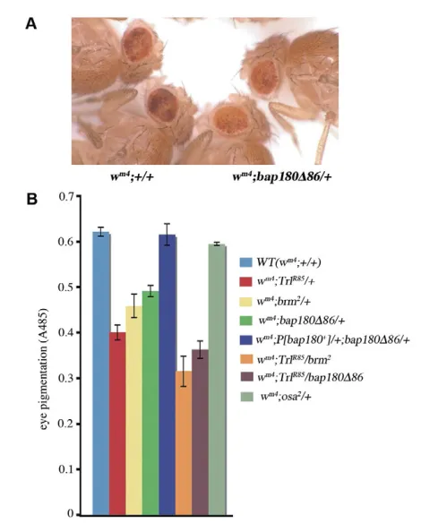
In the wm4 line, where w is juxtaposed with pericentric
heterochromatin, counteraction of the heterochromatin spread at the d1 boundary maintains wexpression (Nakayama et al., 2007). Therefore, the boundary function at d1can be assessed by position effect variegation (PEV). To test whether PBAP complex is involved in the boundary function at d1, we analyzed PEV in wild type and various mutants under the wm4background (Fig. 5). TrlR85
is a deletion allele that is obtained by excision of the P-element in Trl13C. Its deletion removes extensive regions of the Trl
transcription unit and, hence, it is presumed to be a null allele (Farkas et al., 1994). As reported previously (Farkas et al., 1994), PEV was enhanced (i.e. eye pigmentation was reduced) in TrlR85/+. brm2/+ also enhanced PEV. Similarly, PEV was enhanced in bap180Δ86/+ and the enhancement was restored by expressing Polybromo/Bap180 from the transgene. Moreover, in TrlR85and brm2 or TrlR85 and bap180Δ86 trans-heterozygotes, PEV was
enhanced more than in each single heterozygote. As a control, osa2/+ did not affect PEV. These data indicate that the PBAP
complex plays a crucial role in the boundary function at d1together with GAGA factor.
[image:6.612.320.556.60.242.2]Fab-7is the boundary that insulates between iab-6and iab-7 enhancers of Abd-B(Mihaly et al., 1997). Defects in the Fab-7 boundary function results in a homeotic transformation of the male A6 to A5, as revealed by formation of extra bristles on male A6. Previously, we have shown that defects in Trland spt16encoding a FACT subunit cause the A6 to A5 transformation and genetic interaction between Trland spt16on the phenotype (Shimojima et al., 2003). To examine whether PBAP complex is involved in the Fig. 4.SpeI accessibility assays.The assays were carried out in wild type and indicated mutants with qPCR levels relative to undigested control (%). A SpeI recognition sequence is present immediately before (GA)8at d1. A control SpeI site was chosen arbitrarily ~11 kb
downstream of d1. The SpeI incubations were repeated ten times in each line. Data are mean±s.d. Differences between wild type and
[image:6.612.52.297.64.303.2]bap180Δ86or ssmare significant (P<0.001; Welch’s t-test).
Fig. 3. PBAP complex and HIRA are required for H3.3 replacement at chromatin boundaries.ChIP assays were carried out with the anti-FLAG M2 antibody using embryos from wild type or from each mutant line harboring either P[EGFP, hsp83-H3.3-FLAG] orP[EGFP, hsp83-H3-FLAG]transgene. Shown is the ratio of H3.3 to H3 in each line. The experiments were repeated at least four times. Data are mean±s.d. Differences between wild type and each heterozygote (except osa2 heterozygote) or between Trl13Cheterozygote and each trans-heterozygote are significant (P<0.001; Welch’s t-test).
D
E
V
E
LO
P
M
E
N
boundary function at Fab-7, we analyzed frequencies of the A6 to A5 transformation in wild type and various mutants (Table 2). We were unable to find the A6 to A5 transformation in wild type and osa2/+. However, we observed the transformation in TrlR85/+, brm2/+ and Δspt16/+, although the frequency was low. A similar
frequency of the transformation was detected in bap180Δ86/+, and this phenotype was rescued by expressing Polybromo/Bap180 from the transgene. In the bap180Δ86 homozygote, we observed a marked increase in the frequency of this transformation compared with the bap180Δ86 heterozygote and this phenotype was rescued by expressing Polybromo/Bap180 from two doses of the transgene. Moreover, in TrlR85and brm2, TrlR85and bap180Δ86or Δspt16and bap180Δ86 trans-heterozygotes, the frequency of this transformation was higher than in each single heterozygote. These data indicate that PBAP complex functions at the Fab-7boundary together with GAGA factor and FACT.
DISCUSSION
In the previous study, we have shown that the GAGA factor-FACT complex directs the H3.3 replacement at chromatin boundaries in conjunction with the histone chaperone HIRA (Nakayama et al., 2007). The present study demonstrates that PBAP remodeling complex is recruited to the boundaries in a GAGA factor-dependent manner and contributes to the H3.3 replacement at the boundaries.
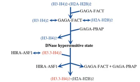
Our study also revealed that Polybromo is responsible for the generation of a DNase HS and that HIRA functions to restore the altered state. Taken together, we propose the following model for the H3.3 replacement at a chromatin boundary (Fig. 6). GAGA factor recruits FACT and PBAP to boundaries carrying multiple GAGA factor-binding sites. FACT displaces a H2A-H2B pair from a nucleosome with the displaced H2A-H2B being anchored by FACT. This facilitates access of PBAP to a H3-H4 tetramer because the tetramer is sandwiched between two H2A-H2B dimers in a nucleosome core. Then PBAP would displace H3-H4. The resulting nucleosome-free region would be a DNase HS and become a target of the HIRA-ASF1 chaperone complex (Tagami et al., 2004) for deposition of H3.3-H4, as proposed by Ray-Gallet et al. (Ray-Gallet et al., 2011). The deposition of H3.3-H4 would mask the DNase HS. Finally, FACT deposits the tethered H2A-H2B pair to reconstitute the nucleosome. The process would be repeated continuously to achieve boundary function. As FACT, Polybromo-containing Brm complex, HIRA and ASF1 are conserved among species from Drosophilato human, a similar mechanism may also operate in mammals.
What is the role of H3.3 replacement in boundary function? It has been reported that H3.3 is associated with active histone modifications such as Lys 4 methylation but tends to avoid Lys 9 methylation (McKittrick et al., 2004). Indeed, we have detected a peak of H3K4 methylation and a dip of H3K9 methylation at the d1boundary (Nakayama et al., 2007). Therefore, the continuous H3.3 replacement at chromatin boundaries can prevent the spreading of specific histone modifications. Alternatively, the intrinsic instability of a nucleosome containing H3.3 (Jin and Felsenfeld, 2007) and the transient disruption of nucleosome structure during H3.3 replacement would generate a nucleosome-free region. This nucleosome gap can serve as a barrier against the spreading of chromatin with specific epigenetic memories.
[image:7.612.52.287.53.345.2]Before isolation of GAGA factor-associated proteins, we anticipated the presence of a huge complex containing GAGA factor, FACT and a chromatin remodeling factor. However, our data suggest the presence of two distinct complexes: the GAGA factor-FACT complex and the GAGA factor-PBAP complex. This means that at least two GAGA factor-binding sites, one for the GAGA FACT complex and the other for the GAGA factor-PBAP complex, are necessary within a boundary to allow Fig. 5. Importance of PBAP complex to counteract
heterochromatin spreading at d1boundary.(A) A typical example of position effect variegation (PEV), demonstrating enhancement in
bap180Δ86/+(right two flies) compared with +/+ (left two flies). (B) PEV assays were carried out by measuring eye pigmentation in male flies of indicated genotypes under the wm4background. The
experiments were repeated at least eight times. Data are mean±s.d. Differences between wild type and each heterozygote, except osa2/+, or between TrlR85/+ and each trans-heterozygote are significant (P<0.001; Welch’s t-test).
Table 2. The PBAP complex is required for boundary function at Fab-7
A6 to A5 transformation/
Genotype total number Percentage
Wild type 0/504 0
TrlR85/+ 19/472 4
brm2/+ 18/452 4
bap180Δ86/+ 27/706 4
P[bap180+]/+;bap180Δ86/+ 0/807 0
bap180Δ86 104/260 40
P[bap180+];bap180Δ86 0/511 0
Δspt16/+ 21/461 5
TrlR85/brm2 36/313 12
TrlR85/bap180Δ86 33/275 12
Δspt16/bap180Δ86 45/305 15
osa2/+ 0/516 0
Frequencies of the A6 to A5 transformation were measured in males of indicated genotypes. Differences between wild type and each single heterozygote, except
osa2/+, or between TrlR85/+ or Δspt16/+and each transheterozygote were significant (P<0.001; Fisher’s exact test).
D
E
V
E
LO
P
M
E
N
[image:7.612.314.564.83.227.2]concerted actions of FACT and PBAP for the H3.3 replacement. This may preclude the H3.3 replacement to occur at a single GAGA factor-binding site that is present frequently within the genome. In good agreement with the idea, chromatin boundaries harbor a cluster of GAGA factor-binding sites that are occupied with the factors. For example, ChIP assays using anti-GAGA factor antibodies showed a prominent signal at d1consisting of (GA)8
compared with other single GAGA factor-binding sites around w (Nakayama et al., 2007). The present study revealed that the level of GAGA factor at Fab-7 HS1 or bxd, which harbor multiple closely spaced GAGA factor-binding sites, was comparable with that at d1(Fig. 2A,B, wild type). Because multiple GAGA factor-binding sites are present in other boundaries [such as Fab-8and Mcpin the Bithoraxcomplex (Busturia et al., 2001; Moon et al., 2005), regulatory elements of the α1-tubulin gene (O’Donnell et al., 1994), and the SF1 insulator of the Antennapediacomplex (Belozerov et al., 2003)], the FACT- and PBAP-dependent H3.3 replacement may also occur at these boundaries. Multiple closely spaced GAGA factor-binding sites are presumably an indicator of the chromatin boundary.
The present study demonstrates that the PBAP complex but not the BAP complex is required for the boundary functions. These two subtypes of Swi/Snf remodeling factor possess overlapping, distinct and counteracting functions (Collins and Treisman, 2000; Moshkin et al., 2007; Carrera et al., 2008). As for the distinct functions, while the BAP complex controls the cell cycle and Wingless target genes (Collins and Treisman, 2000; Moshkin et al., 2007), the PBAP complex regulates metamorphosis and the immune response (Carrera et al., 2008). Whether the H3.3 replacement-mediated boundary function is involved in the regulation of metamorphosis or the immune response remains to be clarified in future studies.
Recent articles have reported involvement of ATP-dependent remodeling factors in deposition of H3.3 into chromatin. First, it has been shown that Chd1 is required for the deposition of H3.3 into decondensing sperm chromatin after fertilization (Konev et al., 2007). Others have demonstrated contribution of ATRX to the deposition of H3.3 at pericentric chromatin and telomeres (Drané et al., 2010; Goldberg et al., 2010). We show here that PBAP is
required for H3.3 replacement at the chromatin boundaries. These data support the idea that distinct ATP-dependent remodelers work for H3.3 deposition at specific locations within the genome (Goldberg et al., 2010). Interestingly, null mutations in Drosophila polybromo/bap180,chd1and hiraexhibit the same phenotype, i.e. viable but female sterile (Bonnefoy et al., 2007; Konev et al., 2007; Carrera et al., 2008), suggesting that the H3.3 deposition at the specific genomic locations is essential for early embryogenesis.
Acknowledgements
We thank Drs J. Treisman, B. Loppin and C. Wu, and the Bloomington Stock Center for providing us with fly lines and antibodies.
Funding
This work was supported by a Grant in Aid for Scientific Research from Ministry of Education, Culture, Sports, Science and Technology (MEXT), Japan. T.S. was supported by the Japanese Society for the Promotion of Science.
Competing interests statement
The authors declare no competing financial interests.
Supplementary material
Supplementary material available online at
http://dev.biologists.org/lookup/suppl/doi:10.1242/dev.083246/-/DC1
References
Ahmad, K. and Henikoff, S.(2002). The histone variant H3.3 marks active chromatin by replication-independent nucleosome assembly. Mol. Cell9, 1191-1200.
Becker, P. B. and Hörz, W.(2002). ATP-dependent nucleosome remodeling.
Annu. Rev. Biochem.71, 247-273.
Bell, A. C., West, A. G. and Felsenfeld, G.(2001). Insulators and boundaries: versatile regulatory elements in the eukaryotic genome. Science291, 447-450.
Belotserkovskaya, R., Oh, S., Bondarenko, V. A., Orphanides, G., Studitsky, V. M. and Reinberg, D.(2003). FACT facilitates transcription-dependent nucleosome alteration. Science301, 1090-1093.
Belozerov, V. E., Majumder, P., Shen, P. and Cai, H. N.(2003). A novel boundary element may facilitate independent gene regulation in the Antennapedia complex of Drosophila. EMBO J.22, 3113-3121.
Bernués, J., Piñeyro, D. and Kosoy, A.(2007). General, negative feedback mechanism for regulation of Trithorax-like gene expression in vivo: new roles for GAGA factor in flies. Nucleic Acids Res.35, 7150-7159.
Bonnefoy, E., Orsi, G. A., Couble, P. and Loppin, B.(2007). The essential role of Drosophila HIRA for de novo assembly of paternal chromatin at fertilization.
PLoS Genet.3, 1991-2006.
Busturia, A., Lloyd, A., Bejarano, F., Zavortink, M., Xin, H. and Sakonju, S.
(2001). The MCP silencer of the Drosophila Abd-B gene requires both Pleiohomeotic and GAGA factor for the maintenance of repression.
Development128, 2163-2173.
Carrera, I., Zavadil, J. and Treisman, J. E.(2008). Two subunits specific to the PBAP chromatin remodeling complex have distinct and redundant functions during drosophila development. Mol. Cell. Biol.28, 5238-5250.
Clapier, C. R. and Cairns, B. R.(2009). The biology of chromatin remodeling complexes. Annu. Rev. Biochem.78, 273-304.
Collins, R. T. and Treisman, J. E.(2000). Osa-containing Brahma chromatin remodeling complexes are required for the repression of wingless target genes.
Genes Dev.14, 3140-3152.
Drané, P., Ouararhni, K., Depaux, A., Shuaib, M. and Hamiche, A.(2010). The death-associated protein DAXX is a novel histone chaperone involved in the replication-independent deposition of H3.3. Genes Dev.24, 1253-1265.
Farkas, G., Gausz, J., Galloni, M., Reuter, G., Gyurkovics, H. and Karch, F.
(1994). The Trithorax-like gene encodes the Drosophila GAGA factor. Nature 371, 806-808.
Goldberg, A. D., Banaszynski, L. A., Noh, K.-M., Lewis, P. W., Elsaesser, S. J., Stadler, S. J., Dewell, S., Law, M., Guo, X., Li, X. et al.(2010). Distinct factors control histone variant H3.3 localization at specific genomic regions. Cell140, 678-691.
Henikoff, S. and Ahmad, K.(2005). Assembly of variant histones into chromatin.
Annu. Rev. Cell Dev. Biol.21, 133-153.
Jack, R. S. and Eggert, H.(1990). Restriction enzymes have limited access to DNA sequences in Drosophila chromosomes. EMBO J.9, 2603-2609.
Jenuwein, T. and Allis, C. D.(2001). Translating the histone code. Science293, 1074-1080.
Jin, C. and Felsenfeld, G.(2007). Nucleosome stability mediated by histone variants H3.3 and H2A.Z. Genes Dev.21, 1519-1529.
[image:8.612.52.286.61.198.2]Kamakaka, R. T. and Biggins, S.(2005). Histone variants: deviants? Genes Dev. 19, 295-316.
Fig. 6. Model for H3.3 replacement at the chromatin boundary.
GAGA factor recruits FACT and PBAP to a chromatin boundary harboring a cluster of GAGA factor-binding sites. FACT interacts with a nucleosome and displaces a H2A-H2B pair while anchoring the displaced pair. This would facilitate PBAP-mediated displacement of an H3-H4 tetramer. The histone-displaced region of DNA would be a DNase HS. Then HIRA-ASF1 complex would deposit H3.3-H4 on the region to restore from the DNase-hypersensitive state. Finally, FACT deposits the tethered H2A-H2B pair to reconstitute the nucleosome.
D
E
V
E
LO
P
M
E
N
Konev, A. Y., Tribus, M., Park, S. Y., Podhraski, V., Lim, C. Y., Emelyanov, A. V., Vershilova, E., Pirrotta, V., Kadonaga, J. T., Lusser, A. et al.(2007). CHD1 motor protein is required for deposition of histone variant H3.3 into chromatin in vivo. Science317, 1087-1090.
Loppin, B., Bonnefoy, E., Anselme, C., Laurençon, A., Karr, T. L. and Couble, P.(2005). The histone H3.3 chaperone HIRA is essential for chromatin assembly in the male pronucleus. Nature437, 1386-1390.
Lorch, Y., Zhang, M. and Kornberg, R. D.(1999). Histone octamer transfer by a chromatin-remodeling complex. Cell96, 389-392.
Maeda, R. K. and Karch, F.(2007). Making connections: boundaries and insulators in Drosophila. Curr. Opin. Genet. Dev.17, 394-399.
McKittrick, E., Gafken, P. R., Ahmad, K. and Henikoff, S.(2004). Histone H3.3 is enriched in covalent modifications associated with active chromatin. Proc. Natl. Acad. Sci. USA101, 1525-1530.
Mihaly, J., Hogga, I., Gausz, J., Gyurkovics, H. and Karch, F.(1997). In situ dissection of the Fab-7 region of the bithorax complex into a chromatin domain boundary and a Polycomb-response element. Development124, 1809-1820.
Mito, Y., Henikoff, J. G. and Henikoff, S.(2007). Histone replacement marks the boundaries of cis-regulatory domains. Science315, 1408-1411.
Mohrmann, L., Langenberg, K., Krijgsveld, J., Kal, A. J., Heck, A. J. and Verrijzer, C. P.(2004). Differential targeting of two distinct SWI/SNF-related Drosophila chromatin-remodeling complexes. Mol. Cell. Biol.24, 3077-3088.
Moon, H., Filippova, G., Loukinov, D., Pugacheva, E., Chen, Q., Smith, S. T., Munhall, A., Grewe, B., Bartkuhn, M., Arnold, R. et al.(2005). CTCF is conserved from Drosophila to humans and confers enhancer blocking of the Fab-8 insulator. EMBO Rep.6, 165-170.
Moshkin, Y. M., Mohrmann, L., van Ijcken, W. F. and Verrijzer, C. P.(2007). Functional differentiation of SWI/SNF remodelers in transcription and cell cycle control. Mol. Cell. Biol.27, 651-661.
Nakayama, T., Nishioka, K., Dong, Y. X., Shimojima, T. and Hirose, S.(2007). Drosophila GAGA factor directs histone H3.3 replacement that prevents the heterochromatin spreading. Genes Dev.21, 552-561.
O’Donnell, K. H., Chen, C. T. and Wensink, P. C.(1994). Insulating DNA directs ubiquitous transcription of the Drosophila melanogaster alpha 1-tubulin gene.
Mol. Cell. Biol.14, 6398-6408.
Ray-Gallet, D., Woolfe, A., Vassias, I., Pellentz, C., Lacoste, N., Puri, A., Schultz, D. C., Pchelintsev, N. A., Adams, P. D., Jansen, L. E. T. et al.(2011). Dynamics of histone H3 deposition in vivo reveal a nucleosome gap-filling mechanism for H3.3 to maintain chromatin integrity. Mol. Cell44, 928-941.
Sarma, K. and Reinberg, D.(2005). Histone variants meet their match. Nat. Rev. Mol. Cell Biol.6, 139-149.
Shimojima, T., Okada, M., Nakayama, T., Ueda, H., Okawa, K., Iwamatsu, A., Handa, H. and Hirose, S.(2003). Drosophila FACT contributes to Hox gene expression through physical and functional interactions with GAGA factor.
Genes Dev.17, 1605-1616.
Tagami, H., Ray-Gallet, D., Almouzni, G. and Nakatani, Y.(2004). Histone H3.1 and H3.3 complexes mediate nucleosome assembly pathways dependent or independent of DNA synthesis. Cell116, 51-61.
Workman, J. L.(2006). Nucleosome displacement in transcription. Genes Dev.20, 2009-2017.
Xiao, H., Sandaltzopoulos, R., Wang, H. M., Hamiche, A., Ranallo, R., Lee, K. M., Fu, D. and Wu, C.(2001). Dual functions of largest NURF subunit NURF301 in nucleosome sliding and transcription factor interactions. Mol. Cell8, 531-543.