Assessment of Transient Ischemic Attack with
Diffusion- and Perfusion-Weighted Imaging
Lucas Restrepo, Michael A. Jacobs, Peter B. Barker, and Robert J. Wityk
BACKGROUND:Diagnosing TIA can be difficult, since evidence of brain ischemia is habit-ually lacking on CT and conventional MR imaging. It has been suggested that patients with acute brain infarction on neuroimaging should be considered stroke cases instead of TIA, regardless of duration of symptoms, implying that optimal diagnostic methods need to be utilized. We therefore postulated that perfusion-weighted MR imaging (PW imaging) would be useful in the diagnosis of TIA.
METHODS:Retrospective analysis of 22 patients with reversible neurologic symptoms last-ing less than 24 hours, assessed with DW and PW imaglast-ing.
RESULTS:MR imaging was abnormal in 15 patients (68%): 12 had abnormal DW imaging, four had both DW and PW imaging defects (all with a mismatch) and three had an isolated PW imaging abnormality. There were no differences in symptom duration, stroke etiology or cardiovascular risk factors between patients with abnormal MR imaging and those with unremarkable scan. Patients with mismatch were more likely to need conventional angiography or other cerebrovascular procedures.
CONCLUSION: The combined use of DW imaging and PW imaging provided evidence of brain ischemia in most patients with clinical diagnosis of TIA. Prospective studies using follow-up MR imaging are required to determine the outcome of affected tissue, as well as the clinical implications of DW–PW imaging abnormalities.
It is difficult to establish that transient neurologic symptoms correspond to brain ischemia, because most patients appear normal neurologically at the time of initial assessment and often relate vague com-plaints. In addition, head CT and conventional brain MR imaging may not provide objective evidence of brain ischemia to support the diagnosis. Although a detailed diagnostic work-up may disclose the pres-ence of diseases associated with ischemic stroke and transient ischemic attack (TIA), such findings do not necessarily prove that the cause of symptoms was indeed brain ischemia. On the other hand, the tradi-tional 24-hour defining limit of TIA has recently been challenged by arguing that a diagnosis of ischemic stroke should be assigned to all patients with evidence of acute brain infarction on neuroimaging, regardless of the duration of symptoms, implying that optimal
diagnostic tools need to be utilized systematically in TIA to rule out acute brain infarction (1).
Although diffusion-weighted (DW) imaging can detect signs of acute brain ischemia in 29 – 67% of patients with TIA (2– 8), limited information regard-ing the added value of perfusion-weighted (PW) im-aging is currently available (9 –15). PW imim-aging does not usually provide quantitative measures of cerebral blood flow, but it does allow rapid evaluation of relative hemodynamics in the same scanning session as DW and conventional MR imaging (13, 16, 17). Most PW imaging defects accompany (13, 16, 17) or even precede (14) the development of DW imaging abnormalities in a substantial proportion of patients with brain ischemia. Therefore, the purpose of the current study was to test the hypothesis that PW imaging would substantiate the diagnosis of brain ischemia in an additional proportion of patients with transient neurologic symptoms.
Methods
Patients
Patients admitted to our hospital from January 1996 to August 2003 with reversible neurologic symptoms lasting⬍24 hours (clinically diagnosed as TIA by one of two neurologists [L.R., R.J.W.]), were retrospectively identified from a com-puter registry. The query retrieved 179 cases, from which 22 Received December 15, 2003; accepted after revision May 18,
2004.
From the Departments of Neurology (L.R., R.J.W.) and Radi-ology (M.A.J., PBB), Johns Hopkins Hospital, Baltimore, Mary-land. Supported in part by a Beginning Grant-in-Aid (Number: 0265450U) from the Mid-Atlantic Affiliate Research Committee of the American Heart Association (to L.R.), grants from the NIH (T32CA09630 to M.A.J and P41RR15241 to P.B.B.) and a gift from the Rogers Wilbur foundation (to R.J.W).
Address reprint requests to Robert J. Wityk MD, Associate Professor of Neurology, Johns Hopkins Hospital, 600 N. Wolfe Street, Phipps 126b, Baltimore, MD 21287.
(12%) had brain MR imaging with DW and PW imaging as part of their diagnostic work-up. The MR imaging protocol and acquisition of clinical data were approved by the local IRB.
None of the patients had treatment with thrombolysis (IV or IA), percutaneous angioplasty, mechanical disruption of the occluding thrombus, or induced hypertension beforethe MR imaging. Clinical and demographic variables, as well as infor-mation regarding symptom onset, were gathered from the chart of every patient. Exact time of MR imaging examination was available for all cases, but documentation of the exact onset of symptoms (i.e., date, hour and minute) was available for 17 patients. The remaining five patients did not have an exact time of symptom onset specified in the chart, although the notes of the attending physician remarked that symptoms lasted less than 24 hours in all of these cases.
MR Imaging Examination
Images were acquired on a 1.5 T GE Signa scanner with echo-planar (EPI) capability (GE Milwaukee, WI). The MR imaging protocol has been detailed elsewhere (16, 17). Briefly, it consists of conventional T1, axial fast-spin-echo T2-weighted and axial fluid-attenuated inversion-recovery (FLAIR), time-of-flight MRA (head and neck), DW and PW imaging. Appar-ent diffusion coefficiAppar-ent (ADC) maps were generated and re-ductions in ADC were used to confirm acute brain ischemia. PW imaging was obtained by using dynamic first-pass bolus tracking of gadolinium with a multi-section axial gradient-echo scan.
Cerebral blood flow (CBF), mean transit time (MTT) and time-to-peak (TTP) maps were generated and qualitatively assessed by an investigator. Volumes of DW imaging and TTP abnormalities were manually delineated by region-of-interest, by using Scion Imaging (Scion Corporation, 1998). The areas of abnormality in cm2on each section were summed, and the sum multiplied by the interslice spacing, to determine the volume in cm3. Hypoperfusion was defined as a region with⬎2.5 seconds delay on TTP maps, compared with a homologous area of the contralateral hemisphere. This threshold is based on evidence suggesting that neurologic function correlates with the volume of brain regions exhibiting a TTP delay of⬎2.5 seconds (16, 17).
We defined DW–PW imaging mismatch as a TTP volume defect greater than its related DW imaging lesion. We chose this threshold because it reflects the reality of our practice: MR imaging is assessed by a team of physicians without immediate access to volumetry. Therefore, the involved team made clinical
decisions based on aqualitativeassessment of TTP sequences. We measured the magnitude of mismatch as (TTP volume -DW imaging volume / TTP volume) ⫻100⫽ % mismatch. According to this definition, the mismatch group does include patients withisolatedTTP abnormalities.
Statistical Analysis
Patients were divided into three groups for comparison: Unremarkable MR imaging, abnormal MR imaging (abnormal DWorPW imaging) and DW imaging–PW imaging mismatch. Differences between clinical characteristics of patients with and without MR imaging abnormalities were explored with Chi-squared and Student’sttest. Differences between more than two variables were analyzed with one-way ANOVA; they were significant atP⬍.05.
Results
Average patient age was 62⫾14 years, and half of
the patients were female. A total of 15 patients (68%) had DW or PW imaging abnormalities. Of these, eight had isolated DW imaging lesions, three had isolated PW imaging defects, and four had both DW and PW imaging changes. Thus, we considered that the last seven patients had a mismatch. The magni-tude of the mismatch was typically large (Range: 44 –100%, average: 84%). The region of restricted
diffusion overlapped with the larger TTP defect
par-tiallyon two of the four patients with both DW and
PW imaging changes (Fig 1). The other two patients with both DW and PW imaging changes had an area of diffusion restriction totally encompassed by the TTP defect. The head CT revealed signs of acute infarct on only one of these patients. Average lesion
volume was 17⫾27 cm3for the DW imaging lesions
and 112 ⫾173 cm3for the PW imaging defects. All
the DW and PW imaging abnormalities involved the anterior circulation of the brain.
There was no difference in the prevalence of car-diovascular risk factors between patients with abnor-mal DW–PW imaging as compared with those with unremarkable scan (table 1). However, patients with
FIG 1. Brain MR imaging of case 1.Left, Initial images show faint DW imaging hy-perintensities in the right temporal lobe (top) and frontoparietal junction (bottom).
mismatch were more likely to have coronary artery
disease (P ⫽ .032) and hyperlipidemia (P ⫽ .045)
than the rest of the patients. All lesions noted on MR imaging were consistent with the focal neurologic symptoms reported by the patients. Hemiparesis was significantly associated with abnormal findings on
MR imaging (i.e., abnormal DW or PW imaging,P⫽
.047). However, other neurologic symptoms were not predictive of MR imaging abnormalities. The dura-tion and number of episodes of transient neurologic dysfunction did not differ between patients with ab-normal DW–PW imaging when compared with those patients with unremarkable scan. The presumptive cause of TIA was large artery atherosclerosis (LAA) in eight patients, cardioembolism in three patients, other cause (aortic atherosclerosis) in three patients and unknown cause in eight cases. There was a sig-nificant association between LAA and mismatch (6/7
patients,P⫽.02). Table 2 provides a summary of the
clinical characteristics of the mismatch cases. Time to MR imaging did not differ between pa-tients with abnormal MR imaging compared with those with unremarkable scan. Mean length of stay in
the hospital was 5⫾3 days for patients with abnormal
DW imaging or PW imaging, 6⫾3 days for patients
with mismatch and 3 ⫾3 days for those with normal
MR imaging, which did not reach statistical differ-ence. None of the patients developed clinical stroke during the hospitalization. None of the mismatch cases reported a stroke after several follow-up visits at the out-patient clinic. Only two patients (both with mismatch) had follow-up MR imaging at the hospital, one showing resolution and the other persistence of the PW imaging defect (cases 1 and 3, respectively; see below). Conventional angiography was performed in 5/7 patients with mismatch, whereas none of the other patients required such procedure. Patients with mismatch on MR imaging were more likely to have a surgical or endovascular procedure than patients
without mismatch (5/7 versus 1/15;P⫽.001). Two of
the patients with mismatch were treated with percu-taneous balloon angioplasty (without stent), two had carotid endarterectomy and one had intracranial– ex-tracranial bypass. The following is a report of three illustrative cases:
Case 1
A 56-year-old gentleman with history of coronary artery disease, hypertension, diabetes and hyperlipid-emia, was noted to develop dysarthria and left hemi-paresis during percutaneous coronary angioplasty. He received heparin, midazolam and fentanyl before the procedure. His blood pressure was 140/70 and the NIH stroke scale score was 11. The neurologic defi-cits, however, completely resolved in 20 minutes. The head CT was unremarkable. Brain MR imaging is shown on Figure 1. Initial MR imaging obtained 5 hours after the neurologic deficits resolved demon-strated faint DW imaging hyperintensities involving the right temporal lobe and frontoparietal junction. The PW imaging exhibited a larger area of relative hypoperfusion involving the right middle cerebral ar-tery territory (perfusion– diffusion mismatch). Fol-low-up MR imaging, obtained 6 days later,
demon-strated slight expansion of the DW imaging
abnormalities but complete resolution of the PW im-aging defects. Intracranial MRA, Carotid duplex and transthoracic echocardiograms were unremarkable. The patient was admitted to the stroke unit, where he had a recurrence of symptoms lasting less than 20 minutes, coinciding with an episode of hypotension. He received intravenous normal saline and all anti-hypertensives were withheld to keep mean arterial
blood pressure ⬎100 mm Hg. Therapy with
[image:3.585.55.535.70.284.2]intrave-nous heparin was also initiated. He remained asymp-tomatic thereafter and was discharged to home on oral aspirin and warfarin. We believe that this case is consistent with an embolic brain infarction mani-fested by TIA.
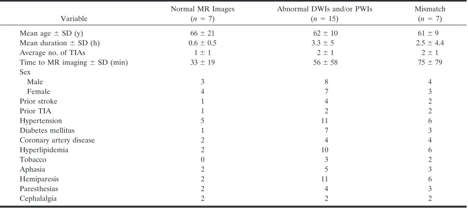
TABLE 1: Clinical and demographic variables
Variable
Normal MR Images (n⫽7)
Abnormal DWIs and/or PWIs (n⫽15)
Mismatch (n⫽7)
Mean age⫾SD (y) 66⫾21 62⫾10 61⫾9 Mean duration⫾SD (h) 0.6⫾0.5 3.3⫾5 2.5⫾4.4 Average no. of TIAs 1⫾1 2⫾1 2⫾1 Time to MR imaging⫾SD (min) 33⫾19 56⫾58 75⫾79 Sex
Male 3 8 4
Female 4 7 3
Prior stroke 1 4 2
Prior TIA 1 2 2
Hypertension 5 11 6
Diabetes mellitus 1 7 3
Coronary artery disease 2 4 4
Hyperlipidemia 2 10 6
Tobacco 0 3 2
Aphasia 2 5 3
Hemiparesis 2 11 6
Paresthesias 2 4 3
TABLE 2: Clinical characteristics of mismatch cases Age (y)/ Sex Duration TIAs vDWI vPWI Echocardiography Carotid Duplex Sonography Angiography MRA MRI Therapy Etiology 64/M 12 h 4 6.1 18.4 TTE: Dilatation L atrium L carotid occlusion Severe L ICA stenosis Absent L ICA Cortical acute infarcts R MCA territory CEA LAA 56/M 20 min 2 14.9 26.8 TTE: Normal Unremarkable Not performed Unremarkable Acute subcortical infarct L MCA territory BP increased, MAP ⬎ 100 mm Hg; IV heparin ⫹ oral aspirin CE 61/M 20 min 2 0 245.7 TEE: Global systolic
dysfunction, inferior-wall akinesis, decreased
[image:4.585.144.447.64.734.2]Case 2
A 61-year-old gentleman was noted to develop cephalalgia, speech difficulties and “confusion” on postoperative day 2 after coronary artery bypass sur-gery (CABG). He underwent sursur-gery because of per-sistent angina after myocardial infarction, despite in-tensive medical therapy. Past medical history was notable for two prior ischemic strokes, known left internal carotid artery (ICA) occlusion despite of bilateral carotid endarterectomy, prior CABG, pe-ripheral vascular disease and hypertension. The neu-rologic examination was remarkable for fluent
apha-sia with difficulty understanding commands.
However, the neurologic deficits completely resolved in 20 minutes. The brain MR imaging, obtained 7 hours after symptoms resolved, is shown on Figure 2. The FLAIR sequence (left, up) reveals two areas of leukomalacia in the right antero-lateral temporal lobe and temporo-occipital junction. The MRA (left, down) demonstrates absent left internal carotid tery with reduced caliber of the middle cerebral ar-tery, which fills out through the anterior communi-cating artery. Large posterior collaterals can also be appreciated. The DW imaging (middle) did not dem-onstrate areas of hyperintensity. However, the PW imaging (right) revealed a large region of perfusion delay affecting most of the left hemisphere. No fol-low-up MR imaging was obtained. The following day, the patient complained of right upper extremity par-esthesia, although his neurologic examination was un-remarkable. He was treated with oral warfarin, while all anti-hypertensive medications were withheld. The patient was discharged to home on postoperative day
number 8. This case is consistent wit a hemodynamic TIA in the setting of ICA occlusion.
Case 3
A 63-year-old gentleman experienced a fall be-cause of sudden weakness on the right leg. He re-ported innumerable previous episodes of right leg weakness, always lasting 15–20 seconds, for almost one year. However, he had never fallen as conse-quence of these. Sometimes the episodes would occur several times per day and feature ipsilateral hand clumsiness. Past medical history was significant for hypertension, hyperlipidemia and diabetes. His phy-sician suspected TIA and treated him with clopi-dogrel and warfarin, which did not ameliorate the symptoms. The neurologic examination was normal. Brain MR imaging and MRA (shown on Fig 3), ob-tained eight days after patient’s last TIA, demon-strated hyperintensities on FLAIR involving the an-terior cerebral artery (ACA) territory bilaterally. Although DW imaging shows hyperintensities in the same locations, the apparent diffusion coefficient was locally increased, suggesting that the lesions were old (i.e., T-2 “shine-through”). PW imaging demonstrates a defect involving the left ACA territory. The MRA shows severe stenosis of the left intracranial ICA and tandem stenosis involving both A1 and A2 segments of the ACAs. MR imaging did not change after ICA angioplasty. A conventional angiography showed high-grade stenosis of the left intracranial ICA, just proximal to the anterior choroidal artery. The left middle cerebral artery filled out from the right side
FIG 2. Brain MR imaging of case 2.
through the anterior communicating artery and A1 segment of the anterior cerebral artery. The patient underwent balloon angioplasty of the left ICA. He remained asymptomatic during his hospital stay and was discharged to home on warfarin, aspirin and ator-vastatin. This case is an example of hemodynamic TIA, in association with severe intracranial stenosis.
Discussion
This study suggests that the addition of DW and PW imaging to the conventional MR imaging proto-col can enhance detection of brain ischemia or hypo-perfusion in patients presenting with transient focal neurologic symptoms. Specifically, TTP maps identi-fied ischemic tissue not detected on conventional or DW imaging sequences in 32% of our patients with TIA. This is relevant because of two main reasons: firstly, it has been suggested that patients with tran-sient focal neurologic symptoms should be diagnosed with ischemic stroke instead of TIA whenever there is evidence of acute brain infarction on neuroimaging, implying that optimal diagnostic methods need to be utilized systematically in the routine diagnostic work-up of TIA (1). Such point of view is endorsed by evidence suggesting that patients with TIA with acute brain infarction may have poorer outcomes than those patients with unremarkable neuroimaging (18). Secondly, transient neurologic symptoms are often vague, causing untoward confusion among practitio-ners regarding the true pathophysiologic nature of the clinical picture. In this study, the diagnostic yield of MR imaging with DW imaging and PW imaging was superior to noncontrast CT, detecting evidence of brain ischemia or hypoperfusion in almost 3/4 of pa-tients. Therefore, an abnormal MR imaging with DW imaging and PW imaging can help the clinician to substantiate the diagnosis of TIA when the symptoms are nonspecific. Despite the advantages of MR
imag-ing over CT, the former is used in less than 5% of TIA cases seen in American emergency rooms (19).
The use of PW imaging permitted the detection of regions of abnormal signal intensity in seven of our patients (four with and three without concomitant DW imaging changes), indicating the presence of tissue at risk for infarction and, theoretically, with a potential for clinical deterioration. Furthermore, MR with PW imaging identified a subset of patients who required conventional angiography, having an impact on patient management. Thus, MR imaging could assist the clinician in the selection of patients that should be admitted to the hospital for close observa-tion, instead of being discharged after a rapid diag-nostic evaluation. This argument is endorsed by our observation that most patients with DW–PW imaging mismatch frequently necessitated surgical or endovas-cular procedures, whereas all controls were treated medically. Although none of the patients in our co-hort sustained an ischemic stroke (regardless of the presence of mismatch), we speculate that the inter-ventions performed on them may have prevented a poor outcome.
TIAs are not benign. Patients presenting with re-versible neurologic defects have a 30% risk of isch-emic stroke within 90 days, particularly during the first 48 hours (20). In addition, there is a 15% annual risk of death and a similarly increased risk of myo-cardial infarction, angina, arrhythmia and congestive heart failure (20, 21). There is abundant evidence suggesting that TIA itself is associated with structural brain damage (2–9). Furthermore, patients with TIA develop more “silent” infarcts and cortical atrophy on follow-up brain MR imaging than age-matched con-trols (22, 23).
Only a few studies have used MR imaging-based cerebral blood flow or perfusion estimates in patients with TIA. The largest study did not detect any blood flow abnormalities, which contradicts our findings (9).
However, the employed technique was a 2D phase-contrast flow measurement, which may not optimally reflect regional brain perfusion. Another study dem-onstrated PW imaging abnormalities (using relative cerebral blood volume, CBF and MTT) in patients with ICA stenosis or occlusion, including four cases presenting with TIA (10). This is in agreement with our findings that PW imaging abnormalities in TIA patients are associated with the presence of large-artery atherosclerosis. Two additional reports have suggested a relationship between delayed TTP and TIA in patients with nonatherosclerotic large-vessel disease, such as moya-moya (11, 12).
Isolated PW imaging changes in TIA have been observed by other investigators (13–15). At least two patients with TIA imaged with DW–PW imaging 2 hours after symptom onset, have exhibited an isolated region of signal intensity abnormality on PW imaging with unremarkable DW imaging (13, 14). Follow-up MR imaging showed an acute infarct on DW imaging in one case (14), whereas the other patient had no subsequent DW or PW imaging abnormalities (13). Another case report described a patient with DW and PW imaging abnormalities that resolved on follow-up MR imaging (15). Our observations add to the liter-ature new cases of TIA imaged with PW imaging, particularly using TTP maps, demonstrating DW–PW imaging mismatch in almost 1/3 of patients.
Our study is limited by its retrospective nature and lack of systematic follow-up neuroimaging. The last constraint prevents a direct assessment of the out-come of tissue with abnormal signal intensity on DW imaging and PW imaging. Although most DW imag-ing abnormalities progress irreversibly to infarcts fea-turing T-2 signal intensity hyperintensity, instances of complete resolution of diffusion restriction have been well documented (2, 5, 8). On the other hand, PW imaging abnormalities may not progress to infarction and resolve spontaneously (13). Nevertheless, clinical indicators can be used as surrogate markers of tissue injury and recovery. For instance, all patients from our cohort returned to their neurologic baseline in less than 24 hours and left the hospital in excellent condition, suggesting that structural brain damage was modest, if any.
In addition, the optimal PW imaging technique is subject of controversy. We utilized TTP, which is widely used and appears to correlate well with other techniques, such as MTT (24). TTP has good trast-to-noise ratio, which is an estimate of the con-spicuity of abnormal signal intensity, facilitating the identification of lesion boundary (24). Another con-straint of this study is a selection bias, since not all patients with TIA managed at our institution rou-tinely undergo diffusion and perfusion MR imaging studies. A prospective study by using DW–PW imag-ing in all patients presentimag-ing with TIA would be needed to better assess the effectiveness of this diag-nostic test.
Although our results endorse previous studies showing that transient hemiparesis is associated with MR imaging abnormalities (4), we could not identify
other clinical characteristics which could predict DW–PW imaging changes. Our patients with abnor-mal DW–PW imaging had more numerous and pro-longed episodes of transient neurologic dysfunction, but this difference did not reach statistical signifi-cance. This suggests that clinical characteristics of TIA may not be useful to triage patients for MR with DW and PW imaging. Although the patients with mismatch had an overrepresentation of coronary ar-tery disease and hyperlipidemia, the number of cases was small. Another issue is whether or not the pres-ence of DW–PW imaging abnormalities herald grim outcomes, but this question transcends the con-straints of the present study.
Conclusion
This retrospective study of patients with TIA shows that MR with DW and PW imaging can improve the documentation of brain ischemia and may have the potential to determine which patients have brain tis-sue at risk for infarction and therefore, may require close observation in the hospital or need aggressive intervention. A prospective study by using follow-up neuroimaging is required to confirm the validity of these suggestions.
Acknowledgement
This article is dedicated to the enduring memory of Carlos J. Abad “Whom the gods love dies young” Plautus, The Two Bacchises.
References
1. Albers GW, Caplan LR, Easton JD, Fayad PB, Mohr JP, Saver JL, Sherman DG, the TIA Working Group.Transient ischemic attack– proposal for a new definition.N Engl J Med2002;347:1713–1716 2. Kidwell CS, Alger JR, Di Salle F, Starkman S, Villablanca P,
Bentson J, Saver JL. Diffusion MRI in patients with transient ischemic attacks.Stroke1999;30:1174 –1180
3. Ay H, Oliveira-Filho J, Buonanno FS, et al.’Footprints’ of tran-sient ischemic attacks: a diffusion-weighted MRI study. Cerebro-vasc Dis2002;14:177–186
4. Crisostomo RA, Garcia MM, Tong DC. Detection of diffusion-weighted MRI abnormalities in patients with transient ischemic attack: correlation with clinical characteristics. Stroke
2003;34:932–937
5. Marx JJ, Mika-Gruettner A, Thoemke F, Fitzek S, Fitzek C, Vu-curevic G, Urban PP, Stoeter P, Hopf HC. Diffusion weighted magnetic resonance imaging in the diagnosis of reversible isch-aemic deficits of the brainstem. J Neurol Neurosurg Psychiatry
2002;72:572–575
6. Kastrup A, Schulz JB, Mader I, et al.Diffusion-weighted MRI in patients with symptomatic internal carotid artery disease.J Neurol
2002;249:1168 –1174
7. Rovira A, Rovira-Gols A, Pedraza S, Grive E, Molina C, Alvarez-Sabin J. Diffusion-weighted MR imaging in the acute phase of transient ischemic attacks.AJNR2002;23:77– 83
8. Lecouvet FE, Duprez TPJ, Raymackers JM, Peeters A, Cosnard G. Resolution of early diffusion-weighted and FLAIR MRI abnormal-ities in a patient with TIA.Neurology,1999;52:1085
9. Bisschops RHC, LJ Kappelle LJ, Mali WPTM, van der Grond J. Hemodynamic and Metabolic Changes in Transient Ischemic At-tack Patients: A Magnetic Resonance Angiography and 1H-Mag-netic Resonance Spectroscopy Study Performed Within 3 Days of Onset of a Transient Ischemic Attack.Stroke2002;33:110 –115 10. Chaves CJ, Staroselskaya I, Linfante I, Llinas R, Caplan LR,
11. Kim SK, Wang KC, Oh CW, Kim IO, Lee DS, Song IC, Cho BK. Evaluation of cerebral hemodynamics with perfusion MRI in child-hood moyamoya disease.Pediatr Neurosurg2003;38:68 –75 12. El-Koussy M, Lovblad KO, Steinlin M, Kiefer C, Schroth G.
Per-fusion MRI abnormalities in the absence of difPer-fusion changes in a case of moyamoya-like syndrome in neurofibromatosis type 1. Neu-roradiology2002;44:938 –941
13. Darby DG, Barber PA, Gerraty RP, et al.Pathophysiological to-pography of acute ischemia by combined diffusion-weighted and perfusion MRI.Stroke1999;30:2043–52
14. Ide C, De Coene B, Trigaux JP, Baudrez V, Ossemann M, Mor-mont E, Laloux P.Discrepancy between diffusion and perfusion imaging in a patient with transient ischaemic attack.J Neuroradiol
2001;28:118 –122
15. Neumann-Haefelin T, Wittsack HJ, Wenserski F, Li TQ, Moseley ME, Siebler M, Freund HJ. Diffusion- and perfusion-weighted MRI in a patient with a prolonged reversible ischaemic neurolog-ical deficit.Neuroradiology2000;42:444 – 447
16. Hillis AE, Wityk RJ, Tuffiash E, Barker BB, Beauchamp NJ, Wityk RJ.Hypoperfusion of Wernicke’s area predicts severity of semantic deficit in acute stroke.Ann Neurol2001;50:561–566
17. Hillis AE, Kane A, Tuffiash E, et al.Reperfusion of specific brain regions by raising blood pressure restores selective language func-tions in subacute stroke.Brain Lang,2001;79:495–510
18. Evans GW, Howard G, Murros KE, Rose LA, Toole JF.Cerebral infarction verified by cranial computed tomography and prognosis for survival following transient ischemic attack. Stroke
1991;22:431– 436
19. Edlow JA, Kim S, Emond JA, et al.US Emergency Department Visits for Transient Ischemic Attack, 1992–2000.Acad Emerg Med
2003;10:432
20. Johnston SC, Gress DR, Browner WS, Sidney S.Short-term prog-nosis after emergency-department diagprog-nosis of TIA. JAMA
2000;284:2901–2906
21. Bravata DM, Ho SY, Brass LM, Concato J, Scinto J, Meehan TP. Long-term mortality in cerebrovascular disease. Stroke
2003;34:699 –704
22. Walters RJ, Fox NC, Schott JM, et al.Transient ischemic attacks are associated with increased rates of global cerebral atrophy.
J Neurol Neurosurg Psychiatry2003;74:213–216
23. Bhadelia RA, Anderson M, Polak JF, et al.Prevalence and asso-ciations of MRI-demonstrated brain infarcts in elderly subjects with a history of transient ischemic attack. The Cardiovascular Health Study.Stroke1999;30:383–388