organic papers
o550
Bond and Parsons C7H9N DOI: 10.1107/S160053680200692X Acta Cryst.(2002). E58, o550±o552Acta Crystallographica Section E Structure Reports Online
ISSN 1600-5368
2,4-Lutidine
Andrew D. Bonda* and Simon
Parsonsb
aDepartment of Chemistry, University of Cambridge, Lensfield Road, Cambridge CB2 1EW, England, andbDepartment of Chemistry, University of Edinburgh, King's Buildings, West Mains Road, Edinburgh EH9 3JJ, Scotland
Correspondence e-mail: [email protected]
Key indicators Single-crystal X-ray study T= 150 K
Mean(C±C) = 0.002 AÊ Rfactor = 0.055 wRfactor = 0.146
Data-to-parameter ratio = 19.7
For details of how these key indicators were automatically derived from the article, see http://journals.iucr.org/e.
#2002 International Union of Crystallography Printed in Great Britain ± all rights reserved
The crystal structure of 2,4-lutidine (2,4-dimethylpyridine, C7H9N) has been determined at 150 (2) K, followingin situ
crystal growth from the liquid. In space group P21/n, the
asymmetric unit comprises a whole molecule. Molecules are linkedviaCÐH N interactions into chains that align to form polar sheets, and adjacent sheets are packed in an antiparallel arrangement.
Comment
As part of a study devoted to improving techniques for determining the crystal structures of substances that are liquid at room temperature, we have reported previously the struc-tures of four of the six lutidine (dimethylpyridine) isomers (Bondet al., 2001; Bond & Davies, 2002a,b,c). In each case, crystals of these low-melting materials were grownin situfrom the liquid, using a relatively simple `zone re®nement' tech-nique (Davies & Bond, 2001). Curiously, we had been unable to obtain a single crystal of 2,4-lutidine, (I), using this approach. A crystal of (I) was eventually grown by Boese's laser-assisted crystallization technique (Boese & Nussbaumer, 1994), using a sample held at 203 K in a 0.5 mm capillary. The crystal was cooled subsequently to 150 K for data collection.
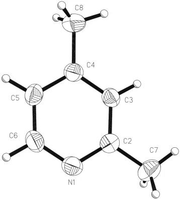
In all of the lutidine isomers where the 4-position of the ring is unsubstituted, polar chains are formed via C4ÐH4 N interactions. In the 3,4-isomer, where the 4-position is substi-tuted, dimers are formedviasimilar interactions from the 2-and 6-positions. In (I), where the 4-position is also substituted (Fig. 1), molecules are linked into chains along [010]viaCÐ H N interactions from the 3-position [H3 N1i = 2.75 AÊ
and C3ÐH3 N1i= 166.8; symmetry code: (i) 1/2ÿx, 1/2+y,
3/2ÿz]. The fact that the 3-position is utilized in (I) would appear to be a consequence of effective packing of the methyl substituents between adjacent chains. The chains in (I) lie in sheets parallel to (100) (Fig. 2), and adjacent sheets are aligned in an antiparallel manner such that the crystal is not macroscopically polar (Fig. 3). Similar arrangements are observed in the 2,5- and 3,5-isomers.
Experimental
The sample (99%) of (I) was obtained from the Aldrich company and used without further puri®cation. A crystal was grown by Boese's laser-assisted crystallization technique (Boese & Nussbaumer, 1994), using a sample held at 203 K in a 0.5 mm capillary. The crystal was cooled subsequently to 150 K for data collection. The length of the cylindrical crystal was not measured, but it exceeded the diameter of the collimator (0.5 mm).
Crystal data
C7H9N
Mr= 107.15
Monoclinic,P21=n
a= 7.4247 (8) AÊ b= 10.9401 (12) AÊ c= 8.2473 (9) AÊ = 107.342 (2)
V= 639.45 (12) AÊ3
Z= 4
Dx= 1.113 Mg mÿ3
MoKradiation Cell parameters from 2543
re¯ections = 2.5±28.9
= 0.07 mmÿ1
T= 150 (2) K Cylinder, colourless 0.25 mm (radius)
Data collection
Bruker SMART APEX diffractometer equipped with Oxford Cryosystems cryostream and OHCD laser-assisted crystallization device (Scienti®c Consulting, Essen, Germany) Thin-slice!scans
Absorption correction: multi-scan (SADABS; Sheldrick, 2001) Tmin= 0.653,Tmax= 1.000
4515 measured re¯ections 1540 independent re¯ections 1133 re¯ections withI> 2(I) Rint= 0.030
max= 28.9
h=ÿ9!9 k=ÿ13!14 l=ÿ11!10
Re®nement
Re®nement onF2
R[F2> 2(F2)] = 0.055
wR(F2) = 0.146
S= 0.94 1540 re¯ections 78 parameters
H-atom parameters constrained
w= 1/[2(F
o2) + (0.0973P)2]
whereP= (Fo2+ 2Fc2)/3
(/)max< 0.001 max= 0.20 e AÊÿ3 min=ÿ0.31 e AÊÿ3
Extinction correction:SHELXL97 Extinction coef®cient: 0.19 (2)
All H atoms were placed geometrically and re®ned with isotropic displacement parameters, with a common parameter assigned to all H atoms of the methyl groups, and a second parameter for H atoms attached to the ring (two displacement parameters in total). Each methyl group was allowed to rotate about its local threefold axis.
Data collection:SMART(Bruker, 1998); cell re®nement:SAINT
(Bruker, 1998); data reduction: SAINT; program(s) used to solve structure: SHELXS97 (Sheldrick, 1997); program(s) used to re®ne structure: SHELXL97 (Sheldrick, 1997); molecular graphics: XP
(Sheldrick, 1993) and CAMERON(Watkin et al., 1996); software used to prepare material for publication:SHELXL97.
Acta Cryst.(2002). E58, o550±o552 Bond and Parsons C7H9N
o551
organic papers
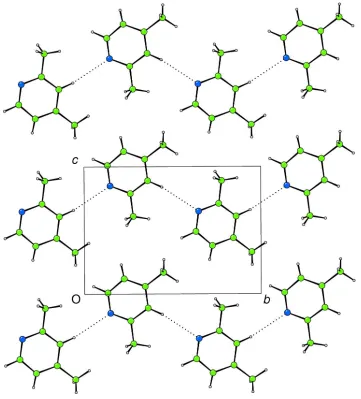
Figure 2
Projection of (I) on to (100), showing molecules linked into chainsvia
CÐH N interactions.
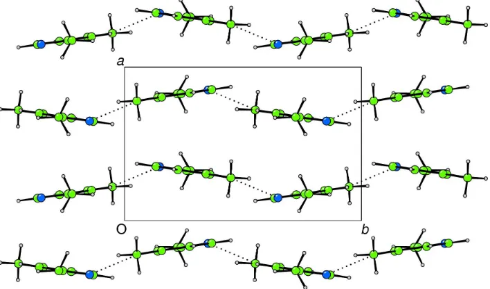
Figure 3
Projection of (I) on to (001), showing sheets aligned in an antiparallel manner.
Figure 1
organic papers
o552
Bond and Parsons C7H9N Acta Cryst.(2002). E58, o550±o552We are grateful to the EPSRC for funding and to Dr John E. Davies (University of Cambridge) for bringing this problem to our attention.
References
Boese, R. & Nussbaumer, M. (1994). Correlations, Transformations and Interactions in Organic Crystal Chemistry, IUCr Crystallographic Symposia, Vol. 7, edited by D. W. Jones & A. Katrusiak, pp. 20±37. Oxford University Press.
Bond, A. D., Davies, J. E. & Kirby, A. J. (2001).Acta Cryst.E57, o1242±o1244.
Bond, A. D. & Davies, J. E. (2002a).Acta Cryst.E58, o5±o7. Bond, A. D. & Davies, J. E. (2002b).Acta Cryst.E58, o326±o327. Bond, A. D. & Davies, J. E. (2002c).Acta Cryst.E58, o328±o330.
Bruker (1998).SMARTandSAINT. Bruker AXS Inc., Madison, Wisconsin, USA.
Davies, J. E. & Bond, A. D. (2001).Acta Cryst.E57, o947±o949. Sheldrick, G. M. (1993).XP. University of GoÈttingen, Germany.
Sheldrick, G. M. (1997). SHELXL97 and SHELXS97. University of GoÈttingen, Germany.
Sheldrick, G. M. (2001).SADABS. Version 2.06. University of GoÈttingen, Germany.
supporting information
sup-1
Acta Cryst. (2002). E58, o550–o552
supporting information
Acta Cryst. (2002). E58, o550–o552 [doi:10.1107/S160053680200692X]
2,4-Lutidine
Andrew D. Bond and Simon Parsons
S1. Comment
As part of a study devoted to improving techniques for determining the crystal structures of substances that are liquid at
room temperature, we have reported previously the structures of four of the six lutidine (dimethylpyridine) isomers
(Bond et al., 2001; Bond & Davies, 2002a,b,c). In each case, crystals of these low-melting materials were grown in situ
from the liquid using a relatively simple `zone refinement′ technique (Davies & Bond, 2001). Curiously, we had been
unable to obtain a single-crystal of 2,4-lutidin,e (I), using this approach. A crystal of (I) was eventually grown by Boese's
laser-assisted crystallization technique (Boese & Nussbaumer, 1994) using a sample held at 203 K in a 0.5 mm capillary.
The crystal was cooled subsequently to 150 K for data collection.
In all of the lutidine isomers where the 4-position of the ring is unsubstituted, polar chains are formed via C4—H4···N
interactions. In the 3,4-isomer, where the 4-position is substituted, dimers are formed via similar interactions from the 2-
and 6-positions. In (I), where the 4-position is also substituted (Fig. 1), molecules are linked into chains along [010] via C
—H···N interactions from the 3-position [H3···N1i = 2.75 Å and C3—H3···N1i = 166.8°; symmetry code: (i) 1/2 - x, 1/2 +
y, 3/2 - z]. The fact that the 3-position is utilized in (I) would appear to be a consequence of effective packing of the
methyl substituents between adjacent chains. The chains in (I) lie in sheets parallel to (100) (Fig. 2), and adjacent sheets
are aligned in an antiparallel manner such that the crystal is not macroscopically polar (Fig. 3). Similar arrangements are
observed in the 2,5- and 3,5-isomers.
S2. Experimental
The sample (99%) of (I) was obtained from the Aldrich company and used without further purification. A crystal was
grown by Boese's laser-assisted crystallization technique (Boese & Nussbaumer, 1994) using a sample held at 203 K in a
0.5 mm capillary. The crystal was cooled subsequently to 150 K for data collection. The length of the cylindrical crystal
was not measured, but it exceeded the diameter of the collimator (0.35 mm).
S3. Refinement
All H atoms were placed geometrically and refined with isotropic displacement parameters, with a common parameter
assigned to all H atoms of the methyl groups, and a second parameter for H atoms attached to the ring (two displacement
supporting information
sup-2
[image:5.610.126.484.70.466.2]Acta Cryst. (2002). E58, o550–o552 Figure 1
supporting information
sup-3
[image:6.610.126.487.70.469.2]Acta Cryst. (2002). E58, o550–o552 Figure 2
supporting information
sup-4
[image:7.610.129.483.74.283.2]Acta Cryst. (2002). E58, o550–o552 Figure 3
Projection of (I) on to (001), showing sheets aligned in an antiparallel manner.
2,4-dimethylpyridine
Crystal data
C7H9N Mr = 107.15 Monoclinic, P21/n a = 7.4247 (8) Å b = 10.9401 (12) Å c = 8.2473 (9) Å β = 107.342 (2)° V = 639.45 (12) Å3 Z = 4
F(000) = 232
Dx = 1.113 Mg m−3 Melting point: 213 K
Mo Kα radiation, λ = 0.7107 Å Cell parameters from 2543 reflections θ = 2.5–28.9°
µ = 0.07 mm−1 T = 150 K
Cylinder, colourless 0.25 mm (radius)
Data collection
Bruker SMART APEX
diffractometer equipped with Oxford Cryosystems cryostream and OHCD laser-assisted crystallisation device (Scientific Consulting, Essen, Germany)
Radiation source: fine-focus sealed tube Graphite monochromator
thin–slice ω scans
Absorption correction: multi-scan (SADABS; Sheldrick, 2001)
Tmin = 0.653, Tmax = 1.000 4515 measured reflections 1540 independent reflections 1133 reflections with I > 2σ(I) Rint = 0.030
θmax = 28.9°, θmin = 3.7° h = −9→9
k = −13→14 l = −11→10
Refinement
Refinement on F2 Least-squares matrix: full R[F2 > 2σ(F2)] = 0.055 wR(F2) = 0.146 S = 0.94 1540 reflections
78 parameters 0 restraints
Primary atom site location: structure-invariant direct methods
supporting information
sup-5
Acta Cryst. (2002). E58, o550–o552 Hydrogen site location: inferred from
neighbouring sites
H-atom parameters constrained w = 1/[σ2(F
o2) + (0.0973P)2] where P = (Fo2 + 2Fc2)/3 (Δ/σ)max < 0.001
Δρmax = 0.20 e Å−3 Δρmin = −0.31 e Å−3
Extinction correction: SHELXL97, Fc*=kFc[1+0.001xFc2λ3/sin(2θ)]-1/4 Extinction coefficient: 0.19 (2)
Special details
Geometry. All e.s.d.'s (except the e.s.d. in the dihedral angle between two l.s. planes) are estimated using the full covariance matrix. The cell e.s.d.'s are taken into account individually in the estimation of e.s.d.'s in distances, angles and torsion angles; correlations between e.s.d.'s in cell parameters are only used when they are defined by crystal symmetry. An approximate (isotropic) treatment of cell e.s.d.'s is used for estimating e.s.d.'s involving l.s. planes.
Refinement. Refinement of F2 against ALL reflections. The weighted R-factor wR and goodness of fit S are based on F2, conventional R-factors R are based on F, with F set to zero for negative F2. The threshold expression of F2 > σ(F2) is used only for calculating R-factors(gt) etc. and is not relevant to the choice of reflections for refinement. R-factors based on F2 are statistically about twice as large as those based on F, and R- factors based on ALL data will be even larger.
Fractional atomic coordinates and isotropic or equivalent isotropic displacement parameters (Å2)
x y z Uiso*/Ueq
N1 0.14857 (14) 0.64758 (9) 0.65190 (14) 0.0394 (3)
C2 0.17370 (16) 0.76169 (10) 0.71317 (15) 0.0334 (3)
C3 0.19828 (16) 0.86006 (10) 0.61580 (15) 0.0345 (3)
H3 0.2174 0.9395 0.6643 0.050 (2)*
C4 0.19522 (16) 0.84361 (10) 0.44918 (15) 0.0353 (3)
C5 0.16838 (18) 0.72517 (11) 0.38610 (16) 0.0392 (4)
H5 0.1652 0.7086 0.2722 0.050 (2)*
C6 0.14654 (19) 0.63256 (11) 0.49059 (18) 0.0427 (4)
H6 0.1287 0.5521 0.4452 0.050 (2)*
C7 0.1742 (2) 0.77769 (13) 0.89345 (16) 0.0474 (4)
H7A 0.0635 0.7370 0.9102 0.103 (3)*
H7B 0.1702 0.8650 0.9186 0.103 (3)*
H7C 0.2892 0.7415 0.9696 0.103 (3)*
C8 0.2206 (2) 0.94818 (12) 0.33953 (18) 0.0545 (4)
H8A 0.2258 1.0252 0.4014 0.103 (3)*
H8B 0.1142 0.9503 0.2352 0.103 (3)*
H8C 0.3384 0.9371 0.3106 0.103 (3)*
Atomic displacement parameters (Å2)
U11 U22 U33 U12 U13 U23
N1 0.0418 (6) 0.0327 (6) 0.0468 (7) 0.0003 (4) 0.0178 (5) 0.0034 (4)
C2 0.0280 (6) 0.0379 (6) 0.0335 (6) −0.0001 (4) 0.0080 (5) 0.0011 (5)
C3 0.0369 (6) 0.0295 (6) 0.0372 (7) −0.0016 (4) 0.0112 (5) −0.0030 (4)
C4 0.0361 (6) 0.0335 (6) 0.0396 (7) 0.0030 (4) 0.0162 (5) 0.0035 (5)
C5 0.0435 (7) 0.0416 (7) 0.0374 (7) −0.0004 (5) 0.0194 (6) −0.0057 (5)
C6 0.0506 (8) 0.0306 (6) 0.0537 (8) −0.0033 (5) 0.0259 (6) −0.0077 (5)
C7 0.0481 (8) 0.0594 (9) 0.0344 (7) −0.0080 (6) 0.0121 (6) 0.0001 (6)
supporting information
sup-6
Acta Cryst. (2002). E58, o550–o552 Geometric parameters (Å, º)
N1—C6 1.3361 (17) C5—H5 0.950
N1—C2 1.3388 (15) C6—H6 0.950
C2—C3 1.3870 (16) C7—H7A 0.980
C2—C7 1.4960 (17) C7—H7B 0.980
C3—C4 1.3794 (17) C7—H7C 0.980
C3—H3 0.950 C8—H8A 0.980
C4—C5 1.3884 (16) C8—H8B 0.980
C4—C8 1.5045 (17) C8—H8C 0.980
C5—C6 1.3706 (18)
C6—N1—C2 116.70 (10) N1—C6—H6 117.7
N1—C2—C3 122.18 (12) C5—C6—H6 117.7
N1—C2—C7 116.29 (11) C2—C7—H7A 109.5
C3—C2—C7 121.52 (11) C2—C7—H7B 109.5
C4—C3—C2 120.64 (10) H7A—C7—H7B 109.5
C4—C3—H3 119.7 C2—C7—H7C 109.5
C2—C3—H3 119.7 H7A—C7—H7C 109.5
C3—C4—C5 116.93 (10) H7B—C7—H7C 109.5
C3—C4—C8 122.13 (11) C4—C8—H8A 109.5
C5—C4—C8 120.94 (11) C4—C8—H8B 109.5
C6—C5—C4 119.01 (11) H8A—C8—H8B 109.5
C6—C5—H5 120.5 C4—C8—H8C 109.5
C4—C5—H5 120.5 H8A—C8—H8C 109.5
N1—C6—C5 124.53 (11) H8B—C8—H8C 109.5
C6—N1—C2—C3 −0.57 (18) C2—C3—C4—C8 179.75 (12)
C6—N1—C2—C7 179.53 (10) C3—C4—C5—C6 0.06 (17)
N1—C2—C3—C4 0.84 (18) C8—C4—C5—C6 179.76 (12)
C7—C2—C3—C4 −179.26 (10) C2—N1—C6—C5 0.05 (19)
C2—C3—C4—C5 −0.55 (17) C4—C5—C6—N1 0.2 (2)
Hydrogen-bond geometry (Å, º)
D—H···A D—H H···A D···A D—H···A
C3—H3···N1i 0.95 2.75 3.6822 (15) 167
![Aqua(4 nitrophthalato)bis[2 (1H pyrazol 3 yl)pyridine]zinc(II) hemihydrate](data:image/gif;base64,R0lGODlhAQABAIAAAP///wAAACH5BAEAAAAALAAAAAABAAEAAAICRAEAOw==)