STUDY OF EGGSHELL MEMBRANE EMBEDDED WITH BIOSYNTHESIZED SILVER NANOPARTICLES
FOR THEIR ANTIBACTERIAL ACTIVITY
*,1
Manasi Gaikwad and
*1,2
Department of Biotechnology Engineering, Kolhapur Institute of Technology, College of Engineering,
ARTICLE INFO ABSTRACT
Facile,
interesting topic in materials chemistry and heterogeneous catalysis. This paper reports an method for the synthesis and immobilization of small silver nanoparticles (AgNPs) at room temperature on natural eggshell membrane (ESM). The eggshell membrane is the clear film lining the eggshell, visible when one peels a boiled egg, which presents inte
used as a unique protein
the synthesis of antimicrobial silver nanoparticle (AgNPs), but the green synthesis is the most emerging method of synth
leaf extracts which act as both reducing and capping agent. Visual color change, UV spectroscopy, scanning electron microscopy and transmission electron microscopy unambiguously iden
presence of AgNPs on ESM. Besides the antibacterial activity of these ESM’s was seen through the zone of inhibitions observed for the test samples against plates of Escherichia coli. Such egg membranes can be used against skin wounds like that in
bacterial infections and the presence of ESM will also help the recovery of the skin as the amino acid composition of the egg membranes is quite close to that of human skin and other human tissues.
Copyright © 2016 Manasi Gaikwad and Shweta Deshpande
permits unrestricted use, distribution, and reproduction in any medium, provided the original work is properly cited.
INTRODUCTION
Silver nanoparticles (AgNPs) with zero, one, or two dimensional nanostructures such as nanoparticles, nanowires, and nanocubes have been the subject of focused researc
their great biocidal potential. Many techniques are known to be available with the evidence of publication for the synthesis of AgNPs. Silver nanoparticles have unique properties which help in molecular diagnostics, in therapies, as well as in dev
are used in several medical procedures. The major methods used for silver nanoparticle synthesis are the physical and chemical methods. The problem with the chemical and physical methods is that the synthesis is expensive and can also have toxic substances absorbed onto them. To overcome this, the biological method provides a feasible alternative. The major biological systems involved in this are bacteria, fungi, and plant extracts. Biological generation of AgNPs, especially using plant extract is known to produce large quantities of
*Corresponding author: Manasi Gaikwad,
Department of Biotechnology Engineering, Kolhapur Institute of Technology, College of Engineering, Kolhapur, India.
ISSN: 0975-833X
Article History:
Received 05th October, 2015 Received in revised form
19th November, 2015
Accepted 08th December, 2015
Published online 31st January,2016
Key words:
Silver nanoparticles,(AgNPs), Eggshell membrane(ESM), Ocimum sanctum (tulsi), Biosynthesis, Characterization, Antibacterial activity.
Citation: Manasi Gaikwad and Shweta Deshpande
antibacterial activity”, International Journal of Current Research,
RESEARCH ARTICLE
STUDY OF EGGSHELL MEMBRANE EMBEDDED WITH BIOSYNTHESIZED SILVER NANOPARTICLES
FOR THEIR ANTIBACTERIAL ACTIVITY
Manasi Gaikwad and
2Shweta Deshpande
Department of Biotechnology Engineering, Kolhapur Institute of Technology, College of Engineering,
Kolhapur, India
ABSTRACT
Facile, efficient, and robust immobilization of metal nanostructures on porous bioscaffolds is an interesting topic in materials chemistry and heterogeneous catalysis. This paper reports an method for the synthesis and immobilization of small silver nanoparticles (AgNPs) at room temperature on natural eggshell membrane (ESM). The eggshell membrane is the clear film lining the eggshell, visible when one peels a boiled egg, which presents inte
used as a unique protein-based biotemplate. Many chemical and physical techniques are available for the synthesis of antimicrobial silver nanoparticle (AgNPs), but the green synthesis is the most emerging method of synthesis. Here the silver nanoparticles were synthesized using
leaf extracts which act as both reducing and capping agent. Visual color change, UV spectroscopy, scanning electron microscopy and transmission electron microscopy unambiguously iden
presence of AgNPs on ESM. Besides the antibacterial activity of these ESM’s was seen through the zone of inhibitions observed for the test samples against plates of Escherichia coli. Such egg membranes can be used against skin wounds like that in burns wherein the AgNPs can prevent the bacterial infections and the presence of ESM will also help the recovery of the skin as the amino acid composition of the egg membranes is quite close to that of human skin and other human tissues.
Manasi Gaikwad and Shweta Deshpande. This is an open access article distributed under the Creative Commons Att use, distribution, and reproduction in any medium, provided the original work is properly cited.
Silver nanoparticles (AgNPs) with zero, one, or two-dimensional nanostructures such as nanoparticles, nanowires, and nanocubes have been the subject of focused research due to their great biocidal potential. Many techniques are known to be available with the evidence of publication for the synthesis of AgNPs. Silver nanoparticles have unique properties which help in molecular diagnostics, in therapies, as well as in devices that are used in several medical procedures. The major methods used for silver nanoparticle synthesis are the physical and chemical methods. The problem with the chemical and physical methods is that the synthesis is expensive and can also have substances absorbed onto them. To overcome this, the biological method provides a feasible alternative. The major biological systems involved in this are bacteria, fungi, and plant extracts. Biological generation of AgNPs, especially
known to produce large quantities of
Department of Biotechnology Engineering, Kolhapur Institute of Technology, College of Engineering, Kolhapur, India.
nanoparticles that are free of contamination and have a well defined size and morphology which is important for diagnostic application (Hutchison et al., 2008; Amit Kumar Mittal 2013). In most of the therapeutic applications, it is the antimicrobial property that is being majorly explored, though the anti-inflammatory property has its fair share of applications. So here we have used a unique green synthesis method of synthesizing AgNPs by using leaf extract of
Ocimum sanctum i.e. Tulsi and eggshell
ESM is bilayered membrane which contains some proteins which are antimicrobial in nature (due to presence of lysozymes). It is used in remedy of some diseases from ancient era. It can be easily obtain from an eggshell which is waste product (ESM History, 2014; Wong
composition of ESM is mainly water
such as collagen (types I, V and X), and amino acids like glycine, alanine and uronic acid. Wong
presence of Type I and Type V p
shell membranes whereas Arias et al., observed the presence of Type X collagen in the egg shell membrane.
Available online at http://www.journalcra.com
International Journal of Current Research
Vol. 8, Issue, 01, pp.25221-25226, January, 2016
INTERNATIONAL
Manasi Gaikwad and Shweta Deshpande, 2016. “Study of eggshell membrane embedded with biosynthesized silver nanoparticles for their
International Journal of Current Research, 8, (01), 25221-25226.
STUDY OF EGGSHELL MEMBRANE EMBEDDED WITH BIOSYNTHESIZED SILVER NANOPARTICLES
Department of Biotechnology Engineering, Kolhapur Institute of Technology, College of Engineering,
efficient, and robust immobilization of metal nanostructures on porous bioscaffolds is an interesting topic in materials chemistry and heterogeneous catalysis. This paper reports an in situ method for the synthesis and immobilization of small silver nanoparticles (AgNPs) at room temperature on natural eggshell membrane (ESM). The eggshell membrane is the clear film lining the eggshell, visible when one peels a boiled egg, which presents interwoven fibrous structure and can be based biotemplate. Many chemical and physical techniques are available for the synthesis of antimicrobial silver nanoparticle (AgNPs), but the green synthesis is the most esis. Here the silver nanoparticles were synthesized using Ocimum sanctum leaf extracts which act as both reducing and capping agent. Visual color change, UV spectroscopy, scanning electron microscopy and transmission electron microscopy unambiguously identifed the presence of AgNPs on ESM. Besides the antibacterial activity of these ESM’s was seen through the zone of inhibitions observed for the test samples against plates of Escherichia coli. Such egg burns wherein the AgNPs can prevent the bacterial infections and the presence of ESM will also help the recovery of the skin as the amino acid composition of the egg membranes is quite close to that of human skin and other human tissues.
is an open access article distributed under the Creative Commons Attribution License, which
nanoparticles that are free of contamination and have a well-defined size and morphology which is important for diagnostic
., 2008; Amit Kumar Mittal et al., 2013). In most of the therapeutic applications, it is the al property that is being majorly explored, though inflammatory property has its fair share of applications. So here we have used a unique green synthesis method of synthesizing AgNPs by using leaf extract of
i.e. Tulsi and eggshell membranes (ESM). ESM is bilayered membrane which contains some proteins which are antimicrobial in nature (due to presence of lysozymes). It is used in remedy of some diseases from ancient era. It can be easily obtain from an eggshell which is waste uct (ESM History, 2014; Wong et al., 1984). The composition of ESM is mainly water-insoluble glycoproteins such as collagen (types I, V and X), and amino acids like glycine, alanine and uronic acid. Wong et al., reported the presence of Type I and Type V proteins in the inner and outer shell membranes whereas Arias et al., observed the presence of Type X collagen in the egg shell membrane.
INTERNATIONAL JOURNAL OF CURRENT RESEARCH
A few known major applications of ESM are as a matrix for the recovery of heavy metals, as a template for hierarchically ordered macroporous materials, and for biosensing of enzyme immobilized Au-ESM42–45 (ESM History, 2014; Wong et al., 1984; Arias et al., 1991; Moulton et al., 2010). ESM possess not only antimicrobial property but also ability to produce metallic nanoparticals
MATERIALS AND METHODS
Preparation of Ocimum sanctum leaf extract
10 g of Ocimum sanctum (Tulsi) leaves were taken. They were surface sterilized and finely chopped. They were then boiled in 50 mL of deionized water and stirred for 1 h. The mixture was then cooled, filtered through filter paper and stored at 40C for further use. (Debajit Borah et al., 2013)
Separation of egg membranes from egg shells
Eggshell membrane is visible as a clear film lining the eggshell, on boiling the egg. Thus 4 eggs were boiled and the ESM was simply peeled off. (Hincke et al., 2000)
Synthesis of AgNP’S
5 mL of the leaf extract was added in 45 mL of 10-3 M silver nitrate (AgNO3) solution. Also 0.35g of separated ESM was
added to reaction mixture. In the reference blank only the 10-3 M AgNO3 solution was replaced by distilled water. The
reaction was carried out at room temperature for 2 to 3 hours. The change of colour from yellow to reddish brown indicates the formation of Ag nanoparticles. (Charusheela Ramteke et
al., 2013)
Analysis of bioreduced AgNP’s
The initial synthesis was monitored by color change at 1st and 3rd hr. UV- vis analysis of the reaction solutions was carried out at room temperature on “Shimadzu UV-1800“double beam UV-visible spectrophotometer at a resolution of 1 nm. UV-VIS spectrophotometric analysis was carried out at 3rd hr using cuvette of path length 10 mm to confirm the presence of AgNPs. The measurements were carried out as a function of reaction time at room temperature. (Jin Zhang et al., 2014) Nano-silver samples for TEM were prepared by placing a drop of silver nanoparticle solution on carbon coated copper grids and allowing for the complete evaporation of water. The shape and size of the formed nano-silver was analyzed from the conventional TEM micrographs recorded at 120kv on JEM 2100, JEOL, Japan. The surface properties of the egg membranes were also studied using scanning electron micrographs performed on high pressure Hitachi SEM S3400N. For the SEM characterization the ESM samples were mounted on a double faced conductive adhesive tape. Images were obtained at an accelerating voltage of 15-kv. (Miao Liang et
al., 2014; Dong Yang et al., 2003)
Assessment of antibacterial activity
In order to examine the antibacterial activity of the AgNPs on selected bacteria (E. coli ), the well diffusion and a mimic of
standard disc diffusion was carried out using egg membranes containing AgNP’s.100ml of nutrient agar (HiMedia) was prepared in sterile flask. A fresh loopful of culture of E.coli was inoculated in the nutrient agar flask under sterile conditions. Pour plate technique was performed for having 6 petri dishes. Well diffusion: Holes were punched on the solidified agar plates using gel punch.45μl of AgNP containing solution was added to the wells. Disc diffusion: A test strip of egg membranes containing AgNPs was placed onto solidified agar plates using sterile forcep .Both the plates were placed in refrigerator for 15mins to facilitate diffusion. Antimicrobial activity was demonstrated by zone of inhibition on and around the test strips.
RESULTS AND DISCUSSION
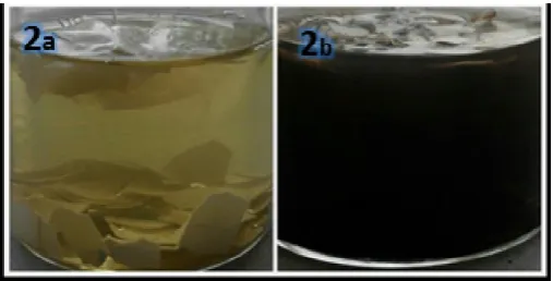
[image:2.595.309.562.345.473.2]Formation of AgNPs by reduction of silver nitrate during exposure to Tulsi leaf extract and ESM can be easily monitored from the change in colour of the reaction mixture. Silver nanoparticles bear a characteristic yellow brown colour due to the excitation of surface plasmon vibrations. The change in color of the reaction mixture from 0th hour and after 3 hours is presented in Figure 1 and 2 respectively, indicating the formation of AgNPs.
Fig. 1. Colour change in reaction mixture at 0 hours. (a) Control-ESM+ 5ml O.sanctum extract+45ml D/W water. (b)Test- Control-ESM+
5ml O.sanctum extract+45ml AgNO3 solution (1mM)
Fig. 2. Colour change in reaction mixture at after 3 hours. ; (a) Control-ESM+ 5ml O.sanctum extract+45ml D/W water. (b)Test-
ESM+ 5ml O.sanctum extract+45ml AgNO3 solution (1mM)
[image:2.595.308.562.524.653.2]the presence of high quantity of antioxidants in the leaves extract. Also the composition of ESM and the aldehyde moieties on its surface can contribute to reduction of silver ions. (Dong Yang et al., 2003) A simultaneous colour change of the membrane from white to yellow was noticed, suggesting the adsorption of Ag⁰ on the membrane surface.
Fig. 3. UV-Vis spectrum image showing peak at 469 nm indicates the formation of AgNPs by aqueous extract of O. sanctum
Figure 3 gives a UV-Vis absorption spectrum for the AgNP samples synthesized using O.sanctum leaf extract and ESM membranes with its peak at 469nm.It is observed that the silver surface plasmon resonance (SPR) occurs at 450 nm and steadily increases in intensity as a function reaction time. The reduction of silver ions was quite rapid. More than 70% of the reaction was complete within 60 minutes of the reaction time. Generally, biosynthetic methods are considered as time consuming when compared with chemical methods.
As given by the studies of Ramtheke et al., reaction time of at least 12 hours is required in plant-mediated nanomaterials synthesis. However, the time consumed in the present study for the reaction to complete is several fold lesser than reported. Such alacrity in reaction time can be the outcome of potent antioxidant activity of the Tulsi extract, which makes the reaction much more efficient than others. (Govindaraju et al., 2010; Mandal et al., 2006; Huang et al., 2007; Ankamwar et
al., 2005; Li et al., 2007; Song and Kim, 2008; Jain et al.,
2009). According to previous literature, this metallic Ag+ reducing ability of plant-extracted polyphenol is attributed to the multiple hydroxyls that can chelate with the Ag+ ions and reduce the chelated Ag+ into Ag0 in situ.
The difference in the peaks as reported in different papers is due to the influence of concentration of reducing agent, size and shape of the nanoparticles formed. The SPR peak of AgNPs are influenced by especially for the NPs with small diameters. Therefore, the morphology and size distribution of AgNPs cannot be deduced simply through the SPR peak, but they should be examined further by TEM analysis. We obtained the absorption maximum at 469 nm which is close to the values of surface plasmon resonance of silver nanoparticles. The plasmon band is broad due to the presence of components in Tulsi extract which are also being read in the
spectrophotometric range (Huang et al., 2010; Bulut and Ozacar, 2009; Guo et al., 2011; Moulton et al., 2010; Dadosh
et al., 2009; Peng et al., 2010). Yet the absorption max at
[image:3.595.43.288.137.312.2]469nm confirms the presence of AgNP’s.
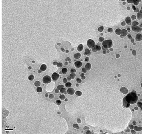
Fig. 4. TEM images of AgNPs synthesized with O.sanctum leaf extract
TEM and SEM were also used to investigate nanoparticle morphology. In the TEM analysis, a heterogeneous population constituting of nanostructures morphologically different in size and shape can be observed. The histogram of particle size reveals dimensions ranging from 5nm to 30nm with a maximum frequency of 15nm.
Fig. 5. Frequency distribution histogram for particle size of AgNPs
[image:3.595.308.561.465.675.2]SEM analysis was performed to characterize the morphology changes of ESM. Figure 6a, b shows the typical SEM images of the natural ESM at different magnifications. The biomembrane exhibited a structurally macroporous network constructed of interwoven and coalescing protein fibers with a diameter ranging from 0.5 to 2.0μm.
ESM was seen with good permeability that allows reactants to contact the inner fibers sufficiently. . The high magnification image of ESM+ O. sanctum presented in Figure 6d and 6e reveals that the surface of the fibers is slightly rougher than that of the natural ESM, indicating the successful grafting of plant extracts and thus some of the biomolecules onto the fiber surface. The inherent interconnected fibrous structure of ESM is still well preserved, and the synthesized AgNPs can also be well observed in Figure 6e and 6f as microdots evenly distributed on the ESM surface.
[image:4.595.311.560.52.167.2]Magnification of image a, c, e = 20μm, Magnification of image b, d, f = 3μm
Fig. 6. SEM images of natural ESM (a, b), O.Sanctum+ESM (c ,d), and AgNPs + O.sanctum + ESM (e, f) at different magnifications
The SEM result which picturised the presence of AgNPs on the surface of ESM’s .This further opened gates to assess the antibacterial activity of AgNPs on impregnated on ESM against bacterial cultures.
Figure 7 shows the well diffusion image of the AgNP solution against a E.coli plate. The assay pictured here give the zones of inhibition and the test is called the Kirby-Bauer Test. (Hirotaka Koga and Takuya Kitaoka, 2010)
Fig. 7. Plates showing diameters of zones of inhibition against E.
coli. (a) AgNPs +O.sanctum + ESM = 2.1 cm ; (b). Tulsi extract+
water control =1cm
It is used clinically to measure the antibiotic resistance. In the present study the zones of inhibition measured for AgNP solution was 2cm and for O. sanctum extract was 1cm respectively. Both the samples are known to possess antibacterial activity, but that seen from AgNP synthesized from the O. sanctum extract is significantly greater than that of
O. sanctum solution. Thus biosynthesized AgNPs are twice
[image:4.595.39.291.260.601.2]more effective than the crude plant extract.
Fig. 8. The zone of inhibition were seen as: (A)Test sample: ESM dipped in ESM + O.sanctum +AgNO3 solution= 2.3cm (B)Control
sample: ESM dipped in ESM + O.sanctum +water = none
The antibacterial properties of the AgNP impregnated ESM’s, from the diameters of inhibition obtained after they were incubated on a E.coli plate for 24 hours at 37⁰C.The ESM dipped in O. sanctum, to surprise gave no zone of inhibition while the ESM’s present in AgNP solution gave a zone of inhibition measuring 2.3cm. This result indicates that the AgNP’s impregnated on the ESM’s are infact responsible for the antibacterial activity and not merely the ESM or O.sanctum extracts adhered to the surface of ESM.
[image:4.595.307.562.326.451.2](Sondi and Salopek-Sondi, 2004) The formation of free radicals by the silver nanoparticles may be considered to be another mechanism by which the cells die (Danilcauk et al., 2006; Kim et al., 2007). Antimicrobial effect of AgNPs is efficiently seen against gram negative E.coli (Lee et al., 2007; Shah et al., 2008; Wang et al., 2008; Zheng et al., 2010) and gram positive B. subtilis. Hence, it is beneficial to use ESM’s with AgNPs in burn treatments, as they are majorly effective against E.coli which is generally found on burn wounds (Abdelrahm et al., 2014). Besides as reported by Debojit et.al the biosynthesized AgNP’s can be also effective against drug resistance bacterial infection.
Advantages of this method of nanoparticle synthesis over other methods include cost effectiveness, easy scale up for large scale synthesis and eco-friendly method for biomedical applications. The major benefit is that the green synthesis using
O.sanctum always takes place extracellularly and the reaction
time is very short compared to that of microbial synthesis. Conclusion
In conclusion, we demonstrated that O.sanctum leaf extract and ESM had reductive properties to synthesize AgNPs. Such ESM’s bear many potential applications where treatment of bacterial infection is a critical task for antibiotics too. Our future scope includes a design of ESM bandage carrying silver nanoparticles that can act against bacterial burn infections. The AgNPs could be tuned through controlling the concentration of AgNO3 and the O. sanctum extract. This method is low-cost,
simple and straightforward for large scale production of AgNPs. More importantly, these prepared AgNPs are known for higher activity and stability in antibacterial activity. The in
vivo tests for wound healing properties of such membranes are
in progress and present a novel way for preliminary treatment of open wounds.
Acknowledgement
Sincere thanks to Dr. Jerald Mahesh Kumar, Ms Jayashree Phukon, Mr. N Sairam for their help, the instrumentation facilitiy, technical staff-Mr. A Harikrishna from “Centre for Cellular and Molecular Biology (CCMB)”,Hyderabad, Telangana, India for his valuable guidance. Special thanks to the faculty of Department of Biotechnology,for letting us pursue our interest.
REFERENCES
Abdelrahman M. Abdelgawad, Samuel M. Hudson, Orlando J. Rojas 2014. Antimicrobial wound dressing nanofibermats from multicomponent (chitosan/ silverNPs/ polyvinylalcohol) systems, Carbohydrate polymers100, 166–178.
Amit Kumar Mittal, Yusuf Chisti, Uttam Chand Banerjee. 2013. Synthesis of metallic nanoparticles using plant extracts. Biotechnology Advances, 31:346–356.
Ankamwar, B., Damle, C., Ahmad, A. and Sastry, M. 2005. “Biosynthesis of gold and silver nanoparticles using Emblicaofficinalis fruit extract, their phase transfer and transmetallation in an organic solution,” Journal of
Nanoscience and Nanotechnology, Vol. 5, no. 10, pp.
1665–1671.
Arias, J. L., Fernandez, M. S., Dennis, J. E. and Caplan, A. I. 1991. Collagens of the chicken eggshell membranes.,
Connect Tissue Res., , 26, 37–45.
Bulut, E. and Ozacar, M. 2009. Rapid facile synthesis of silver nanostructure using hydrolyzable ,Tannin. Ind. Eng. Chem.
Res., 48, 5686−5690.
Charusheela Ramteke, Tapan Chakrabarti, Bijaya Ketan Sarangi and Ram-Avtar Pandey, Research Article 2013. Synthesis of Silver Nanoparticles from the Aqueous Extract of Leaves of Ocimum sanctum for Enhanced Antibacterial Activity., Journal of Chemistry,
Volume 2013, Article ID 278925.
Dadosh, T. et al., 2009. Synthesis of Uniform Silver Nanoparticles with a Controllable Size. Mater. Lett., 63, 2236−2238.
Danilcauk, M., Lund, A., Saldo, J., Yamada, H. and Michalik, J. 2006. Conduction electron spin resonance of small silver particles. Spectrochimaca. Acta., Part A.63,189–191 Debajit Borah, Priyadarshini Deka, Piyalee Bhattacharjee ,
Anupam Changmai, R.N.S. Yadav. 2013. “Ocimum sanctum mediated silver nano particles showed better antimicrobial activities compared to citrate stabilized silver nano particles against multidrug resistant bacteria.” Journal
of Pharmacy Research, 7: 478-482
Dong Yang, Limin Qi and Jiming Ma, 2003.Hierarchically ordered networks comprising crystalline ZrO2 tubes through sol–gel mineralization of eggshell membranes, J.
Mater. Chem, 13, 1119–1123
ESM History
,
ESM Technologies. Retrieved 13 June 2014. Govindaraju, K. et al., 2010. Biogenic silver nanoparticles bySolanum torvum and their promising antimicrobial activity,
Journal of Biopesticides, Vol.3, no.1, pp.394-399.
Guo, J., Wu, H., Liao, X. and Shi, B. 2011. Facile synthesis of size-controlled silver nanoparticles using plant tannin grafted collagen fiber as reductant and stabilizer for microwave absorption application in the Whole Ku Band. J.
Phys. Chem., C, 115, 23688−23694.
Hincke, M. T. Gautron, J. Panheleux, M. Garcia-Ruiz, J. McKee, M. D. and Nys, Y. 2000. Identification and localization of lyzosome as a component of eggshell membranes and eggshell matrix, Matrix Biol., 19, 443. Hirotaka Koga and Takuya Kitaoka 2010. On-Paper Synthesis
of Silver Nanoparticles for AntibacterialApplications, Silver Nanoparticles, David Pozo Perez (Ed.), ISBN: 978-953-307-0285
Huang, J. Li, Q. Sun, D. et al., 2007. “Biosynthesis of silver and gold nanoparticles by novel sundried Cinnamomumcamphora leaf,” Nanotechnology, Vol. 18, no. 10, Article ID 105104
Huang, X., Wu, H., Liao, X. and Shi, B. 2010. One-step, size-controlled synthesis of gold nanoparticles at room temperature using plant tannin, Green Chem., 12, 395−399. Hutchison, J.E. et al., 2008. Greener nanoscience: a proactive
approach to advancing applications and reducing implications of nanotechnology. ACS Nano., 2:395–402. Jain, D., Kumar Daima, H., Kachhwaha, S. and Kothari, S. L.
microbial activities,” Digest Journal of Nanomaterials and
Biostructures, Vol. 4, no. 3, pp. 557–563.
Jin Zhang, Guangwen Duan, Fu Yunzhi and Yucang Zhang 2014. Preparation of the egg membrane bandage contained the antibacterial AgNPs. Materials Characterization, doi: 10.1016/j.matchar.2014.07.003
Kim, J.S., Kuk, E., Yu, K., Kim, J.H., Park, S.J., Lee, H.J., Kim, S.H., Park, Y.K., Park, Y.H., Hwang, C.Y., Kim, Y.K., Lee, Y.S., Jeong, D.H. and Cho, M.H. 2007. Antimicrobial effects of silver nanoparticles. Nanomedicine, 3,95–101 .
Lee, H.Y., Park, H.K., Lee, Y.M., Kim, K. and Park, S.B. 2007. A practical procedure for producing silver nanocoated fabric and its antibacterial evaluation for biomedical applications. Chemical Communications, 28, 2959-2961, ISSN 1359-7345.
Li, S. Shen, Y. Xie, A. et al., 2007. “Green synthesis of silver nanoparticles using Capsicum annuum L. extract,” Green
Chemistry, Vol. 9, no. 8, pp. 852–858.
Mandal, D. et al., 2006. The use ofmicroorganisms for the formation of metal nanoparticles and their application,
Applied Microbiology and Biotechnology, Vol.69, no.5,
pp.485-492.
Miao Liang, Rongxin Su, Renliang Huang, Wei Qi, Yanjun Yu, Libing Wang, and Zhimin He, 2014. Facile in Situ Synthesis of Silver Nanoparticles on Procyanidin-Grafted Eggshell Membrane and Their Catalytic Properties. ACS
Appl. Mater. Interfaces, 6:4638−4649
Moulton, M. C., Braydich-Stolle, L. K., Nadagouda, M. N., Kunzelman, S., Hussain, S. M. and Varma, R. S. 2010. Characterization and Biocompatibility of “Green” Synthesized Silver Nanoparticles using Tea Polyphenols.
Nanoscale., 2, 763−770
Moulton, M. C., Braydich-Stolle, L. K., Nadagouda, M. N., Kunzelman, S., Hussain, S. M. and Varma, R. S. 2010. Synthesis, Characterization and Biocompatibility of Green Synthesized Silver Nanoparticles using Tea Polyphenols.
Nanoscale, 2, 763−770.
Peng, S., McMahon, J. M., Schatz, G. C., Gray, S. K. and Sun, Y. 2010. Reversing the Size-Dependence of Surface Plasmon Resonances. Proc. Natl. Acad. Sci., U.S.A., 107, 14530−14534.
Shah, M.S.A.S., Nag, M., Kalagara, T., Singh, S. and Manorama, S.V. 2008. Silver on PEG-PUTiO2 polymer nanocomposite films: An excellent system for antibacterial applications. Chemistry of Materials, 20, 7, 2455-2460, ISSN 0897-4756.
Sondi, I. and Salopek-Sondi, B. 2004. Silver nanoparticles as antimicrobial agent: a case study on E. coli as a model for Gram-negative bacteria. J. Colloid Interface Sci., 275, 177– 182.
Song, J.Y. and Kim, B. S. 2008. “Biological synthesis of bimetallic Au/Ag nanoparticles using Persimmon (Diopyros kaki) leaf extract,” Korean Journal of Chemical
Engineering, Vol. 25, no. 4, pp. 808–811.
Sukumaran Prabhu and Eldho K Poulose, 2012. “Silver nanoparticles: mechanism of antimicrobial action, synthesis, medical applications, and toxicity effects” International Nano Letters.
Wang, Y., Du, G., Liu, H., Liu, D., Qin, S., Wang, N., Hu, C., Tao, X., Jiao, J., Wang, J. and Wang, Z.L. 2008. Nanostructured sheets of Ti-O nanobelts for gas sensing and antibacterial applications. Advanced Functional
Materials, 18, 7, 1131-1137, ISSN 1616-301.
Wong, M. Hendrix, M. J. C. von der Mark, K. Little, C. and Stern, R. 1984. Collagen and Eggshell Membrane of Hen
Dev. Biol., 104, 28–36.
Zheng, B., Qian, L., Yuan, H. and Xiao, D. 2010. Preparation of gold nanoparticles on eggshell membrane and their biosensing application, Talanta: 82(1):177-83