LASERS
*Mensudar, R., Anuradha, B., Mitthra, S. Vellore Ravi Anupriya, Chandra Mohan Anusha, and
Department of Conservative Dentistry & Endodontics, Sree Balaji Dental College & Hospital, Bharath University,
Narayanapuram, Pallikaranai, Chennai
ARTICLE INFO ABSTRACT
Dentistry has incorporated a lot of newer technological advancement to benefit both dentist and patients. In this modern technological
potentially profitable arena. Currently, various types of laser are available based on their applications. This article explains the basic principle, mechanism and interactions of laser and a
dentistry.
Copyright©2017, Mensudar et al. This is an open access distribution, and reproduction in any medium, provided
INTRODUCTION
The word LASER stands for “Light Amplification By
Stimulated Emission Of Radiation”. The application of lasers is almost in every fields of human endeavour
science and technology to business and entertainment over the
past few years.In 1960, Theodore Maiman, developed the first
working laser or maser device. Maser is like laser which denotes “Microwave amplification of stimulated emission of radiation”. From the ruby crystal a deep red colour is emitted. Over the next few years, researchers studied various possible applications of this visible laser energy. (Robert A. Convissar 2004) Later studies from 1970s to 1980s found newer other laser devices, such as CO2 and neodymium YAG (Nd:YAG), which had better tissue interaction with dental hard tissues.
(Robert A. Convissar, 2004) The purpose of the article is to
highlight the various laser applications developed in dental practice and its clinical application.
Classification of laser: (Donald J.Coluzzi, 2004;
George, 2009)
I: According to ANSI and OHSA:
CLASS I: These are low powered lasers that are safe to use, e.g. Laser beam pointer.
*Corresponding author: Mensudar, R.
Department of Conservative Dentistry & Endodontics, Sree Balaji Dental College & Hospital, Bharath University, Narayanapuram, Pallikaranai, Chennai – 600100, Tamil Nadu, India.
ISSN: 0975-833X
Article History:
Received 09th July, 2017 Received in revised form 22nd August, 2017
Accepted 17th September, 2017 Published online 31st October, 2017
Citation: Mensudar, R., Anuradha, B., Mitthra, S.
59805-59809.
Key words:
Laser, Laser physics, Tissue interaction, Endodontic applications.
RESEARCH ARTICLE
LASERS – AN ASSET TO ENDODONTICS!!!
*Mensudar, R., Anuradha, B., Mitthra, S. Vellore Ravi Anupriya, Chandra Mohan Anusha, and
Panneer Selvam Victoria
Dentistry & Endodontics, Sree Balaji Dental College & Hospital, Bharath University,
Narayanapuram, Pallikaranai, Chennai – 600100, Tamil Nadu, India
ABSTRACT
Dentistry has incorporated a lot of newer technological advancement to benefit both dentist and patients. In this modern technological era, lasers offer dentist a door to its rewarding, high tech and potentially profitable arena. Currently, various types of laser are available based on their applications. This article explains the basic principle, mechanism and interactions of laser and a
dentistry.
access article distributed under the Creative Commons Attribution License, the original work is properly cited.
word LASER stands for “Light Amplification By Stimulated Emission Of Radiation”. The application of lasers is almost in every fields of human endeavour from medicine, science and technology to business and entertainment over the In 1960, Theodore Maiman, developed the first working laser or maser device. Maser is like laser which denotes “Microwave amplification of stimulated emission of ruby crystal a deep red colour is emitted. Over the next few years, researchers studied various possible
Robert A. Convissar,
Later studies from 1970s to 1980s found newer other and neodymium YAG (Nd:YAG), which had better tissue interaction with dental hard tissues. The purpose of the article is to highlight the various laser applications developed in dental
Donald J.Coluzzi, 2004; Roy
CLASS I: These are low powered lasers that are safe to use,
Conservative Dentistry & Endodontics, Sree Balaji Dental Narayanapuram, Pallikaranai, Chennai
CLASS II A: These are low powered lasers that are hazardous only when viewed directly for l
He-Ne lasers.
CLASS II B: Low powered visible lasers that are hazardous when viewed for more than 0.25 seconds.
CLASS III A: Medium powered lasers that are normally
hazardous if viewed for less than 0.25 seconds without magnifying optics.
CLASS III B: Medium powered lasers that are hazardous if viewed directly.
CLASS IV: These are high powered lasers (> 0.5 W) that produce ocular skin and fire hazards.
II: Based on the wavelength of the beam:
Ultraviolet rays:140-400 nm
Visible light:400-700 nm
Infrared:700 to microwave spectrum.
III) Based on the type of laser medium used:
1. Gas laser.
Helium Cadmium.
International Journal of Current Research
Vol. 9, Issue, 10, pp.59805-59809, October, 2017
Mensudar, R., Anuradha, B., Mitthra, S. et al. 2017. “Lasers – An asset to Endodontics!!!”, International Available online at http://www.journalcra.com
*Mensudar, R., Anuradha, B., Mitthra, S. Vellore Ravi Anupriya, Chandra Mohan Anusha, and
Dentistry & Endodontics, Sree Balaji Dental College & Hospital, Bharath University,
600100, Tamil Nadu, India
Dentistry has incorporated a lot of newer technological advancement to benefit both dentist and era, lasers offer dentist a door to its rewarding, high tech and potentially profitable arena. Currently, various types of laser are available based on their applications. This article explains the basic principle, mechanism and interactions of laser and also its application in
License, which permits unrestricted use,
CLASS II A: These are low powered lasers that are hazardous only when viewed directly for longer than 1000 seconds, e.g.
Low powered visible lasers that are hazardous when viewed for more than 0.25 seconds.
Medium powered lasers that are normally hazardous if viewed for less than 0.25 seconds without
Medium powered lasers that are hazardous if
These are high powered lasers (> 0.5 W) that produce ocular skin and fire hazards.
II: Based on the wavelength of the beam:
400 nm 700 nm
700 to microwave spectrum.
III) Based on the type of laser medium used:
INTERNATIONAL JOURNAL OF CURRENT RESEARCH
Helium neon.
Krypton
Carbon monoxide.
Argon.
Nitrogen.
Carbon dioxide.
2. Solid laser.
Ruby
Rhodamine
Erbium.
Neodynium.
3. Liquid laser.
Liquid dye
Water vapour
4. Electronic laser
Semiconductor
Diode
VI) Based on the type of delivery system:
Flexible hollow wave guide/articulating arms.
Glass fibre optic cable
V) Based on laser modes:
Continuous mode
Gated mode
Pulsed mode
VI) Based on type of interaction with tissue:
Contact laser
Noncontact laser
VII) Based on type of application:
a) Soft tissue lasers-low power about 1 w
Helium Neon
Gallium neon
Gallium arsenide
Gallium aluminium arsenide
b) Hard tissue laser-high power about 3 w or more
Argon laser
CO2 laser
Nd; YAG laser
Er; YAG laser
Er; YSG laser
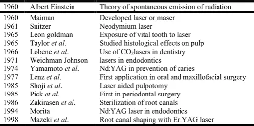
[image:2.595.34.292.674.802.2]History
Table 1. Evolution of laser in dental filed are tabulated below
1960 Albert Einstein Theory of spontaneous emission of radiation
1960 Maiman Developed laser or maser
1961 Snitzer Neodymium laser
1965 Leon goldman Exposure of vital tooth to laser
1965 Taylor et al. Studied histological effects on pulp
1966 Lobene et al. Use of CO2lasers in dentistry
1971 Weichman Johnson lasers in endodontics
1974 Yamamoto et al. Nd:YAG in prevention of caries
1977 Lenz et al. First application in oral and maxillofacial surgery
1985 Shoji et al. Laser aided pulpotomy
1985 Pick et al. First in periodontal surgery
1986 Zakirasen et al. Sterilization of root canals
1994 Morita Nd:YAG laser in endodontics
1998 Mazeki et al. Root canal shaping with Er:YAG laser
Laser physics
The three common principles of laser arm monochromatic,
coherence and collimation. (Roy George, 2009; Theodore Maiman, 1960)
Monochromatic: The light produced by the laser will be of a
characteristic wavelength. If the light is produced in the visible spectrum (400-750nm), it will be seen as a beam of intense colour. This property enables the laser to attain high spectral power density.
Coherence: The light which leaves as laser is all perfect in one
phase unlike the normal light source, their individual contributions are summated and reinforce each other. A large amount of energy is lost out of phase waves, that cancel each other in an ordinary light source.
Collimation: The laser light beam is collimated to achieve
parallelism on leaving the laser aperture, the laser beam collimation, is an important property for good transmission through the delivery systems.
Laser emission modes and delivery systems
Dental laser can be used both in contact mode or non-contact mode. When the laser tip touches directly the target tissue it is known as contact mode. Whereas in non-contact mode, the laser tip is kept at a distance a few millimetres to centimetres away from the target tissue. (Husein, 2006) Laser emits light energy in three different modes.
1. Continuous mode: The beam is emitted continuously
at one power level for the length of the time operator presses the foot switch.
2. Gated – pulse mode: Similar to a strobe light, the laser
energy is switched on and off by opening and closing the mechanical shutter in front of the beam path of a continuous wave every few milliseconds.
3. Pulsed mode: Large energies are emitted for a short
time, of a few milliseconds usually, followed by a long time when the laser is off.
Lasers utilize different delivery systems, depending on wavelength and the access required at the terminal target tissue. (Husein, 2006; Convissor and Coluzzi, 2004) These include;
A. Articulated arms: these have joints made of tubes that
allow the arm to bend at the joints where a mirror reflects a beam into centre of the next tube without touching the inner surface of the tube.
B. Hollow waveguide: It is a flexible hollow tube that has
an interior mirror that reflects the laser energy along this tube and exits through handpiece. These are much thinner than the articulated arms.
C. Glass fiberoptic cable: It is even more flexible than a
wave guide, smaller in diameter with sizes ranging from 200 to 600 microns. Quartz optical fibre is encased in a resilient sheath.
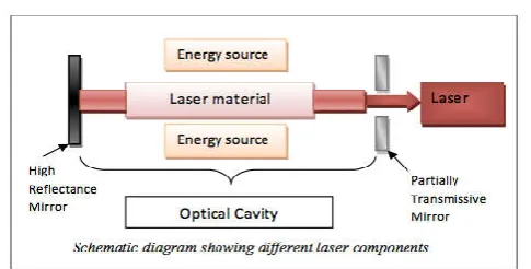
How laser works
Figure 1. Schematic diagram showing laser components
1.Optical cavity:
Parallel mirrors
Lasing medium
2. Pump energy source 3. Cooling system
1. Optical Cavity: Itis the core and centre device. It consists
of molecules, chemical or compounds elements and is known as lasing / active medium. Based on the nature of active medium, it may be gas, crystal or solid semiconductor, the laser is named. Argon and CO2 are the two active gaseous medium lasers used in dentistry. Others are solid state semiconductor wafers which are made with multiple layers of metals such as indium, gallium, aluminium, and arsenic or solid rods of garnet crystal with various combinations of yttrium, aluminium, scandium and gallium and then doped with the elements of chromium, neodymium, or erbium. (Walsh, 2003) Another component of optical cavity is two parallel mirrors that are placed on either side of the lasing medium. These parallel mirrors, excite the photons to bounce off and then re-enters the active medium to activate the release of more photons. Thus, the mirrors collimate the light that is photons exactly perpendicular and make them re-enter the active medium, while those off axis leave the lasing process. (Walsh, 2003) Usually, one mirror is totally reflective while the other mirror is partially transmissive and the light that escapes first through the mirror becomes the laser beam.
2: Pump energy source: It is provided by flash lamp or
electrical coil which pumps the high energy radiation into the active medium.
3. Cooling System: Most of the remaining energy is converted
into heat hence it is necessary to provide some form of cooling, which is provided by water. Thus, finally photons generate a collimated, coherent, and monochromatic laser beam of light.
Laser tissue interaction and its effect
When the laser beam is aimed at tissue the energy reaches the biological interface and any one of four interactions will take place; reflection, transmission, scattering, or absorption. (Walsh, 2003)
Absorption – Specific molecules in tissue known as
chromophores absorb the photons. The light energy is then converted into other forms of energy to perform the task.
Reflection – Sometimes the laser beam bounces off the surface
with no penetration or interaction. Reflection is usually an
undesired effect, but a useful example of reflection is found when Erbium lasers reflect off titanium allowing for safe trimming of gingiva around implant abutments.
Transmission – The laser energy pass through superficial
tissues to interact with deeper tissues. The deeper penetration is seen with Nd:a YAG and diode laser is an example of tissue transmission.
Scattering – Once the laser energy enters the target tissue it
will scatter in various directions. This phenomenon is usually not helpful, but can help with certain wavelengths biostimulative properties.
Effect of laser irradiation
Photochemical interaction includes
Biostimulation
Photodynamic therapy
Photothermal interactions includes
Photoablation
Photopyrolysis
Photomechanical interactions includes
Photodisruption / photodissociation
Photoacoustic interactions
Photoelectrical interactions includes
Photoplasmolysis
There are five important types of biological effects that can occur once the laser photons enter the tissue: fluorescence,
photothermal, photodisruptive, photochemical, and
photobiomodulation. (Guttenberg, 2004; Merchant, 2007)
Fluorescence: When active carious lesion is exposed to
655nm visible wavelength of the Diagnodent diagnostic device, the amount of fluorescence correlates the size of lesion which in turn is useful in diagnosing and treating early carious lesions.
Photothermal: When chromophores absorb the laser energy,
heat is generated. This heat helps to perform tissue incision or coagulating blood. Photothermal interactions predominate when most of the soft tissue procedures are performed with dental lasers. Photothermal ablation effect occurs when CO2 lasers are used on teeth. Most of the procedures produce heat and utmost care must be taken to avoid any thermal damage to the tissues.
Photodisruptive effects (or photoacoustic): Hard tissues are
tissue, in which their laser energy is predominantly absorbed. Hence bone and tooth are not vaporized but pulverized through the photomechanical ablation process. This shock waves result in the distinct popping sound that is heard at the time of erbium laser use. Thermal damage is not produced, as no residual heat is created when used properly, particularly when the concept of thermal relaxation is considered.
Photochemical: It occurs when photon energy causes a
chemical reaction. These reactions are involved in some of the beneficial biostimulation effects.
Photobiomodulation or Biostimulation It known as Low
Level Laser Therapy (LLLT) and its the ability of lasers to enhance healing, increase circulation, reduce edema, and minimize pain. Various other studies have also demonstrated effects such as increased collagen synthesis, fibroblast proliferation, increased osteogenesis, enhanced leukocyte phagocytosis. The exact mechanism of these effects is not clear but it is stated that it occur mostly through photochemical and photobiological interactions within the cellular matrix and mitochondria. Biostimulation is mainly used to reduce postoperative discomfort and also to treat recurrent herpes or aphthous stomatitis. Is another term used to describe this phenomenon. When laser heats oral tissues certain reversible or irreversible changes occur. Irreversible effects such as denaturation or carbonization result in thermal damage that cause inflammation, pain, and edema. (Frentzen and Koort, 1990)
The thermal effects of laser irradiation are:
Temperature < 60º C
Tissue hyperthermia
Enzymatic changes
Edema
Temperature > 60º C
Protein denaturation
Temperature > 100º C
Tissue ablation
Super heating
Clinical application of lasers in Endodontics
I: Desentization of hypersensitive dentin
After drying the dentin, the laser tip is placed in direct contact with the tooth surface, which is then irradiated for 30 seconds to one minute. HeNe lasers, pulsed Nd:YAG lasers and CO2 lasers are used. (Gerschman et al., 1994; Luciana Chucre and Sebastião Luiz, 2004) Sodium fluoride paste or petroleum jelly is coated on tooth surface and then it is exposed to CO2 lasers to prevent occurrence of pain and carbonization of tooth surface by laser.
II: Application of laser in pulp therapy
Deep cavities, hypersensitive cavities and cavity that require sedative treatment are some of the indications for this treatment. It helps to produce mediated effect,
pulpal analgesia, seals the dentinal tubules and reduces permeability. On using pulsed Nd:YAG laser, it is necessary to combine the application of black ink to tooth surface and air spray cooling to prevent dental pulp damage resulting from laser energy. (Luciana
Chucre and Sebastião Luiz, 2004) When using
CO2lasers, dental tissue must not be exposed to high energy for long periods of time.
In case of direct pulp capping, high success rate is due to hemostasis, disinfection, sterilization, carbonization and stimulatory effect of dental pulp cells. It causes scar formation in the irradiated area due to thermal effects, which preserve the pulp from bacterial invasion. (Luciana Chucre and Sebastião Luiz, 2004; Gerschman
et al., 1994) In addition laser minimizes the formation
of hematoma between pulp tissue and the calcium hydroxide dressing allowing an intimate contact between the exposed pulp and the dressing.
Vital pulp amputation, it is one of the highly anticipated laser treatments in because this treatment offer amputation of the pulp tissue at satisfactory level. A CO2 laser is used for pulpal hemostasis, after which pulp amputation is done with excavator or a bur. (Goldman et al., 1964; Klein et al., 2005)
Root canal cleaning and shaping helpsin elimination of
microorganisms. Thin optical laser fibre [Nd:YAG, Er,Cr:YSGG, argon, or diode) delivers laser beam to the root canal system. (Mensudar, 2014; Gerry Ross and Alana Ross, 2004) Following biomechanical instrumentation, additional cleansing is performed with the help of these lasers.
Root Canal Irrigation in Combination with laser in case
of slightly curved canals as well as wide root canals. The pulsed Nd:YAG laser, Er:YAG laser, or Er,Cr:YSGG laser are used. Solution such as 5.25% sodium hypochlorite or 14 % ethylenediaminetetra-acetic acid (EDTA) must be used along with lasers for effective irrigation.
It helps to sterilize and disinfect the infected root canals by effectively killing microorganisms. Pulsed Nd:YAG, argon, diode, CO2, Er:YAG are used.
It can be used for root Canal Obturation by melting the
Gutta percha with Laser heat energy followed by
vertical condensation method. Nd:YAG laser can be used for obturation of root canals. It is very time consuming thus it is not practical to use.
Other applications
Use of hard tissue laser: (Sumez et al., 2002; Stern and Sognnaes, 1964; Gimbel, 2000)
Class I, II, III, IV and V cavity preparation
Caries removal
Hard tissue surface roughening and etching
Enameloplasty, excavation of pits and fissures for placement of sealants
Access to a root canal.
Root canal preparation including enlargement
Root canal debridement and cleaning
Pulpotomy as an adjunct to root canal therapy.
Apicoectomy – amputation of the root end.
Uses of Soft tissue laser: (Pick and Colvard, 1993; Walsh, 2003; Guttenberg, 2004; Merchant, 2007)
Incision, excision, vaporization, ablation and
coagulation of oral soft tissues
Exposure of unerupted teeth (Operculectomy)
Fibroma removal
Incision of soft tissue to raise a flap
Frenectomy, Vestibuloplasty, and frenotomy
Gingival troughing for crown impressions,
gingivectomy or gingivoplasty and gingival incision and excision.
Hemostasis.
Implant recovery.
Incision and drainage of abscesses.
Treating conditions like leukoplakia, oral
papillectomies, canker sores, herpetic and aphthous ulcers.
Sulcular debridement - removal of diseased or inflamed
soft tissue from the periodontal pocket
Removal of granulation tissue from bony defects and curettage of the extraction sockets and periapical area during apical surgery.
Laser safety
An adequate safety policy for managing and controlling the risks arising from the use of laser equipment has to be established. The following are the safety measures to be followed during laser use in dental practice. (Parker, 2007)
Fire and electrical control measures
Environment protection / control airborne
contamination
Laser protection advisor/laser safety officer
Laser safety features / procedural safety measures
Eye protection / personal protective equipment
Test firing
Local rules
Training.
Conclusion
Use of Laser in dentistry has proven to be beneficial in treating numerous dental conditions and also serves as a therapeutic tool in tissue management. The dynamics of laser energy beams pose general risks to non-oral tissues and the immediate surrounding environment and hence necessary safety measures have to be devised to safeguard staff and patients who are involved in dental treatment using lasers. The high quality state of art has been employed in various procedures such as in diagnosis, early caries detection, pulpal blood flow using laser Doppler flowmetry, root canal disinfection and many more applications. It is not only important to realize the various potential uses, it is also necessory to select proper wavelength, to know the laser tissue interactions, its applications, limitations and safety issues.
REFERENCES
Convissor. R.A. and Coluzzi.D J. 2004. The biological rationale for the use of lasers in dentistry. Dental clinics of North America, 48(4):771-794.
Donald J.Coluzzi. 2004. Fundamentals of dental lasers: science
and instruments. Dental Clinics of North America,
148:751-770.
Frentzen.M. and Koort.H.J. 1990. Lasers in dentistry: new possibilities with advancing Technology. IDJ, 40:323-332. Gerry Ross and Alana Ross. 2004. Photobiomodulation: An
Invaluable Tool for all Dental Specialties. Int Endod J., 3:1-15.
Gerschman J A, Ruben J, Gebart-Eaglemont J. 1994. Low level laser therapy for dentinal tooth hypersensitivity. Aust Dent J., 39:353-7.
Gimbel CB. 2000. Hard tissue laser procedures. Dental Clinics of North America, 44(4):931-953.
Goldman L, Hornby P, Meyer R and Goldman B. 1964. Impact of the laser on dental caries. Nature, 203:417.
Guttenberg. S.A. 2004. Laser Physics and tissue interaction;
Oral & Maxillofacial clinics of North America. 16:143-147.
Husein, 2006. Applications of Lasers in Dentistry: A Review.
Arch of Oro Sci., 1:1-4.
Klein AL, Rodriguez LK, Eduardo CP, Nobre dos Santos M, Cury JA. 2005. Caries inhibition around composite restorations by pulsed carbon dioxide laser application. Eur J Oral Sci., 113:239-44.
Luciana Chucre and Sebastião Luiz. 2004. Clinical evaluation of dentin hypersensitivity treatment with low intensity
Gallium- Aluminium-Arsenide laser– AsGaAl. J Appl Oral
Sci., 12(4):267-72.
Mensudar R. 2014. Photodynamic therapy. World Journal of Medical Science, 10(2):139-142.
Merchant.N. 2007. Laser in conservative dentistry and endodontics. IDRR, 25-26.
Parker S. 2007. Laser regulation and safety in general dental practice. Br Dent J., 202:527-531.
Pick RM. and Colvard MD. 1993. Current status of lasers in soft tissue dental surgery. J Periodontal., 64(7):589-602. Robert A. Convissar. 2004. The biologic rationale for the use
of lasers in dentistry. Dental Clinics of North America, 148:771-794.
Roy George. 2009. Laser in dentistry-Review. Int J of Dent Clin., 1(1):13-19.
Stern RH. and Sognnaes RF. 1964. Laser beam effect on dental hard tissues. J Dent Res., 43:873.
Sumez S, Orhan M, Sumez A. 2002. Laser etching of enamel for direct bonding with an Er, Cr:YSGG hydrokinetic laser system. Am J Orthod Dentofacial Orthop., 122:649-56. Theodore Maiman, 1960. Stimulated optical radiation in ruby.
Nature, 187:493-494.
Walsh, L.J. 2003. The current status of laser application in dentistry. Australian Dental Journal, 143-154.