IMPACT OF THE NON-STRUCTURAL 2 PROTEIN ON RESPIRATORY SYNCYTIAL VIRUS PATHOGENESIS
Rachael Marie Liesman
A dissertation submitted to the faculty of the University of North Carolina at Chapel Hill in partial fulfillment of the requirements for the degree of Doctor of Philosophy in the
Department of Microbiology and Immunology.
Chapel Hill 2013
Approved by:
Raymond Pickles
Nancy Raab-Traub
William Goldman
Mark Heise
ii © 2013
iii ABSTRACT
Rachael Marie Liesman: Impact of the Non-structural 2 Protein on Respiratory Syncytial Virus Pathogenesis
(Under the direction of Raymond Pickles)
Respiratory syncytial virus (RSV) infection is the major cause of bronchiolitis in
young children. The factors contributing to increased propensity of RSV-induced distal
airway disease compared to other commonly encountered respiratory viruses are unknown.
Using a model of the well-differentiated airway epithelium, we characterized the
consequences of RSV infection of ciliated epithelial cells, the primary cellular targets of RSV
infection in vivo. These studies show that RSV infection results in cell rounding and
degradation of the cilia apparatus, followed by active extrusion of infected cells from the
epithelium, eventually resulting in a decline in the ability of the airway epithelium to perform
mucociliary transport. Using recombinant respiratory viruses, we attribute these
consequences to the RSV non-structural 2 (NS2) protein. Using parainfluenza virus 3 (PIV3)
to deliver and express RSV NS2 in the ciliated epithelium of hamster airways, we assessed
the impact of NS2 on respiratory viral pathogenesis. These studies identified the RSV NS2
protein as a unique viral genetic determinant for RSV-induced pathogenesis, resulting in two
distinct effects in vivo. First, NS2 promoted epithelial cell extrusion and accelerated
clearance of whole lung virus titers, presumably by clearing virus-infected cells from the
airway mucosa. Second, epithelial cell extrusion promoted by NS2 resulted in accumulation
of detached, pleomorphic epithelial cells in the narrow diameter bronchiolar airway lumen,
iv
early and robust neutrophilic influx, which may contribute to distal airway obstruction and
constriction. These studies reveal a novel consequence of RSV infection of the airway
epithelium, where NS2-promoted epithelial cell shedding and morphologic changes
accelerate viral clearance but also cause acute distal airway obstruction. NS2-promoted
epithelial cell shedding in the distal airways and the resulting obstruction of these airways
represent a mechanism that may explain why RSV is the dominant virus causing
bronchiolitis in young children. We identify for the first time NS2 as a pathogenesis factor for
v
To my parents, Aaron and Joyce, and my brother, Adam. I will forever be thankful for your
vi
ACKNOWLEDGMENTS
I would like to first thank my advisor, Dr. Ray Pickles, for his mentorship and
guidance throughout graduate school. I would also like to thank the members of my thesis
committee, Drs. Nancy Raab-Traub, Barb Sherry, Mark Heise, and Bill Goldman, for their
encouragement, guidance, and insight. I would also like to acknowledge and thank my
earliest scientific mentor, Dr. Loni Walker at Illinois Wesleyan University, for introducing me
to the world of scientific research and for her guidance, encouragement, and enthusiasm for
teaching.
Many thanks to the members of the Pickles lab, especially Dr. Meg Scull, Dr. Nikki
Worthington, Susan Burkett, Mike Tarpley, and Dr. Liqun Zhang, for their scientific
contributions to this project, as well as for their advice, encouragement, and camaraderie. I
would like to acknowledge and thank the members of the Department of Microbiology and
Immunology and the Cystic Fibrosis Center, including the Directors and teams of the Tissue
Procurement and Cell Culture Core, the Morphology and Morphometry Core, and the
Michael Hooker Microscopy Facility, for their help. I would like to especially thank Dr. Robert
Tarran for his guidance, advice, and friendship, and the members of the Tarran lab, who
became my de facto lab mates, for their scientific support, advice, and camaraderie.
During my time in Chapel Hill, I have made so many wonderful friends and am
thankful for the advice, laughter, and support that each of them has given at one time or
vii
Finally, I would like to thank my family for their support and confidence in me
throughout graduate school. My parents, Aaron and Joyce, are my biggest fans and their
love, guidance, advice, and support is invaluable to me. My brother, Adam, has been a
wonderful source of motivation, laughter, and friendship. I would also like to thank my Aunt
Judy, my cousin Emily, and my entire extended family for their support, love, and
encouragement. My family is an essential source of strength and love and I am incredibly
viii
TABLE OF CONTENTS
LIST OF TABLES ... xi
LIST OF FIGURES ... xii
LIST OF ABBREVIATIONS AND SYMBOLS ...xiii
CHAPTER I. INTRODUCTION ... 1
Epidemiology and Disease Burden ... 1
Clinical Features of RSV Infection ... 2
Treatment Strategies and Vaccine Development ... 4
RSV Virology ... 6
Attachment and Entry: G Glycoprotein ... 6
Immune Evasion Strategies: NS Proteins ... 8
Human Airway Epithelia ... 10
Airway Defense Mechanisms Protective Barrier ... 12
Epithelial Cell Response ... 12
Mechanical Clearance ... 16
Cellular Immune Response ... 18
Dissertation Objectives ... 19
II. THE ROLE OF THE G GLYCOPROTEIN IN EFFICIENT RSV INFECTION OF HUMAN CILIATED AIRWAY CELLS ... 22
Overview ... 22
ix
Results ... 24
Requirement for RSV G in infection of human airway epithelial cultures... 24
Impact of G phenotype on infection of HAE ... 25
Development and characterization of HEp2-V cell line ... 26
Discussion ... 28
Materials and Methods ... 32
III. THE RSV NS2 PROTEIN PROMOTES CYTOPATHIC EFFECT OF COLUMNAR CILIATED AIRWAY EPITHELIAL CELLS FOLLOWING RSV INFECTION ... 42
Overview ... 42
Introduction ... 43
Results ... 45
Kinetics of RSV replication in HAE ... 45
IFN and cytokine message following RSV infection ... 46
A distinctive morphologic change in RSV-infected columnar airway cells ... 47
Shedding of epithelial cells from RSV-infected HAE ... 48
Effects of RSV infection on mucociliary transport ... 50
Cell rounding is unique to RSV among a number of common paramyxoviruses ... 51
Columnar cell rounding is associated with expression of the NS2 gene ... 52
Expression of NS2 by recombinant PIV3 mimics RSV-induced cell morphology ... 53
NS2 expression adversely affects lumenal surface structures and functions of ciliated cells ... 54
IFNβ and cytokine changes in HAE infected by PIV3, PIV3-NS1, and PIV3-NS2 ... 55
Discussion ... 56
x
IV. RSV NS2 PROTEIN PROMOTES EPITHELIAL CELL SHEDDING AND DISTAL
AIRWAY OBSTUCTION ... 83
Overview ... 83
Introduction ... 84
Results ... 87
Effects of PIV3-NS2 on the infection of the hamster nasal cavity and lower airways ... 87
Neutrophil dominated inflammation in PIV3 and PIV3-NS2 infected animals ... 89
Accumulation of shed cells in the distal airway of PIV3-NS2 infected hamsters ... 90
Accumulation of infected cells in the distal airways of RSV infected infants ... 91
Discussion ... 92
Materials and Methods ... 100
V. CONCLUSIONS... 115
xi
LIST OF TABLES Table
xii
LIST OF FIGURES Figure
2.1 RSV requires the G protein for efficient infection of HAE ...36
2.2 Infection of HAE with RSV amplified in Vero cells or HEp2 cells ...37
2.3 Infection of HAE with RSV NS deletion mutants ...38
2.4 Characterization of HEp2-V cells ...39
3.1 RSV-GFP infection, replication, and clearance in HAE cultures ...68
3.2 IFN and cytokine production in HAE following infection with RSV-GFP ...69
3.3 Morphologic and structural changes in RSV-infected ciliated columnar cells ...71
3.4 Ciliated cell shedding and loss of cilia activity during RSV-GFP infection of HAE ...73
3.5 RSV-induced ciliated cell rounding is unique to RSV infection and due to expression of the RSV NS2 protein ...75
3.6 RSV-induced cell rounding is ablated by mutations within the NS2 protein ...77
3.7 Expression of RSV NS2 in HAE ciliated cells using PIV3 results in infected cell rounding ...79
3.8 Expression of RSV NS2 by PIV3 in HAE cultures mimics RSV-induced cytopathology at the apical surface of ciliated cells ...80
3.9 IFN and cytokine changes following infection of HAE with PIV3, PIV3-NS1, or PIV3-NS2 ...82
4.1 PIV3-NS2 causes ciliated cell rounding in the hamster nasal respiratory epithelium in vivo ... 106
4.2 Accelerated clearance of virus-infected cells in hamsters infected by PIV3-NS2 .... 107
4.3 Accelerated clearance of viral load in hamsters infected by PIV3-NS2 ... 109
4.4 Neutrophilic inflammation in distal airways of PIV3-NS2 infected hamsters ... 110
4.5 Shedding of cells infected by PIV3-NS2 but not PIV3 into hamster lower conducting airways ... 112
xiii
LIST OF ABBREVIATIONS AND SYMBOLS
CBF cilia beat frequency
club cell formerly known as Clara cell
CXCL10 C-X-C motif chemokine ligand 10; also known as IP-10
dsDNA double-stranded DNA
GFP green fluorescent protein
HMPV human metapneumovirus
ICAM-1 intercellular adhesion molecule 1; also known as CD54
IFN interferon
IL-1β interleukin 1 beta
IL-6 interleukin 6
IL-8 interleukin 8; also known as CXCL8
IRF3 IFN regulatory factor 3
IVIG intravenous immunoglobulin
JAK Janus kinase
MAVS mitochondrial antiviral signaling
MCT mucociliary transport
MOI multiplicity of infection
NBF neutral buffered formalin
NF-κB nuclear factor kappa B
NS1 RSV non-structural 1 protein
NS2 RSV non-structural 2 protein
PFA paraformaldehyde
xiv pi post-inoculation
PIV3 parainfluenza virus 3
PIV3-NS1 parainfluenza virus 3 expressing respiratory syncytial virus non-structural 1
PIV3-NS1/2 parainfluenza virus 3 expressing respiratory syncytial virus non-structural 1 and non-structural 2
PIV3-NS2 parainfluenza virus 3 expressing respiratory syncytial virus non-structural 1
RANTES regulated upon activation, normal T cell expressed and secreted; also CCL5
RIG-I retinoic acid-inducible gene I
RLR RIG-I-like receptor
RSV respiratory syncytial virus
SeV Sendai virus
STAT signal transducer and activator of transcription
TCID50 50% tissue culture infectious dose
TEER transepithelial electrical resistance
TLR Toll-like receptor
TNFα tumor necrosis factor alpha
ZO-1 zonula occludens 1 protein
CHAPTER I INTRODUCTION
Epidemiology and Disease Burden
Human respiratory syncytial virus is the most important etiological agent of
respiratory disease in children worldwide and by age 2, over 95% of individuals have
experienced at least one RSV infection (1, 2). Seasonal infections of RSV generally begin in
autumn and, in the US, range in duration from 13 – 23 weeks (3). Infection of adults and
children most frequently results in upper respiratory tract illness (URTI) causing common
cold-like symptoms, which may progress to lower repiratory tract illness (LRTI).
It has recently been estimated that 34 million new cases of RSV-associated lower
airway disease occur globally in children less than 5 years of age, resulting in 66,000 –
199,000 deaths per year (4). The majority of these deaths occur in underdeveloped
countries with limited access to standardized healthcare. Each year in the United States, an
estimated 1.5 million outpatient visits are attributable to RSV infections in children <5 years
old, resulting in 75,000-125,000 hospitalizations, of which 1.5% require admission to the
pediatric intensive care unit (5-7). Average length of hospital stay for infants diagnosed with
RSV bronchiolitis is 3.5 days (8-10) and hospitalization due to RSV-associated bronchiolitis
results in total estimated costs of up to 700 million US dollars annually (11). RSV infections
clearly represent a significant economic and health burden both globally and in the United
2 Clinical Features of RSV Infection
RSV infection results in a range of symptoms, varying from upper respiratory tract
illness characterized by rhinorrhea to severe lower respiratory tract illness characterized by
labored breathing necessitating oxygen supplementation. Although RSV infection often
causes only URTI, 1 to 2% of infected individuals progress to severe lower respiratory tract
infection, which can result in hospitalization with bronchiolitis being the most common
clinical diagnosis (5, 12). RSV-associated bronchiolitis is known to be more severe and
results in prolonged hospitalization compared to non-RSV associated bronchiolitis, resulting
in a mortality rate of 0.01 – 0.5% annually (6, 9, 10, 13, 14). Studies of young children found
that RSV infection results in twice as many visits to the emergency department, six times as
many hospitalizations, and nine times as many deaths compared to seasonal influenza
infections (15, 16). The most important risk factor for the development of RSV bronchiolitis is
very young age (< 6 months). Other risk factors include congenital heart defects,
immunodeficiency, neurodevelopmental deficits, familial history of atopy, chronic lung
disease, and bronchopulmonary dysplasia (7, 9, 17, 18). Low body weight at time of
hospitalization is most significantly associated with all measures of severe disease, with
premature infants being the largest at-risk population (19). Despite identification of
numerous factors associated with elevated rates of hospitalization, ~50% of RSV-associated
hospitalizations occur in healthy children born at term with no additional identifiable risk
factors (17).
Natural infection by RSV does not induce lifelong immunity to reinfection and
recurring infections are common despite no significant antigenic change of the virus (1).
RSV infection among the elderly and immunocompromised populations is recognized as a
significant health problem. In community-dwelling US adults over age 65, RSV is the
etiological agent in 3-15% of incidences of acute respiratory illness per year (20-23). Among
3
identified as RSV-induced illness and, in one study, 18% of RSV-infected patients were
admitted to the intensive care unit (24, 25). RSV associated mortality rates are higher in the
elderly population than among young children (16). Additionally, RSV causes increased
morbidity and mortality in immunocompromised patients, particularly in transplant patients
(especially lung and bone marrow transplants) and patients with leukemia. In nosocomial
outbreaks among bone marrow transplant patients, RSV has been detected in 2 – 20% of
patients, resulting in a pneumonia-associated mortality rate of 55 – 80% (26-28). Aggressive
treatment using aerosolized ribavirin and intravenous immunoglobulin (IVIG) is frequently
initiated at the onset of upper respiratory tract illness, although this treatment regime is
costly and cumbersome (29, 30). Recently, clinical efficacy and tolerance of oral ribavirin
has demonstrated, representing a promising, cost-effective therapeutic strategy for RSV
infection in immunocompromisd patients (31, 32).
Although the majority of infants infected by RSV eventually resolve infection and
disease, history of RSV infection can be associated with long-term abnormalities in
pulmonary function, including asthma, airway hypersensitivity, and recurrent wheezing
(33-35). It is well accepted that severe LRTI is associated with an increased risk of wheezing,
though a causal relationship to RSV infection remains controversial. An RSV-neutralizing
antibody treatment (Palivizumab) used as prophylaxis for RSV infection also reduces
recurrent wheeze in preterm infants, implicating RSV infection as an important cause of
post-bronchiolitis wheeze (36, 37). Risk factors for development of wheeze following RSV
infection include severity of disease as well as allergic risk factors, such as familial history of
atopy or asthma, and predisposition to airway hypersensitivity (33, 35, 38-40). The
association of severe RSV disease to long-term airway morbidity further highlights the need
4 Treatment Strategies and Vaccine Development
Despite the significant impact of RSV bronchiolitis on infant health, therapeutic
treatment options to reduce the severity of distal airway disease are limited to poorly
efficacious anti-viral and anti-inflammatory therapies, supplemental oxygen, or mechanical
ventilation. There remains a significant clinical need to reduce the severity of RSV-induced
distal airway disease in infants and young children as well as elderly and
immunocompromised populations.
Treatment with the RSV-neutralizing monoclonal antibodies palivizumab or
motavizumab caused a slight reduction in viral shedding, yet no clear clinical benefit has
been demonstrated (41-43). Prophylactic use of these antibodies, however, has shown to
significantly reduce the severity of disease, although treatment as prophylaxis relies on
expensive monthly injections and is therefore restricted to at-risk populations (14, 44-48).
Ribavirin is the only anti-viral drug currently approved for treatment of RSV infection.
Although exhibiting anti-viral efficacy in tissue culture models and experimentally infected
animals (49), studies of clinical use of ribavirin in RSV-infected infants and young children
have failed to show significant reduction in disease outcome (50-53). Due to efficacy
concerns, difficulty of delivery (aerosol), and potential side-effects in neonates, ribavirin is
not currently used for routine clinical treatment of RSV in infants and young children (54).
Clinical efficacy, however, has been shown for treatment of RSV infection in
immunocompromised patients and nebulized, oral, or intravenous ribavirin treatment
remains widely used, despite concerns for tolerance and safety (31, 32, 55).
In the absence of efficacious viral therapies, bronchodilators and
anti-inflammatory steroids remain widely used to dampen the anti-inflammatory consequences of
RSV bronchiolitis despite sufficient evidence these treatments provide no clinical benefit.
Meta-analysis of corticosteroid use in hospitalized non-intubated patients demonstrated only
5
shown no clinical effect on hospital admission rates, concentration of proinflammatory
cytokines in tracheal aspirates, or length of mechanical ventilation (57-59). A more
comprehensive, multi-center placebo controlled trial demonstrated no effect of
dexamethasone on clinical outcome of disease (60). In spite of these studies, corticosteroid
treatment for RSV infection is still used in conjunction with bronchodilators.
Despite decades of study and an obvious need, licensed RSV vaccines are currently
unavailable. Because of the early age of first RSV infection, vaccination should occur within
the first weeks of life. The likely need for multiple doses at such an early age further
complicates the development of a pediatric RSV vaccine. In the 1960s, a
formalin-inactivated RSV vaccine (FI-RSV) was developed and evaluated. Though immunogenic, this
vaccine demonstrated poor protection against natural RSV infection and vaccinated
individuals experienced immune-mediated enhancement of disease resulting in
hospitalization of nearly 80% of infected vaccinated individuals, including two fatalities (61).
The failure of this vaccine has been attributed to an aberrant immune response to FI-RSV
that did not result in the production of neutralizing antibodies and induced an immune
response inherently different to that induced by a natural RSV infection (61-63). As a result,
inactivated whole-virus vaccine development has largely been abandoned and current
vaccine development strategies are focused on live-attenuated vaccines, subunit vaccines,
and virus-like particle vaccines. Biologically-derived RSV vaccines, such as vaccines based
on cold-passaged viruses and temperature sensitive mutants, have been evaluated clinically
but discontinued due to insufficient attenuation in infants (64). Live-attenuated vaccines
were revisited with the introduction of RSV reverse genetics technology, and novel
combinatorial mutations in several different RSV genes have yielded promising vaccine
6 RSV Virology
RSV is classified within the family Paramyxoviridae, subfamily Pneumovirinae, and
has a negative-sense, single-stranded RNA genome with 10 genes encoding 11 proteins.
RSV encodes for 3 transmembrane glycoproteins: the fusion protein F, the attachment
protein G, and the small hydrophobic protein SH. The matrix M protein is essential for the
structure and assembly of the virion. The RNA genome associates with 4 proteins within the
virion: the nucleoprotein N, the phosphoprotein P, the transcription processivity factor M2-1,
and the large polymerase protein L. M2-2 is encoded by a small ORF within the M2 mRNA
and may play a role in RNA synthesis. Two non-structural proteins, NS1 and NS2, are
encoded by the virus and are not packaged within the virus particle. Replication occurs in
the cytoplasm and the RNA genome is transcribed sequentially to produce 10 separate 5’
capped mRNAs. Each individual gene begins with a highly conserved gene start signal that
initiates mRNA synthesis and ends with a semi-conserved gene end signal that directs
termination and polyadenylation of the mRNA. Dissociation of the viral polymerase after the
gene end signal results in a gradient of transcription, whereby genes proximal to the 3’
region are transcribed first and at a greater abundance than genes more distal to the leader
sequence. RSV assembly and budding occur at the plasma membrane resulting in spread of
infection.
Attachment and Entry: G Glycoprotein
The RSV glycoprotein (G) and fusion protein (F) mediate attachment and entry of
RSV to the target cell. RSV binding and entry is a two-step process, whereby the virion first
attaches to the cell membrane followed by a fusion event. In cell line models of infection, the
F protein is sufficient for binding and entry of RSV and, although the G protein enhances
attachment and syncytia formation, G is not considered required for entry (70-73).
7
RSV F and G protein. A specific cellular receptor for RSV has yet to be identified, though the
discovery of interactions between the cell surface nucleolin protein and RSV F suggest
promising avenues of research (74, 75). G-mediated attachment to cultured cells is
dependent on glycosaminoglycans (GAGs) on the cell surface (76, 77). GAGs are repeating
disaccharide units of hexuronic acid and hexosamine that form unbranched polysaccharide
chains. Heparan sulfate (HS) is the GAG most commonly associated RSV entry into HEp2
cells, and G-mediated attachment likely involves electrostatic interaction between G and HS,
though requirements for GAG structural elements have been noted (76-79). Although
interactions between G and HS have been well documented, HS is not detectable on the
surface of primary well-differentiated models of the human airway epithelium (80, 81).
Several other GAGs have been detected on the surface of the airway epithelium, including
keratan sulfate, chondroitin / dermatan sulfate, and hyaluronan (82), though it is unclear
what the role of GAGs are for attachment of RSV to target cells in the human airway
epithelium.
A small hydrophobic region near the N-terminus of the G protein serves as a
membrane anchor, resulting in insertion of G into the cell membrane and incorporation into
the viral envelope. G is an extensively glycosylated protein, with the external component of
the protein containing 4 predicted N-linked glycosylation sites and 24-25 predicted O-linked
glycosylation sites (83). N-linked carbohydrate groups are added first in the ER, increasing
to size of G from 32 kDa to an apparent 45 kDa processing intermediate form, which are
then converted to complex sugars in the trans-Golgi network. O-linked oligosaccharides are
also added in the Golgi compartment and O-linked sugars are estimated to comprise ~50%
of the total mature 90 kDa form of G (84, 85). The glycosylation profile is cell-type specific,
as G produced in different cells lines exhibits different electrophoretic mobility (86, 87). Due
8
having a “mucin-like” structure, which may help shield the virion from antibody recognition
(84).
Translation from a second initiation site within the G open reading frame results in a
truncated G that is further cleaved to completely lack the membrane anchor, and this
truncated protein is secreted (88, 89). Secreted G (sG) is rapidly secreted in cell culture,
comprising 80% of total G protein released within 24h post inoculation (88). While the
contributions of sG in productive infection remains to be fully elucidated, evidence indicates
a role for immune evasion and antibody decoy (90, 91).
Immune Evasion Strategies: NS Proteins
RSV encodes 2 non-structural proteins, NS1 and NS2, at the 3’ beginning of the
genome immediately following the leader sequence. These genes are expressed to high
levels immediately upon infection, with both proteins being detectable 5h post-infection of
A549 cells (92). NS1 and NS2 have primary roles as type I and III interferon (IFN)
antagonists and deletion of one or both of these genes from recombinant RSV resulted in a
large increase in type I IFN expression and signaling in infected human epithelial cell
cultures and macrophages (93, 94). The magnitude of increase was greater following
deletion of the NS1 gene as compared to deletion of NS2, though deletion of both genes
produced the greatest effect (93). Co-immunoprecipitation studies indicate these proteins
form a heterodimer, although expression of both proteins together does not appear to be
essential to function (95). The mechanisms by which these proteins inhibit type-I interferon
are complex, involving both coordinated and independent functions.
The signal transduction pathways involving IRF3 activation are significant targets for
antagonism by both NS1 and NS2. IRF3 is a major interferon regulatory factor which, upon
activation, dimerizers and transports to the nucleus to activate transcription at IFN-response
9
RIG-I-like receptor (RLR) activation, including TLR3 and RIG-I which are known to activate
upon RSV infection (96, 97). NS2 has been shown to bind RIG-I, preventing interactions
between RIG-I and MAVS to block signal transduction and prevent activation of IRF3 (98).
Additionally, expression of either NS1 or NS2 resulted in a decrease in TRAF3 protein
levels, which is downstream of MAVS along the signal transduction pathway leading to IRF3
activation (95). In the same study, NS1, but not NS2, was shown to decrease activation of
IKKε, a protein kinase involved in activation of IRF3 (95).
Type I IFN-mediated signal transduction is also strongly suppressed by RSV through
the JAK/STAT pathway. Both NS1 and NS2 have been shown to decrease protein levels of
STAT2 (99, 100), likely via the proteosomal degradation pathway. Indeed, NS1 can function
as an E3 ligase due to the presence of an elongin C and cullin 2 binding domain, resulting in
ubiquitination and degradation of STAT2, and mutation of this domain results in degradation
of the NS1 protein and attenuation of the virus (101, 102). Additional studies, however,
indicate a primary role of NS2 in degradation of STAT2, where expression of both NS1 and
NS2 together enhanced STAT2 degradation over NS2 expression alone, though expression
of NS1 had no effect on STAT2 degradation (95, 99, 100).
RSV also inhibits activation of the type III IFN pathway in human epithelial cells and
macrophages (93). The observed effects of NS1 and NS2 on type III IFN induction were
similar to those demonstrated for type I IFN induction (93). The induction of the type I and III
pathways occurs through different receptors, though both receptors signal through the
JAK/STAT pathway and utilize STAT2 (103), which may account for similar effects of NS1
and NS2 on type I and III IFN induction. Type III IFN receptors are expressed primarily in
epithelial cell types, suggesting a potentially important role for immune regulation in
pulmonary epithelial cells (103).
Pro-survival roles via activation of NF-kB by the NS proteins have also been
NF-10
kB (104). Knockdown of NS1 or NS2 using siRNA also resulted in a reduction in several
anti-apoptotic functions, including activated forms of NF-kB and phosphorylation of AKT,
leading to early apoptosis (92). Suppression of apoptosis by the NS proteins also occurs in
Vero cells which lack IFN-α and –β genes, suggesting anti-apoptotic functions of the NS
proteins may be IFN independent (92).
Predictably, deletion of the NS1 or NS2 genes from a recombinant RSV results in
reduced titer in cultured interferon-competent cell lines (93). In seronegative chimpanzees,
NS1 and NS2 deletion mutants are highly attenuated (105-107), further emphasizing the
role of NS1 and NS2 in RSV replication and pathogenesis.
Human Airway Epithelia
The human respiratory epithelium is the primary target of RSV infection. The
conducting airways extend from the nose to the terminal and respiratory bronchioles and
serve as a protective physical barrier between the external environment and host tissues as
well as controlling the temperature and humidity of inspired air. The alveolar regions occupy
the greatest internal surface area of the lung and facilitate gas exchange.
The cartilaginous conducting airways are lined by a pseudostratified ciliated
epithelium consisting of basal, ciliated, and secretory cell types. The columnar cell
superficial epithelium attaches to a sublayer of cuboidal basal epithelial cells, which serve as
an attachment point to the basement membrane and function as progenitor cells (108, 109).
Ciliated cells are the predominant columnar cell-types throughout the conducting airways
(110). Goblet cells can extend into but not distal to the bronchiolar regions and secrete
mucus into the airway essential for mucociliary clearance. Club (Clara) columnar cells are
located predominately in the distal airways in humans, although regional distribution of this
11
noxious inhalants and maintaining distal airway defense by secreting proteases,
anti-microbial peptides, surfactants, anti-proteases, and anti-inflammatory proteins (113).
In vitro models of well-differentiated airway epithelia indicate that RSV primarily
infects columnar ciliated cells while sparing goblet and basal cells (114, 115). RSV infection
of ciliated epithelial cells, but not basal cells, was noted throughout the respiratory tract of
RSV-infected individuals post-autopsy and in calves experimentally infected with bovine
RSV (116, 117). In the smaller, non-cartilaginous airways, RSV infection was also noted in
ciliated columnar and cuboidal cells. In the larger airways, infection was evident in
non-contiguous clumps of epithelium, while circumferential airway infection was often noted in
smaller airways (117). Infection did not cause overt cytopathogenesis, as intact
RSV-positive epithelial layers are found in vivo and airway epithelial models note no obvious
deterioration of the epithelium (115, 117-119). In polarized models of epithelial cells, RSV
sheds from the apical surface of infected cells into the apical lumenal space with no
infectious virus detectable in the basolateral compartment (8, 114, 115).
Due to constant contact with the external environment, including insults from
environmental toxicants, allergens, and pathogens, the respiratory mucosa employs a
multitude of innate defense mechanisms, both physical and biological, to preserve the
integrity of the epithelium and ensure efficient gas exchange. Epithelial cells play a critical
role in regulating the immunological homeostasis of the airway. These cell types are also
are common targets of respiratory viruses and these viruses have evolved diverse
mechanisms to subvert the inter- and extracellular levels of innate airway defense. Our
current knowledge of innate airway defense mechanisms and the methods by which RSV
12 Airway Defense Mechanisms: Protective Barrier
The columnar airway epithelium, existing at the interface of the external environment
and the internal milieu, serves a critical role in airway defense by providing a physical barrier
to systemic penetration of pathogens crucial to maintaining lung sterility. Barrier integrity is
established and maintained by tight junction proteins, the most apically located of the
epithelial junction complexes. Occludin, claudin, and junctional adhesion molecule (JAM)
proteins form tight junctions, in addition to the family of scaffold PDZ-domain expression
proteins, including zonula occludens (ZO)-1 (120). Tight junctions primarily serve to inhibit
water and solute flow through intercellular spaces between cells and to establish the polarity
of columnar epithelial cells (121-123).
Studies of the effects of RSV infection on membrane barrier integrity have largely
concluded that RSV does not have a gross cytopathic impact on barrier integrity of the
epithelial membrane. Infection of well-differentiated models of the columnar airway
demonstrate no changes in staining intensity or location of ZO-1, a protein integral to tight
junctions of airway epithelial cells (114). Measurements of transepithelial electrical
resistance (TEER), an index of tight junction and barrier integrity, in well-differentiated
cultures infected with RSV show only modest decreases in TEER compared to control,
indicating no significant compromise of barrier integrity (124, 125).
Airway Defense Mechanisms: Epithelial Cell Response
The ciliated cell is the site of first contact with invading pathogens and acts as both a
target and an effector cell to mediate the cellular response to viral infection. This response
includes production of anti-viral and pro-inflammatory cytokines but may lead to the initiation
of apoptosis and eventual extrusion of the infected cell from the epithelium into the airway
13
Interferon: Interferon (IFN) production and release is an integral component to the
cellular antiviral response. Three classes of IFN have been described and are designated
type I, type II, and type III. Type I inteferons include IFN-α, IFN-β, and IFN-ω and are the
major class of IFN expressed in mammalian cells. Type II IFN, or IFN-γ, is produced by
natural killer cells and activated T cells. Type III interferons include the IFN-λ family and
signal via downstream pathways similar to the type I IFN signal transduction pathway,
though the type III IFN receptor is restricted to epithelial cell types (103). Ciliated columnar
cells signal through both the type I and type III IFN, though the type I IFN signal transduction
pathway is thought to be more important. Secretion of type I IFN from virally infected cells
acts in both autocrine and paracrine fashion by binding the type I IFN receptor at the cell
surface and stimulating signaling through the JAK/STAT pathway, whereby STAT signaling
complexes translocate to the nucleus to initiate transcription at numerous IFN-stimulated
response elements (ISREs). The RSV NS proteins subvert type I IFN signaling via
interactions with the JAK/STAT arm of the signaling pathway and IFN-α/β secretion is
significantly reduced or absent in infants and adults infected with RSV (93, 95, 99, 100,
126-128). Interestingly, a recent study identified type III IFN as the predominant IFN secreted in
nasal epithelial cells infected with RSV and demonstrated suppression of viral replication in
the presence of IFN-λ (129).
Cytokines: Virally infected epithelial cells also release a diverse array of cytokines
and chemokines that function as both pro- and anti-inflammatory mediators to modulate the
pulmonary immune response. Production of cytokines following RSV infection has been
characterized in epithelial cell lines and well-differentiated models of the airway epithelium,
as well as in vivo in nasal aspirates, bronchoalveolar lavages, and blood from RSV-infected
infants.
IL-1β, IL-6, and TNF-α release mediate a wide range of proinflammatory processes
14
infected epithelial cells and well-differentiated models of RSV infection (130, 131) and is
detectable in infants experiencing RSV bronchiolitis (118, 132). Elevated IL-1β levels in
nasopharyngeal secretions have been directly associated with severity of RSV disease
(133). TNF-α is increased in tissue culture models of infection as well as infants with
RSV-induced bronchiolitis (104, 118, 132, 134-136). IL-6 is also detected in cell lines and airway
epithelium models infected with RSV (114, 124, 131, 137) and in nasophyngeal secretions
and bronchoalveolar lavage fluid from RSV-infected infants (118, 132, 134-136, 138).
Cytokine secretion is an important mechanism for recruitment of immune effector
cells to sites of infection. IL-8 and CXCL10 are a major chemoattactants for neutrophils and
are produced by both epithelial cells and macrophages in response to RSV infection (104,
114, 124, 131, 137, 139, 140). IL-8 levels are elevated in serum, nasophyngeal secretions,
and bronchoalveolar lavage samples from infants with severe RSV bronchiolitis (118, 132,
138, 141-143). Elevated IL-8 levels in nasophyngeal aspirates are also associated with RSV
disease severity (144). RANTES is a major chemoattractant for eosinophils and expression
is upregulated following RSV infection of cell lines (104, 145) and airway models of infection
(114, 124, 131, 140), and is detectable in RSV-infected infants (138, 143, 146).
The list of cytokines reviewed in this section is not exhaustive and many other
cytokines have been detected in studies of RSV infection of cell lines, airway models, animal
models, and humans. Emphasis was placed on cytokines secreted from airway epithelial
cells, cytokines associated with leukocyte recruitment at early times after infection, and
cytokines associated in increased severity of RSV disease.
Club cell secretory proteins: In human airways, club (Clara) cells are located in the
terminal-respiratory bronchiolar regions and are the major type of non-ciliated secretory cells
in the bronchial region, comprising10 – 25% of cells in the human epithelia (113, 147, 148).
RSV-positive club cells have been observed in the distal airways of calves, lambs, and
15
replication (116, 117, 149). Club cells participate in the innate defense of the airway by
secreting several anti-inflammatory proteins, including CC10 (also known as CCSP,
secretoglobin, and uteroglobin), surfactant protein A A), and surfactant protein-D
(SP-D). Damage to club cells is associated with exacerbated lung injury and inflammation after
exposure to irritants (150). Few studies have investigated the role of club cells in the context
of RSV infection. CC10- and SP-D-deficient mice, when challenged with RSV, show
increased viral persistence and lung inflammation (151, 152). SP-A and SP-D gene
expression was increased in RSV-infected neonatal lambs, suggesting an important role for
club cells during RSV infection (153).
Apoptosis and cell extrusion: Though infected epithelial cells initiate numerous
anti-viral and pro-inflammatory signaling cascades, cell death can be the final outcome of
viral infection. Cell shedding induces anoikis, a process defined as induction of apoptosis by
disruption of cell-cell interactions (154, 155). Induction of anoikis serves to limit both viral
replication and cytokine release, ridding the epithelium of cytokine-producing infected cells
to dampen pro-inflammatory signals (156, 157). Furthermore, regulation of cell death and
shedding helps to preserve the integrity of the epithelial barrier and reduce damage caused
by infiltrating immune cells (158). Shed cells are then available for clearance from the airway
lumen by mucociliary clearance mechanisms.
Anoikis and cell shedding are likely consequences of RSV infection of the airway
epithelium. Increased numbers of cells are detected in the apical compartment of RSV
infection of well-differentiated airway epithelial cell cultures, suggesting that infected cells
are sloughed from the epithelium (114, 124, 125). Sloughed, antigen positive cells have
been noted in the airway lumen of experimentally infected animals and, in addition to
inflammatory debris, these cells contribute to airway obstruction and narrowing of the airway
lumen, suggesting a potential role for cell shedding in RSV pathogenesis (116, 153, 159,
16
documented RSV-antigen positive epithelial cells within the airway lumen (117, 118, 161).
Despite the frequent appearance of RSV-infected cells in the airway lumen, the process of
airway epithelial cell death and shedding following RSV infection has yet to be fully
characterized and the importance of this process in development or limitation of
RSV-associated disease is unknown.
Airway Defense Mechanisms: Mechanical Clearance
Several types of mechanisms mediate clearance of airway debris following viral
infection, including immune-cell mediated and mechanical clearance, and these
mechanisms work in tandem to facilitate clearance of viral infection and resolution of
disease. In immune-cell mediated clearance, macrophages, dendritic cells, T cells and
neutrophils migrate to the airway parenchyma to kill and clear infected cells via
phagocytosis. Mechanical clearance includes both mucociliary clearance and cough
clearance mechanisms. Cough clearance, including cough and sneeze, occurs when
sensory nerves imbedded in the tracheobronchial region are stimulated, either by
mechanical or inflammatory stimulation (162, 163). During periods of viral replication and
shedding, coughing or sneezing likely aids the dissemination of viral particles to subsequent
hosts.
Pathogens that settle on the airway surface encounter a complex mesh of mucins,
decorated with a diverse library of carbohydrate side chains that are able to interact
non-specifically with particles within the respiratory tract. Mechanical clearance of mucus serves
to clear material trapped in the mucus gel via mucociliary transport. Mucociliary clearance is
dependent upon tight regulation of the viscoelastic properties of the mucin layer and rapid,
unidirectional, coordinated cilia beat, resulting in an “escalator” function to clear particles
deposited in the mucus layer from the small airways to the proximal airways and larynx
17
ability of the host to clear respiratory pathogens. Maintenance of a hydrated airway surface
is crucial to the function of cilia and individuals with diseases characterized by depletion in
airway surface liquid volume, such as cystic fibrosis (CF), suffer impaired cilia beat. The
dehydrated and static mucus layer may then adhere to the epithelial cell surface, resulting in
impaired mucociliary clearance and contributing to increased bacterial infections and airway
obstruction (168-170). Individuals primary ciliary dyskinesia (PCD), characterized by defects
in cilia beat and coordination, experience increased respiratory tract infection and sinusitis
resulting from a significant reduction in cilia dependent mucus clearance (171, 172). Cough
clearance remains normal in these patients, possibly accounting for the less severe disease
pathogenesis of PCD compared to CF (173).
Pathogenic effects of RSV infection on cilia structure and function have been noted
in well-differentiated models of the human airway epithelium, animal models of infection, and
infants naturally infected with RSV. RSV infection of airway models results in an overall
decrease of cilia, as determined by β-tubulin staining, and increase in ciliary dyskinesia
(114, 174, 175). RSV-infected ciliated cells demonstrate abnormal beat pattern as early as
24 hours pi and uninfected cells show no changes in cilia structure or function, suggesting
that cilia degradation is a direct effect of RSV infection, rather than resulting from release of
a soluble factor (175). Studies of calves infected with bovine RSV noted disruption of the
cilia apparatus, where infected ciliated cells have fewer intact cilia than uninfected cells,
basal bodies are disorganized within infected ciliated cells, and ciliary fragments are present
in the airway lumen (116). Nasal biopsies from infants with RSV-associated bronchiolitis
showed decreased numbers of ciliated cells within the epithelium, increased numbers of
cells detached from the epithelium, and abnormalities in ultrastructural features of cilia as
compared to uninfected controls (176).
Disruptions in cilia function may impair mucociliary clearance of shed cells and
18
reduced pulmonary function are characteristic of acute obstructive disease and are
associated with severe RSV disease in both infants and animals models (5, 177). Effective
mechanical clearance mechanisms are important for clearance of shed cells and debris in
the distal airways of infants or animals experiencing RSV infection. Impairment of
mucociliary clearance may contribute to the distal airway obstruction noted in experimental
models of RSV disease, which likely contributes to early pulmonary function changes
following RSV infection. Although the clinical implications of RSV-mediated disruptions in
mucociliary clearance are not yet defined, the inability to clear infection may be associated
with more severe disease. In otherwise healthy infants with naturally occurring RSV
infection, inability to clear RSV, as measured by higher viral loads in nasal washes at later
timepoints, was associated with prolonged hospitalization, increased disease severity, and
increased risk of requiring intensive care in RSV infected infants (19). In experimentally
infected adults, viral load correlated with symptom severity, intranasal cytokine secretion,
and nasal mucus output, further highlighting the importance of viral clearance on disease
outcome (178).
Airway Defense Mechanisms: Cellular Immune Response
In addition to mediating infection through both biological and physical mechanisms,
the columnar airway epithelium plays a major role in recruitment of inflammatory cells that
contribute to the antiviral response of the airway. Neutrophils are the most common
leukocyte associated with RSV bronchiolitis and, in RSV infected infants, neutrophils
represent 80 – 93% of cells in upper airway aspirates (nasalphyrangeal aspirates) and 76 –
83% of cells in lower airway lavage fluid (bronchial lavage) (179-182). Neutrophilic infiltrates
have also been noted in animal models of RSV infection. Histological examination of calves
infected with bovine RSV demonstrated neutrophils associated and occasionally fused with
19
infection has also demonstrated neutrophil influx into the airway lumen following infection
with RSV, with neutrophils located below the basement membrane as well as within the
infected airway lumen (153, 183, 184). IL-8, the major chemoattractant of neutrophils, is
released by airway epithelial cells in vitro and is detectable in prenatal lambs and infants
infected with RSV (143, 184-187).
To arrive in the airway lumen, neutrophils leave systemic circulation and must
migrate across the respiratory mucosa (188, 189). Adherence to the epithelium is mediated
by interaction between CD11b/CD18 on the surface of neutrophils with ICAM-1 on the apical
surface of epithelial cells and is important in neutrophil mediated cytotoxicity and
phagocytosis (190, 191). Upregulation of ICAM-1 has been noted in epithelial cells infected
with RSV and increased neutrophil adherence has been demonstrated on epithelial cell
monolayers infected with RSV (191-194). Release of soluble mediators following
neutrophil-epithelial cell interactions, such as neutrophil elastase and matrix metalloproteinases,
contribute to neutrophil mediated epithelial damage. Although neutrophils are present in
large numbers following RSV infection, the impact of the dual functions of viral clearance
and epithelial damage on development and severity of RSV-mediated disease is not yet
completely understood.
Dissertation Objectives
Respiratory syncytial virus is the most frequent cause of bronchiolitis and severe
lower respiratory tract illness in infants and children. Despite being first identified in 1955,
treatment options to reduce severity of disease remain limited and no vaccine currently
exists. Although epidemiologic risk factors of RSV disease have been well characterized, an
understanding of the underlying mechanisms of severe disease remains elusive.
20
underlying viral and epithelial processes responsible for initiation of bronchiolitis remain to
be explored.
Tissue culture cell line models have provided valuable information on RSV virology
and pathogenesis, however, these cells are non-polarized and may not accurately reflect
interaction of RSV with the polarized, pseudostratified airway epithelium, the host target of
RSV infection. Rodent models of RSV infection, especially mice, show limited
permissiveness to RSV lower airway infection and pathological and immunological changes
can differ from clinical observations of RSV infected infants. Infection with closely related,
species-specific pneumoviruses such as bovine RSV (bRSV) or ovine RSV (oRSV) has
provided valuable information on pathology, but these studies are severely limited by
expense, space, and ethical concerns. Autopsy and biopsy studies of human RSV infection
are often confounded by co-morbidity with additional respiratory illness (e.g. viral or bacterial
coinfection or underlying genetic disease) and treatment (e.g. mechanical ventilation) that
greatly impact histopathological findings of these studies. As a result, we currently have a
poor understanding of the pathogenic mechanisms of lower respiratory tract infection that
lead to severe disease outcomes. The use of a well-differentiated polarized airway model of
RSV infection is integral to understanding the response of ciliated airway epithelial cells to
RSV infection. Connecting mechanisms of epithelial cell pathogenesis to pathogenic
findings of early RSV infection in an in vivo model will contribute to an understanding of the
early features of RSV mediated disease.
The purpose of this dissertation was to explore interactions between RSV infection
and ciliated airway epithelia. The overarching goals of these studies were to understand the
interaction of the RSV virion with the ciliated cell, determine the consequences of RSV
infection of the ciliated cell, track the fate of ciliated cells following RSV infection, and
explore the role of the NS2 protein in RSV infection of ciliated cells. To that end, we utilized
21
hamster model of infection. We demonstrate the importance of ciliated cells in mediating
RSV infection, as the ciliated cell upregulated pro-inflammatory and anti-viral modulators
and participated in controlled extrusion from the airway epithelium to clear viral infection
while maintaining the integrity of the epithelium. We identify a novel function of the NS2
protein to cause morphological rounding of columnar epithelial cells upon infection with
RSV. Using an in vivo model, we demonstrate two consequences of NS2 induced cell
rounding and shedding. In the larger airways, NS2 enhances clearance of viral titer and
infected cells from the airway. In the smaller airways, NS2-mediated cell rounding and
shedding causes acute bronchiolar obstruction, which may play a role in initiation of
CHAPTER II
THE ROLE OF THE G GLYCOPROTEIN IN EFFICIENT RSV INFECTION OF HUMAN CILIATED AIRWAY CELLS
2.1 Overview
The G glycoprotein on the surface of human respiratory syncytial virus serves as an
attachment protein to enhance infectivity of RSV in cell line monolayers. Here, we
demonstrate a requirement for the G protein for efficient RSV infection of a primary,
well-differentiated model of the human airway epithelium (HAE). We also describe differences in
electrophoretic mobility of G produced in different cell lines, where virions grown in Vero
cells primarily display a 55 kDa G protein, likely representing a processing intermediate,
while virions grown in HEp2 cells display a 90 kDa protein. A novel, 180 kDa form of G is
identified from virions grown in ciliated cells of HAE. Vero-cell grown virus infected HAE
600-fold less efficiently than HEp2 grown virus, suggesting an important role G glycosylation and
processing in infection of ciliated cells. Finally, we report on the generation of a HEp2 cell
line that expresses a viral IFN antagonist and cannot respond to IFN. This HEp2-V cell line
supports replication of attenuated viruses to high yields and practical applications of this
reagent are discussed.
__________________________
23 2.2 Introduction
Human respiratory syncytial virus (RSV) is a negative-sense, single-stranded RNA
virus in the family Paramyxoviridae, subfamily Pneumovirinae. RSV causes
mild-to-moderate upper respiratory disease, which can progress to severe or fatal lower respiratory
tract disease primarily in infants and the elderly population. Currently no licensed vaccine is
available and treatment generally consists of anti-viral and anti-inflammatory therapies with
limited efficacy and supportive therapy.
RSV expresses three glycoproteins on the virion surface. The large glycoprotein (G)
plays a role in attachment (195), the fusion (F) glycoprotein mediates virion fusion to the
target cell membrane (196), and the small hydrophobic (SH) glycoprotein may attenuate
apoptosis (197). Of these glycoproteins, only the F protein is absolutely required for infection
of cell-lines, although the G protein enhances infection and syncytia formation (77, 198). G
functions by attaching to glycosaminoglycans (GAGs) on the surface of immortalized cells
(76, 199, 200). GAGs are repeating disaccharide units of hexuronic acid and hexosamine
that form unbranched polysaccharide chains and are expressed on the surface of most
mammalian cells. Several types of GAGs have been characterized and heparan sulfate (HS)
is the most important GAG for RSV infection of HEp2 cells (78, 79).
The G protein is a type II integral membrane protein with a cytoplasmic N-terminus
and an extracellular C-terminal ectodomain. A small hydrophobic region near the N-terminus
serves as a membrane anchor. The ectodomain of G contains several predicted N- and
O-linked glycosylation sites. The unglycosylated protein is predicted to be 32 kDa in size and is
modified by addition of N-linked sugars while in the endoplasmic reticulum, increasing the
predicted size of G to 45 – 60 kDa (84). G proteins of this size are likely partially
glycosylated processing intermediates and are found in cell lines and on virions at low
24
carbohydrates result in a predicted 84-92kDa fully maturated protein (201). Given the
extensive glycosylation of the G protein, it is unsurprising that the carbohydrate groups
influence virus infectivity (85, 202) and cell-type specific glycosylation of G has been
documented to affect the electrophoretic mobility of the G protein (86, 87).
In the present study, we determine the requirement for G for efficient infection of a
primary, well-differentiated human airway epithelial culture (HAE) and show that deletion of
G impairs infectivity and spread of RSV. Next, we examine virus produced in HEp2 and Vero
cells, both of which are commonly used to amplify RSV in the laboratory. We show that
ability to infect HAE cultures differs greatly based upon cell line used to amplify virus.
Biochemical comparison of virus grown in these two cell lines reveals a smaller form of the
RSV G protein in virions produced by Vero cells. These results demonstrate the importance
of the G protein and suggest that the cell lines used to produce a virus can alter its
infectivity. Because Vero cells are necessary for growth of RSV mutants, yet virions derived
from Vero cells are less able to infect HAE, we produced a HEp-2 cell line stably expressing
an interferon antagonist and show that RSV mutants grow to high titers in this cell line.
2.3 Results
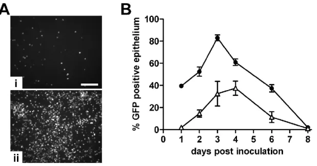
Requirement for RSV G in infection of human airway epithelial cultures
We and others have demonstrated that the RSV glycoprotein (G) is not required for
infectivity in tissue culture cell lines (70). Because deletion of G has been shown to
attenuate RSV in vivo (203), we determined the requirement of G for RSV infection of a
primary well-differentiated model of the human airway epithelium (HAE). RSVΔG infected
fewer numbers of cells at 24 hours pi compared to RSV and analysis of percentage of
GFP-positive epithelium demonstrated a 20-fold decrease in initial infection of HAE (Figure 2.1).
25
titers at a later time compared to RSV (4 days vs 3 days, respectively) and infecting only
37% of the surface epithelium at the time of peak infection compared to 83% GFP positive
epithelium at peak infection with RSV (Figure 2.1B).
Impact of G phenotype on infection of HAE
Post-translational processing and glycosylation is important for the attachment
functions of G and processing intermediates ranging from an immature 45 kDa form to the
fully O-glycosylated, mature 90 kDa form of G have been described in infected cells (86, 87).
To assess the size of G on virus grown in HAE, we purified virus from apical washes of HAE
infected with RSV and compared the electrophoretic mobility to virus grown in HEp2 or Vero
cells. As predicted, the mature 90 kDa form of G was detectable on HEp2-derived virus.
Vero-derived virus, however, primarily contained the 55 kDa G protein. Western blotting of
HAE-grown virions reveals a novel form of G migrating at approximately 180 kDa, in addition
to a smaller amount of the 90 kDa form found in HEp2-derived virus (Figure 2.2A). This
larger form of G is likely either a dimer of the 90 kDa form found in HEp2 cells or represents
additional post-translational modification and more extensive glycosylation unique to the
maturation of G in ciliated cells.
Differences in post-translational modification of G have been noted in in cell lines to
affect the attachment function of G and infectivity of RSV (85, 202). HEp2 and Vero cell lines
are most frequently used to amplify RSV, although the size of G is smaller from
Vero-derived virus compared to HEp2-Vero-derived virus. To assess if this difference in G impacts RSV
infection of HAE, we infected HAE cultures with equal inoculums of RSV amplified in HEp2
cells or Vero cells. Initial infection of HAE with Vero-derived RSV was significantly
decreased compared to HEp2-derived RSV (Figure 2.2B). At 1 day pi, <1% of the HAE
surface infected with Vero-derived RSV was GFP positive, compared to 40% GFP positive
26
round of infection, however, Vero-derived RSV replicated and spread with kinetics similar to
HEp2-derived RSV. Although attenuated at 1 day pi, Vero-derived RSV reached infection
levels almost equal to HEp2-derived RSV, resulting in ~70% GFP positivity of the HAE
apical surface at the time of peak infection compared to ~80% GFP positive epithelium at
the time of peak infection with HEp2-derived RSV (Figure 2.2B). Similar infection kinetics
following the first round of infection is expected, as the form of G on the virion surface of
virus produced by HAE cells would be similar, regardless of the cell line used to amplify the
original inoculum.
Development and characterization of HEp2-V cell line
Vero cells, which lack the genes encoding IFN-α and –β, are frequently used to grow
viruses containing attenuating mutations. RSV encodes two major IFN antagonist proteins,
NS1 and NS2, and deletion of either or both of these genes results in severe attenuation of
the virus in cell lines (104). Similarly, infection of HAE with these viruses results in severe
attenuation of replication and spread (Figure 2.3). RSVΔNS1 infection results in only 1-4%
of GFP positive apical surface area during 8 days of infection, while 16% of the airway
surface was GFP positive at peak infection of RSVΔNS2. In contrast, RSV infected and
replicated efficiently in HAE, resulting in 73% GFP positive surface area at 3 days pi, the
time of peak infection.
Vero cells are commonly used to amplify the RSVΔNS1 and RSVΔNS2 mutant
viruses to sufficiently high titer for study. However, we have demonstrated that amplification
of virus in Vero cells results in initial attenuation of infection. We have therefore identified a
need for a cell line capable of producing virus with the mature form of G while also able to
amplify RSV lacking the type I IFN antagonism genes to high titers. To this end, we stably
transformed HEp2 cells with the IFN antagonist V protein from SV5. HEp2 cells stably
27
IFN signal transduction. V was detected at high levels in whole cell lysates of HEp2-V cells,
confirming robust expression (Figure 2.4A). To characterize type I IFN signal transduction in
HEp2-V cells, we measured IFNβ gene message levels following treatment of HEp2 and
HEp2-V cells with exogenous recombinant IFNβ for 24 hours. Exogenous IFNβ treatment
stimulated a 8-fold increase in IFNβ message in HEp2 cells, while no changes were
detected in HEp2-V cells (Figure 2.4B). To assess the type I IFN response of HEp2-V cells
to viral infection, we infected HEp2 and HEp2-V cells with Sendai virus (SeV), a virus known
to stimulate a robust type I IFN response. SeV infection resulted in a 30-fold increase IFNβ
gene expression in HEp2 cells compared to mock inoculated cells (Figure 2.4C). Type I IFN
secretion following SeV infection was also measured by bioassay (Figure 2.4D). HEp2 cells
secreted high levels of type I IFN 24h following SeV inoculation, while no significant
increases in IFN secretion from SeV-infected HEp2-V cells was detected. SeV infection
resulted in equal numbers of infected cells in both HEp2 and HEp2-V cells (data not shown).
We therefore conclude that stable expression of V in HEp2 cells significantly reduces the
type I IFN response of this cell line.
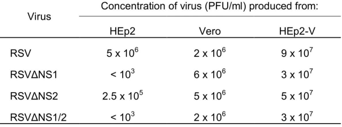
RSV and RSV deleted for the NS1 and NS2 genes, individually (RSVΔNS1 and
RSVΔNS2) and together (RSVΔNS1/2), were amplified in HEp2 cells, HEp2-V cells, and
Vero cells and concentration of virus recovered, expressed as plaque forming units (PFU)
per ml, was assessed for each cell line (Table 2.1). In HEp2 cells, RSVΔNS2 replicated to
titers 1.5 logs lower than RSV. RSVΔNS1 and RSVΔNS1/2 grew to even lower titers and
>103 PFU/ml was recovered in HEp2 cells. In Vero cells, all viruses were amplified to
roughly equal levels, producing 2-6 x10⁶ PFU/ml. In HEp2-V cells, all viruses were amplified
to ~1-log greater concentration compared to amplification in Vero cells. RSV was recovered
at the highest titer in HEp2-V cells (9x10⁷ PFU/ml). Greater than 4 logs more virus was
28
HEp2 cells. The ability to amplify attenuated RSV mutants to high titers in a cell line that
produces a G protein conducive to efficient initial infection will allow for future studies with
attenuated RSV mutants in HAE. Importantly, these findings confirm that inactivation of the
type I IFN response is essential for productive RSV infection and that the NS1 and NS2
proteins are required for attenuation of this response.
2.4 Discussion
The RSV glycoprotein (G) and fusion protein (F) mediate attachment and entry of
RSV to the target ciliated cell. RSV binding and entry is a two-step process, whereby the
virion first attaches to the cell membrane followed by a fusion event. In cell line models of
infection, the F protein is sufficient for binding and entry of RSV and, although the G protein
enhances attachment, G is not considered required (71). In HAE, we demonstrated that
RSV deleted for the G protein is significantly attenuated for initial infection and subsequent
spread throughout the culture, suggesting a heightened requirement for RSV G for efficient
in vivo infection. G-mediated attachment to cultured cells is dependent on
glycosaminoglycans (GAGs) on the cell surface, most notably heparan sulfate (HS), and
likely involves electrostatic interaction between G and GAGs, though requirements for GAG
structural elements have been noted (76-79). Although interactions between G and HS have
been well documented, HS is not detectable on the surface of primary well-differentiated
models of the human airway epithelium (81), suggesting that G may interact with other
GAGs present at the apical cell surface, such as keratan sulfate, or that GAG binding is not
important for the attachment function of G in vivo.
In these studies, biochemical characterization of G on the surface of virus produced
by Vero cells, HEp2 cells, and HAE cultures demonstrated different electrophoretic mobility
29
cell-types. G contains 7 N-linked glycosylation sites and 24-25 O-linked glycosylation sites
and it has been estimated that approximately half of the final 90 kDa molecular mass of
mature G is due to O-linked glycosylation (85). Cell-type specific differences in
electrophoretic mobility of G have been documented and associated with differences in
glycosylation patterns (86, 87). We found that the 55 kDa form of G is predominant on virus
derived from Vero cells and likely represents a processing intermediate, although differential
cleavage of G may also result in this form. The 90 kDa protein on virus derived from HEp2
cells is the mature, fully glycosylated form. Interesting, these studies reveal a larger form of
G on virus produced by ciliated cells in HAE cultures. This 180 kDa form of G may be a
homodimer of the same form of G found on HEp2-grown virus. An unglycosylated region in
G contains four cysteines held together by disulfide bonds followed by a predicted
heparin-binding domain (204-206). Dimerization via this region may make the G homodimer resistant
to reduction during Western blot analysis, allowing for electrophoretic mobility in the 180
kDa range. Alternatively, the 180 kDa form of G may have additional or more extensive
O-linked carbohydrate chains added during processing of G in the ciliated cell.
Maturation of G is associated with infectivity in cell lines (76, 85). Similarly, we found
that the size of G greatly impacted RSV infectivity of HAE cultures, as viruses containing
mature G derived from HEp2 cells are more infectious than viruses containing the smaller
form of G derived from Vero cells. The impact of the increased size of G from viruses grown
in HAE was not evaluated and future studies will aim to determine the glycosylation pattern
of G and infectivity of virus from HAE.
Vero cells are the only cell line approved by the World Health Organization for
production of live-attenuated or inactivated vaccines. However, we found that infectivity HAE
with RSV grown in Vero cells was significantly reduced compared to that of RSV grown in