Original Research Article
Vascular malformations: a hospital based study
Komanapalli Sunilkumar
1, Karthik Aithal
2, Unmesh Kumar Jena
2, Uma Prasad
1,
Bhagyalakshmi Atla
1*, P. V. Sudhakar
2INTRODUCTION
Vascular anomalies are a diagnostic and therapeutic challenge which requires inter disciplinary management.1
Vascular malformations may arise in any of the vascular beds present in the human body. These lesions vary in location, type, and clinical severity. Broadly they are of two types; vascular tumours and vascular malformations, both are rare diseases.
During embryogenesis, normal blood vessels are derived from two processes: 1) Vasculogenesis (denovo
production in the embryo; the process through which endothelial precursors align themselves to form primitive blood vessels. 2) Angiogenesis (development of new vessels); angiogenesis, a term that implies the formation of new microvessels from differentiated endothelium. An error in both or either one of these processes results in malformed vessels. Vascular malformations occur in less than 1% of the population.
Vascular malformations often result from abnormal development in arteries, veins and lymph vessels. They can be congenital or acquired. Congenital vascular
1Department of Pathology, 2Deprtment of Plastic surgery, Andhra Medical College, Visakhapatnam, Andhra Pradesh,
India
Received: 29 June 2019
Accepted: 17 July 2019
*Correspondence:
Dr. Bhagyalakshmi Atla,
E-mail: [email protected]
Copyright: © the author(s), publisher and licensee Medip Academy. This is an open-access article distributed under the terms of the Creative Commons Attribution Non-Commercial License, which permits unrestricted non-commercial use, distribution, and reproduction in any medium, provided the original work is properly cited.
ABSTRACT
Background: Vascular anomalies comprise a widely heterogeneous group of lesions. Diagnosis and management of these lesions present challenges to the surgeons, radiologists and histopathologists. Accurate classification of these lesions results in appropriate therapy. Aim of the study is to study the role of histopathology and histochemical stain in the diagnosis of vascular malformation
Methods: The present study is a hospital based observational study on vascular malformations over a period of three years from 2016 to 2018 done in department of Pathology and Plastic Surgery at tertiary care centre, Visakhapatnam.
Results: Out of 107 specimens of vascular anomalies received, 72 cases were vascular neoplasms, 35 were vascular malformations. Majority of the vascular malformations were seen in the cervicofacial region (43%) followed by extremities (37.1%). Most common malformation in the present study was arterio venous malformations (60%) followed by venous malformations (22.8%). Verhoeff’s VanGieson stain demonstrated discontinuity of internal elastic lamina in cases of arteriovenous malformation and helped in differentiating the lesions from capillary malformation and vascular tumors.
Conclusions: Histopathology, special histochemical stains along with imaging features can be used to reduce diagnostic difficulties and in helping proper management of vascular malformations.
Keywords: Arteriovenous malformations, Histopathology, Vascular malformation, Verhoeff’s VanGieson stain
malformations usually present at birth, grow proportionally with age, and are considered to be localized defects of vascular morphogenesis without endothelial hyperplasia & do not expand rapidly. On the contrary haemangiomas grow by mainly endothelial hyperplasia. They grow rapidly with age and involutes overtime.
Sometimes vascular malformations are seen in older people due to fluid or blood accumulating in poorly formed veins or lymphatic channels forming connections between arteries and veins (acquired malformations).2
Vascular malformations can be affected by hormonal changes during puberty and pregnancy. Vascular malformations are subdivided into capillary, venous, lymphatic, arterio-venous and combined malformations, depending on their dominant vasculature. Most of the vascular anomalies are sporadic and some familial. They usually present as painful swelling and some with bleeding. Vast spectrum of diagnostic modalities, ranging from conventional X-ray, ultrasound , colour Doppler, , CT with 4D imaging ,MRI as well as angiography play important role in diagnosing these lesions.Vascular malformations based on the flow are subcategorized into; Slow-flow (Capillary, Lymphatic, Venous) and Fast-flow (Arterial arterial (aneurysm, stenosis, ectasia), arteriovenous fistulae (AVF), arteriovenous malformation (AV).3
Vascular malformations require treatment to improve appearance, alleviate pain, swelling and bleeding. Children with corrected malformation can achieve high levels of performance at school. Appropriate treatment depends on correct classification and diagnosis of vascular anomalies, which is based on established national and international classification systems, recommendations and guidelines.4
Precise diagnosis of childhood vascular anomalies is challenging, and requires careful correlation of clinical findings, diagnostic imaging, histopathology and genetic analysis. Treatment for vascular malformations depends on the type of malformation. Laser therapy is usually effective for capillary malformation, embolisation for arterial malformation, venous malformation and lymphatic malformation with sclerosing therapy. Histopathology remain an important element in the complete evaluation of many vascular anomalies.5
Technical advances and vascular immunisations such as D2-40, PROX1, and vascular endothelial growth factor receptor 3, S100 immunostain which distinguish lymphatics from arteries and veins, have been of immense help in daily practice. Arteriovenous vascular malformations and hemangiomas are benign vascular lesions that are difficult to distinguish from one another clinically. Hemangiomas and arteriovenous malformations (AVMs) can be distinguished from each other histologically on the basis of absence or presence of
arteriovenous structures and Verhoeff’s Vangeson stain for elastin plays an important role.6
The present study aims at knowing the prevalence of vascular malformations in our Institute and role of histopathology with elastic stains in diagnosing these lesions which is cost effective.
METHODS
The present study is a hospital based observational study conducted in the department of pathology for a period of three years from 2016 to 2018.
Inclusion criteria
All the resected specimens with clinical and radiological diagnosis of vascular malformations received from department of plastic surgery were included in the study.
Exclusion criteria
Vascular tumors and recurrent lesions of vascular malformation were excluded from the study.
Methodology
The clinical and radiological information was noted. Gross features of the specimens were recorded and representative sections were taken from the specimen and processed routinely. In all the cases the sections were stained with H&E stain and special stain (Verhoeff’s VanGieson) for elastic fibers.
The lesions were classified adopting the recent ISSVA classification for vascular anomalies Approved at the 20th ISSVA Workshop, Melbourne, April 2014, last revision May 2018 (Table 1).7 Microscopic features were
correlated with clinico radiological features and results were tabulated. Statistical methods: The data was recorded in MS Excel sheet and percentage distribution was calculated.
RESULTS
Total number of vascular anomalies received in the department was 107 cases. 72 cases (67.2 %) were vascular neoplasms and 35 (32.7 %) cases were vascular malformations.
Out of 35 cases majority of the cases were seen between 21-30 years age group (31.4%) with male preponderance. (54.2%) (Table 2).
malformation (25%), lymphatic malformation (66.66%) and capillary malformation (50%). Overall correlation
percentage of 65.71% (Table 4).
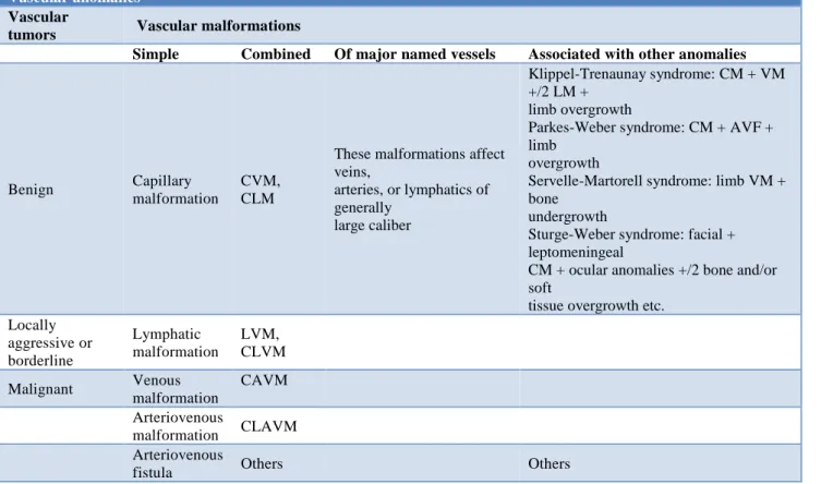
Table 1: ISSVA classification for vascular anomalies Approved at the 20th ISSVA Workshop, Melbourne, April
2014, last revision May 2018.
Vascular anomalies
Vascular
tumors Vascular malformations
Simple Combined Of major named vessels Associated with other anomalies
Benign Capillary malformation
CVM, CLM
These malformations affect veins,
arteries, or lymphatics of generally
large caliber
Klippel-Trenaunay syndrome: CM + VM +/2 LM +
limb overgrowth
Parkes-Weber syndrome: CM + AVF + limb
overgrowth
Servelle-Martorell syndrome: limb VM + bone
undergrowth
Sturge-Weber syndrome: facial + leptomeningeal
CM + ocular anomalies +/2 bone and/or soft
tissue overgrowth etc. Locally
aggressive or borderline
Lymphatic malformation
LVM,
CLVM
Malignant Venous malformation
CAVM
Arteriovenous
malformation CLAVM
Arteriovenous
fistula Others Others
Table 2: Distribution of vascular malformation cases according to age and sex (n=35).
Age
distribution Sex distribution
Total number of cases
(%)
Male Female
1-10 5 1 6 17.1%
11-20 4 6 10 28.5%
21-30 6 5 11 31.4%
31-40 1 4 5 14.2%
41-50 - - - -
51-60 2 - 2 5.7%
61-70 1 - 1 2.8%
Total 19 (54.2 %)
16
(45.7%) 35 100 Age wise distribution of various vascular malformations are; arteriovenous malformations were more common in the age group 21 to 40 years (71.42%), venous malformation, lymphatic malformation, capillary malformation and combined lymphovenous malformation was common in the age group 1 to 20
years (Table 5). Site was distribution of various vascular malformations; 42.85% of vascular malformations were common in cervicofascial region and 37.14% of lesions common in extremities.
Arteriovenous malformations were more common in cervicofascial region (47.61%) and venous malformation was more common on the extremities (50%). Lymphatic, capillary malformations were more common on cervicofascial region (Table 6).
Table 3: Distribution of vascular malformations on histopathology-35.
Type of malformation on
histopathology Total %
Arterio venous malformations 21 60% Venous malformation including
Glomovenous malformation 08 22.8% Lymphatic malformations 03 8.5% Capillary malformation 02 5.7% Combined lymphovenous malformation 01 2.8%
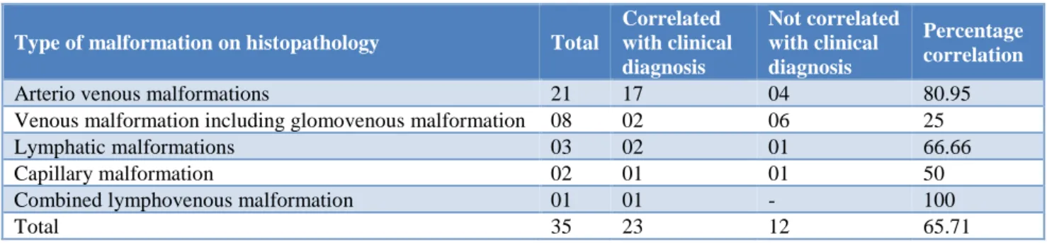
Table 4: Correlation between clinical diagnosis and histopathology in various vascular malformations-35.
Type of malformation on histopathology Total
Correlated with clinical diagnosis
Not correlated with clinical diagnosis
Percentage correlation
Arterio venous malformations 21 17 04 80.95
Venous malformation including glomovenous malformation 08 02 06 25
Lymphatic malformations 03 02 01 66.66
Capillary malformation 02 01 01 50
Combined lymphovenous malformation 01 01 - 100
Total 35 23 12 65.71
Table 5: Age wise distribution of various vascular malformations-35.
Type of malformation on histopathology Total 1-20 years 21-40 years 41-60 years 61-80 years
Arterio venous malformations 21 03 15 02 01
Venous malformation including Glomovenous
malformation 08 08 - - -
Lymphatic malformations 03 02 - - -
Capillary malformation 02 02 - - -
Combined lymphovenous malformation 01 01 - - -
Total 35 16 16 02 01
Table 6: Site was distribution of various vascular malformation-35.
Type of malformation on histopathology Total Cervico fascial Extremities Others
Arterio venous malformations 21 10 09 2 (Scalp)
Venous malformation including
Glomovenous malformation 08 01 04
1 (scalp) 2 (trunk)
Lymphatic malformations 03 02 - 01 (scrotum)
Capillary malformation 02 02 - -
Combined lymphovenous malformation 01 - - 01 (chest)
Total 35 15 13 07
DISCUSSION
Vascular malformations are characterized by vascular dysmorphogenesis and normal endothelial turnover. They never regress and require medical and surgical treatment. Haemangioma are benign vascular tumors, characterized by endothelial hyperplasia and rapid growth. They respond to corticosteroids. If vascular malformation is misdiagnosed as hemangioma, it will lead to treatment failure and a possible recurrence. Therefore, correct diagnosis of both these lesions is essential.
Arteriovenous malformation
Arteriovenous malformations (AVMs) involve both arteries and veins bypassing capillaries. They are fast-flow vascular malformations which most commonly affect head and neck and central nervous system. Congenital AVMs are present at birth but become
apparent during adolescence or late childhood. AVMs are mostly sporadic but can be part of syndromes.
On histopathology AVMs are composed of malformed arteries and veins where nonuniform myxoid degeneration occurs within the walls of vessels and Verhoeff’s VanGieson stain highlights the disrupted elastic lamina. These lesions can be associated with scattered proliferating capillaries; pyogenic granuloma-like proliferation; infantile hemangioma-granuloma-like proliferation (GLUT-1 negative); and pseudo-kaposiform proliferation. Recent studies have shown the presence of increased intralesional nerves in vascular malformations as an additional tool in diagnosing vascular malformations.8-11
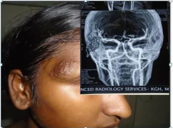
nodular lesions. One case of AV malformation on the right side of face presented as irregular palatal nodule and CT angiogram showed feeding vessels from temporal branch. (Figure 1).
Figure 1: AV malformation involving right side of face showing irregular pulsatile nodular lesion, inset
showing CT angiogram - feeding vessels from temporal branch.
On gross examination the lesion was nodular, cut section showing grey brown cyst like spaces filled with hemorrhagic material. Microscopy showed tortuous arteries with thickened smooth muscle layer and large thick-walled veins, surrounded by nerve bundle (Figure 2). Three cases showed thrombosis and fibrosis of the veins. In 19 cases (90.47%) nerve bundles around the vessels was seen on histopathology which helped in diagnosing these lesions. Verhoff’s VanGieson elastic special stain was used to demonstrate discontinuity of internal elastic lamina confirming malformed nature of the vessel (Figure 3).
Figure 2: Photomicrograph showing Arterio venous malformation (H&E, 100X) with nerve bundles
(Arrow).
Venous and glomuvenous malformation (VM)
VM demonstrates large, widely dilated venous channels with irregular shapes and sizes, haphazardly arranged in
the dermis and subcutis.The muscle tissue is arranged concentrically around the malformed vessels except for malformations in joints, head and neck region, ankle and genitourinary regions. The malformed vessels may surround cutaneous nerves, normal vessels and sometimes dissect through muscle. Luminal thrombi with organization and papillary endothelial hyperplasia (Masson’s phenomenon) are common. Phleboliths or fibromyxoid nodules may be associated with the vessel wall. The endothelial cells are small, flattened without significant nuclear atypia. Pan-vascular markers such as CD31 highlight the lesional cells of VM, while GLUT-1 and lymphatic markers such as D2-40/Podoplanin are negative. Ki-67 labelling rate is low. Glomuvenous malformations (GVMs) represent a subset of VMs which are histologically characterized by multiple layers of glomus cells lining these malformed veins. These glomus cells are immunoreactive to smooth muscle actin and often positive for vimentin and desmin.8-11
Figure 3: Photomicrograph showing Verhoffs Van Geison stain with discontinuity of internal elastic
lamina (arrow) (400X).
Figure 4: Venous malformation involving left forearm showing non pulsatile nodules. Inset showing CT
angiogram with venous sinuses and phleboliths.
malformation involving the left forearm showed non pulsatile nodules with CT angiogram showing venous sinuses and phleboliths (Figure 4).
On gross examination there was irregular gray brown masses ranging from 2.5 to 4.5cm, cut section showed cyst like spaces filled with haemorrhage surrounded by gray tan areas. Microscopically there were dilated venous channels of varying sizes filled with blood and some of the vessels were thrombosed. Out of 7 cases 6 cases showed vessels surrounding cutaneous nerves (Figure 5).
Figure 5: Photomicrograph showing venous malformation, (H&E,100X). Inset showing Nerve
bundle (400X).
In the present study there was a single case of Glomovenous malformation, which was located on the right arm of a female aged 16 years. The nodules were black in colour with purple colour papules. On microscopy the lesion showed squamous epithelium, sub epithelium showing multiple layers of glomus cells lining the malformed veins. Individual cells were small with scanty cytoplasm with punched out nucleoli (Figure 6).
Lymphatic malformation
Lymphatic malformations (LMs) most commonly present in the head and neck region. They are two types; macrocystic when individual channels are > 1cm (i.e., cystic hygroma), microcystic when <1 cm, though combined lesions are more common. Lymphatic malformations can be localized or generalized. Microcystic lymphatic malformation is the localized form within the papillary dermis. On histopathology there are thin-walled, dilated irregular channels containing pale eosinophilic amorphous material, sometimes with lymphocytes, histiocytes and red blood cells. The overlying epidermis usually shows verrucous hyperplasia and hyperkeratosis. Malformed lymphatic channels can also be seen in the reticular dermis and subcutis. Lymphoid aggregates with germinal center formation and plasma cells may be seen in deeper aspects. The macrocystic LM show thicker vessel walls with increased fibrous tissue, myofibroblasts, and few smooth muscle cells. Immunostains for lymphatic markers are helpful in
differentiating LM from other malformations. PROX1, VEGFR-3, D2-40 (podoplanin) and LYVE-1all label lymphatic endothelium and CD34 is focally positive or absent. 8-11
Figure 6: Photomicrograph showing Glomovenous malformation, (H&E 100X), inset showing Glomus
cells (400 X).
In the present study out of 3 cases, 2 cases were from the neck region and other from scrotal region. Grossly received skin covered mass, cut section showed thin walled cyst like spaces, cyst wall thickness ranging from 0.3 to 0.6 cm. Microscopically these were composed of thin-walled, dilated irregular channels which were empty in 3 cases, one case showing pale eosinophilic amorphous material with few lymphocytes, embedded in fibro fatty tissue surrounded by nerve bundle (Figure 7).
Figure 7: Photomicrograph showing lymphatic channels of varying sizes filled with lymphocytes and
proteinaceous material (H&E,100X). Inset showing nerve bundle (400X).
Capillary malformation
have flat endothelium and a pericyte layer. In adulthood, facial lesions may develop intralesional pyogenic granulomas, show epidermal changes and thickened vessels. Abundant fibrous tissue may be seen within the subcutis and fascial layers.8-11
In the present study two cases of capillary malformations were seen on the face, presented with reddish macular lesions. Grossly the lesion was 1 to 1.5 cm skin covered bit. Microscopically dilated thin-walled vessels resembling capillaries were seen in the upper dermis surrounded by fibrous tissue. Nerve bundles were seen associated with these malformed capillaries (Figure 8).
Combined lympho venous malformation
Lymphovascular malformation shows numerous both thin walled channels, filled with blood and pale eosinophilic secretions suggestive of vascular and lymphatic channels with anastomosis. The intervening stroma shows lymphoid follicles. Immunohistochemistry is positive for both endothelial (CD 31 positive) and lymphatic channels (D2-40 positive).12
Figure 8: Photomicrograph showing capillary malformation (100X), inset showing nerve bundle
(400X).
In the present study a case of lymphovenous malformation was reported which presented as a lesion
on the chest region in a male aged 3 yrs. On gross examination the lesion was skin covered measuring 4x3 cm,cut section showing multiple dilated cyst like spaces some are filled with dark brown material (Figure 9). Microscopy showed varying sizes lymphatic channels filled with lymphocytes and eosinophilic material surrounded by large dilated venous sinuses filled with blood. There is no communication between them with absence of nerve bundles (Figure 10).
Figure 9: Gross photograph of combined Lymph venous malformation, showing dilated cyst like spaces
beneath the skin.
Figure 10: Photomicrograph showing lymphatic channels filled with eosinophilic material large dilated
venous sinuses filled with blood without any connection. Inset showing nerve bundle (H&E,100X).
Table 7: Comparison of present study with other study.
Study Gender
Male Female Most common malformation Most common Location
Makhija LK,
et al 52.6% (306/588) 47.9% (282/588)
Venous malformation 53.7% (316/588)
Extremities 49.6% (292/588) Present study 54.2% (19/35) 45.7% (16/35) Arterio venous malformation
60% (21/35)
Cervico facial region 43% (15/35)
Sadick M et al, in their study documented venous malformations as the commonest vascular anomaly
malformation syndromes (6%) and capillary malformations (4%).3 In the study by Makhija LK et al
the most common malformation was venous malformation when compared to the present study being arteriovenous malformation
(Table 7).13
In the study by Karmacharya RM et al venous malformation constituted 35.29% of lesions, capillary malformation was 15.69% and A-V malformation was 27.45%.14
In the present study arteriovenous malformation constituted 60% of lesions, followed by venous malformation including Glomovenous malformation (22.8%), Lymphatic malformations (8.5%), capillary malformation (5.7%) and combined lymphovenous malformation (2.8%).
In cases of arteriovenous malformations when subjected to special stain Verhoff’s VanGieson elastic special stain shows discontinuity in the internal elastic membrane and helps in differentiating hemangiomas from arteriovenous malformations.15, 16
In the present study two cases with clinical diagnosis of vascular tumor and two cases with diagnosis of soft tissue tumor turned out to be arteriovenous malformation on combined H&E stain and special stain with Verhoff’s VanGieson.
CONCLUSION
Vascular malformations should be differentiated from vascular tumors to reduce the risk of treatment failure and recurrence. Combining imaging techniques, histopathology and histochemical stains reduce diagnostic difficulties and help in appropriate management. Histopathological examination helps in subcategorising various vascular malformations. The presence of the intra-lesional nerves can be useful to distinguish between arteriovenous malformation and vascular tumors even on Hematoxylin and Eosin stained sections. Use of elastic stain helps in accurately discriminating between arteriovenous malformations and vascular tumors by clearly outlining arteries and arterioles which is cost effective, hence the need for all the vascular malformations undergoing histopathological examination.
Funding: No funding sources Conflict of interest: None declared
Ethical approval: The study was approved by the Institutional Ethics Committee
REFERENCES
1. Lee BB, Do YS, Yakes W, Kim DI, Mattassi R, Hyon WS. Management of arteriovenous
malformations: a multidisciplinary approach. J Vascular Surg. 2004;39(3):590-600.
2. Joshua A.Cox, Erica Bartlett and Edward I. Lee. Vascular Malformations: A Review. Semin Plast Surg 2014;28(2):58-63.
3. Sadick M, Mueller-Wille R, Wildgruber M, Wohlgemuth WA. Vascular anomalies (part I): classification and diagnostics of vascular anomalies. InRöFo-Fortschritte auf dem Gebiet der Röntgenstrahlen und der bildgebenden Verfahren 2018;190(9):825-835. ©Georg Thieme Verlag KG.
4. Greene AK, Orbach DB. Management of arteriovenous malformations. Clin Plast Surg. 2011;38(1):95-106.
5. Lee BB, Laredo J. Classification of congenital vascular malformations: the last challenge for congenital vascular malformations. Phlebol. 2012;27(6):267-9
6. Miller DD, Gupta A. Histopathology of vascular anomalies: update based on the revised 2014 ISSVA classification, 35, 3. 2016;35(3):137-46.
7. Wassef M, Blei F, Adams D, Alomari A, Baselga E, Berenstein A, Burrows P, Frieden IJ, Garzon MC, Lopez-Gutierrez JC, Lord DJ. Vascular anomalies classification: recommendations from the International Society for the Study of Vascular Anomalies. Pediatr. 2015;136(1):e203-14.
8. Adegboyega PA, Qiu S. Hemangioma versus vascular malformation: presence of nerve bundle is a diagnostic clue for vascular malformation. Arch Pathol Lab Med. 2005;129(6):772-5.
9. Meijer Jorna LB, Breugem CC, DeBoer OJ, Ploegmakers JP, Vander Horst CM, Vander Wal AC. Presence of a distinct neural component in congenital vascular malformations relates to the histological type and location of the lesion. Human Pathology. 2009; 40(10):1467-73.
10. Enjolras O. Ch 104: Vascular Malformations. In: Bolognia JL, Jorizzo JL, Schaffer JV, eds. Dermatology. 3rd Ed. China: Elsevier/Saunders;
2011: 1711-1728.
11. Gupta A, Kozakewich H. Histopathology of vascular anomalies. Clin Plastic Surg. 2011;38(1):31-44.
12. Mitakshara Sharma, Varuna Mallya, Nita Khurana, Praveen Kumar and Rajan Duggal. Lymphovascular Malformation-A Report of Two Cases. J Clin Diagn Res.2017;11(5): ED03-ED04
13. Makhija LK, Bhattacharya S. Management of vascular anomalies: Review of institutional management algorithm. Indian journal of plastic surgery: official publ Assoc Plastic Surg Ind. 2017;50(2):193.
14. Karmacharya RM, Shrestha BK, Shrestha B, Devbhandari M, Tuladhar SM. Vascular anomalies: Presentation and response to medical and surgical management, our experiences in the last 5 years. Ind J Vasc Endovasc Surg. 2019;6(1):10.
and mast cell density. Ind J Pathol Microbiol. 2014;57(2):191.
16. Girard C, Graham JH, Johnson WC. Arteriovenous hemangioma (arteriovenous shunt). A clinicopathological and histochemical study. J Cutaneous Pathol. 1974;1(2):73-87.