Original Research Article
Liver function tests as a prognostic indicator in
Pediatric Intensive Care Unit (PICU)
Akshatha Mallikarjuna*, Ashwani K. Sood
INTRODUCTION
Hepatocytes perform numerous and vital roles in maintaining homeostasis and health. Frank liver failure is incompatible with life, and the functions of the liver are too complex and diverse to be sub served by a mechanical pump, dialysis membrane , or concoction of infused hormones, proteins and growth factors.1 The
severity of the liver disease may be reflected in clinical signs (the occurrence of encephalopathy, variceal hemorrhage, worsening jaundice, apparent shrinkage of liver mass owing to massive necrosis, or onset of ascites) or in biochemical alterations (hypoglycemia, hyperammonemia, electrolyte imbalance, continued hyperbilirubinemia, marked hypoalbuminemia, or a
prolonged pro thrombin time (PT) or INR that is unresponsive to parenteral administration of vitamin K).2
Critically ill patients are most carefully observed and monitored in the indoor hospital setting with many tests being performed on sequential basis. In critically ill patients with multiple organ failure liver dysfunction is often overshadowed or missed.3 Hepatic injury in ICU
can emerge either as a rapid primary episode caused by an acute reduction in perfusion after shock, hemorrhage, resuscitation or low output septic shock or as a late-onset form of hepatic injury emerging secondarily to multiple septic episodes and medical treatment strategies.4 New
onset of liver dysfunction is a frequent finding in critical ill patients and significantly contributes to increased
ABSTRACT
Background: Frank liver failure is incompatible with life. Severity of liver disease is reflected in clinical signs or biochemical alterations. Hepatic dysfunction indicates poor outcome in critically ill patients, but no large systematic investigation into its exact incidence and prognostic relevance has been performed in different population groups.
Methods: All PICU admissions during the study period were included. Pattern of admission LFT (Liver Function Tests) was reported. Relationship between clinical events and patient outcome with the LFT panel on day 1 and day 3 were reported. Association between final outcome and day 7 LFT was explored.
Results: Deranged AST (Aspartate Transaminase), ALT (Alanine Transaminase), Bilirubin, Albumin, PT (Prothrombin time) and INR (International Normalized Ratio) on Day1 and 3 had significant association with increased requirement of inotropic and ventilatory support as well with increased mortality. LFT parameters on day 7 showed significant correlation between normal AST, ALT, bilirubin, PT and INR with improved survival.
Conclusions: LFT's should be essential part of biochemical profile and doctors caring for sick patients in peripheral hospitals should consider for early referral of patients to higher centers based on LFT parameters for the advanced care they deserve.
Keywords: Critically ill children, LFT, Prognosis
Department ofPediatrics, Indira Gandhi Medical College, Shimla, Himachal Pradesh, India
Received: 19 February 2018
Accepted: 28 March 2018
*Correspondence:
Dr. Akshatha Mallikarjuna,
E-mail: [email protected]
Copyright: © the author(s), publisher and licensee Medip Academy. This is an open-access article distributed under the terms of the Creative Commons Attribution Non-Commercial License, which permits unrestricted non-commercial use, distribution, and reproduction in any medium, provided the original work is properly cited.
morbidity and mortality. Early recognition and subsequent therapy of the underlying conditions are still the therapeutic cornerstones.5
Hepatic dysfunction is traditionally considered to indicate poor outcome in critically ill patients, but no large systematic investigation into its exact incidence and prognostic relevance has been performed in different population groups like children, adolescents and adults.6
As there is paucity of data regarding hepatic dysfunction in patients admitted in PICU we have done a prospective study to evaluate the prevalence, patterns and significance of liver function tests in critically ill patients admitted in PICU.
METHODS
A hospital based prospective study was conducted in a tertiary care hospital in North India from June 2012 to May 2013.
Objectives of this study were to evaluate the prevalence and patterns of deranged liver function tests (LFT’s) at admission in critically ill children and to study the correlation of LFT’s to the morbidity and mortality in critically ill children admitted in PICU.
Inclusion criteria
All critically ill children between 1-17 years age group admitted in Paediatric Intensive Care Unit were included.
Exclusion criteria
Any children with pre-existing conditions that would have deranged their baseline liver function such as
• Chronic / acute primary liver disease • Children < 1yr of age
• Metabolic disorder
• Obesity
• Readmission in ICU
The study was approved by the Institutional Ethics Committee (IEC). A written informed consent was taken from the parent (mother/father/legal guardian) before enrolment into the study.
The liver function tests of each patient admitted in PICU were requested on days 1, 3 and 7 of admission as a part of biochemical ICU profile during the study period and recorded on a predesigned pro forma. Day 1, 3 and 7 were chosen to analyse LFT pattern distributed over the sickness period. The LFT panel included ALT, AST, ALP, serum bilirubin, serum albumin, PT and INR. Age wise normal reference range for liver function tests from standard books were used in the study to analyse the values.7,8 The pattern of LFT on the day of admission to
PICU was reported. The relationship between clinical factors (that is requirement of inotropic support or ventilator support or usage of antibiotics for more than 3 days) during the first 48 hours of ICU stay and the final outcome of the patient were compared with the LFT panel on day 1 and day 3 of hospitalization. The association between day 7 LFT and final outcome of patients was also explored in the patients who survived until day 7.
Statistical analysis
Using Chi-square analysis. A p value of <0.05 was taken as significant.
RESULTS
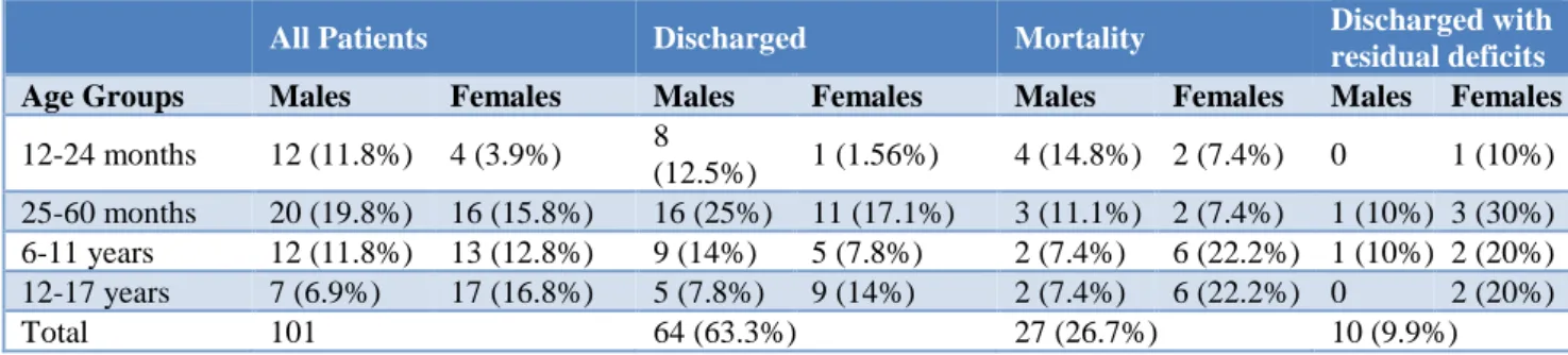
During the study period, 101 patients aged 1-17 years admitted to PICU were included.
50.49% (51) were males and 49.5% (50) were females. Majority of the patients belonged to 2-5 years age group (35.6%). 7-day mortality was 25.7% (27/101) (Table 1).
Table 1: Demographics and clinical data for patients admitted in PICU between June 2012 to May 2013.
All Patients Discharged Mortality Discharged with
residual deficits Age Groups Males Females Males Females Males Females Males Females
12-24 months 12 (11.8%) 4 (3.9%) 8
(12.5%) 1 (1.56%) 4 (14.8%) 2 (7.4%) 0 1 (10%) 25-60 months 20 (19.8%) 16 (15.8%) 16 (25%) 11 (17.1%) 3 (11.1%) 2 (7.4%) 1 (10%) 3 (30%) 6-11 years 12 (11.8%) 13 (12.8%) 9 (14%) 5 (7.8%) 2 (7.4%) 6 (22.2%) 1 (10%) 2 (20%) 12-17 years 7 (6.9%) 17 (16.8%) 5 (7.8%) 9 (14%) 2 (7.4%) 6 (22.2%) 0 2 (20%)
Total 101 64 (63.3%) 27 (26.7%) 10 (9.9%)
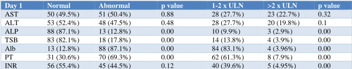
The pattern of LFT on the day 1 of admission depicted abnormal PT and serum albumin levels in large proportion of children however, most of the values were between 1-2 times the upper limit of normal. Serum
bilirubin was normal in most of the patients on admission, however liver enzymes were equally distributed btween the normal and abnormal groups (Table 2).
Table 2: Distribution of LFT values on day 1 of hospital admission.
Day 1 Normal Abnormal p value 1-2 x ULN >2 x ULN p value
AST 50 (49.5%) 51 (50.4%) 0.88 28 (27.7%) 23 (22.7%) 0.32
ALT 53 (52.4%) 48 (47.5%) 0.48 28 (27.7%) 20 (19.8%) 0.1
ALP 88 (87.1%) 13 (12.8%) 0.00 10 (9.9%) 3 (2.9%) 0.00
TSB 83 (82.1%) 18 (17.8%) 0.00 14 (13.8%) 4 (3.9%) 0.00
Alb 13 (12.8%) 88 (87.1%) 0.00 84 (83.1%) 4 (3.96%) 0.00
PT 31 (30.6%) 70 (69.3%) 0.00 62 (61.3%) 8 (7.9%) 0.00
INR 56 (55.4%) 45 (44.5%) 0.12 40 (39.6%) 5 (4.95%) 0.00
#Abnormal values are values greater than upper limit of normal for age; 1-2 x ULN is 1 to 2 times the upper limit of normal; >2 x ULN is greater than two times the upper limit of normal; TSB: Total serum bilirubin; Alb: Serum Albumin; *Abnormal serum albumin is value of serum albumin less than lower limit of normal for age
Analysing individual components of LFT on day 1 and 3 of admission showed deranged AST, ALT, serum bilirubin, serum albumin, pro thrombin time and INR had significant association with increased requirement of both
inotropic and ventilatory support during the hospital stay as well as with increased mortality rate. Only alkaline phosphatase did not show a significant association with requirement of ventilatory/inotropic support and neither with the risk of mortality (Table 3 and Table 4).
Table 3: Association of Day1 LFT with clinical events and outcome of children admitted in PICU.
LFT Invasive ventilation P
value Inotropic support
P
value Death
P value Normal Abnormal Normal Abnormal Normal Abnormal
AST 1 12 (24%) 26 (50.9%) 0.00 26 (52%) 40 (78.4%) 0.00 8 (16%) 19 (37.25%) 0.01 ALT 1 13 (24.5%) 25 (52%) 0.04 27 (50.9%) 39 (81%) 0.01 8 (15%) 19 (39.5%) 0.00 ALP 1 29 (32.9%) 9 (69.2%) 0.01 55 (62.5%) 11 (84.6%) 0.21 18 (20.9%) 9 (68.2%) 0.00 TSB 1 22 (26.5%) 16 (88.8%) 0.00 49 (59%) 17 (94.4%) 0.00 15 (18%) 12 (66.6%) 0.00 Alb1 1 (7.6%) 37 (43%) 0.01 5 (35.7%) 61 (70.1%) 0.01 0 27 (30.6%) 0.01 PT 1 2 (6.4%) 36 (51.4%) 0.00 14 (45.1%) 52 (74.2%) 0.00 1 (3.2%) 26 (37.1%) 0.00 INR 1 9 (16%) 29 (64.4%) 0.00 30 (53.5%) 36 (80%) 0.00 8 (14.28%) 19 (42.2%) 0.00 #AST 1, ALT 1, ALP 1, TSB 1, S. Alb1, PT 1, INR 1-AST, ALT, ALP, TSB, S. Alb, PT, INR on day1 of admission to hospital; TSB: Total serum bilirubin; Alb: Serum Albumin
Table 4: Association of Day3 LFT with clinical events and outcome of children admitted in PICU.
LFT Invasive ventilation P
value Inotropic support
P
value Death
P value Normal Abnormal Normal Abnormal Normal Abnormal
AST2 8 (17.7%) 23 (48.9%) 0.00 20 (44.4%) 37 (78.7%) 0.00 2 (4.4%) 16 (47%) 0.00 ALT2 11 (22%) 20 (47.6%) 0.00 23 (46%) 34 (80.9%) 0.00 4 (8%) 14 (33.3%) 0.00 ALP2 26 (31.7%) 5 (50%) 0.29 49 (59.7%) 8 (80%) 0.3 13 (15.8%) 5 (50%) 0.02 TSB2 20 (25.9%) 11 (73.3%) 0.00 44 (57.1%) 13 (86.6%) 0.03 9 (11.68%) 9 (60%) 0.00 Al 2 3 (13.6%) 28 (40%) 0.02 8 (36.3%) 49 (70%) 0.00 0 18 (25.7%) 0.00 PT2 3 (10.3%) 28 (44.4%) 0.00 14 (48.2%) 43 (68.2%) 0.02 0 18 (31%) 0.00 INR2 8 (15.6%) 23 (56%) 0.00 23 (45%) 34 (82.9%) 0.00 2 (3.9%) 16 (39%) 0.00
$AST2, ALT2, ALP2, TSB2, Alb2, PT2, INR 2-AST, ALT, ALP, TSB, Alb, PT, INR on day 2 of admission to hospital; TSB: Total
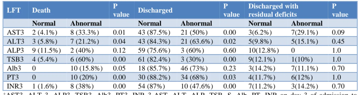
LFT parameters followed until day 7 of admission showed, a significant correlation between normal AST, ALT, serum bilirubin, pro thrombin time and INR on day
7 with a good outcome in these patients. However, those patients who were discharged with some residual deficits (neurological deficits) showed a varied result (Table 5).
Table 5: Association of Day 7 LFT with clinical outcome of children admitted in PICU.
LFT Death P
value Discharged
P value
Discharged with residual deficits
P value Normal Abnormal Normal Abnormal Normal Abnormal
AST3 2 (4.1%) 8 (33.3%) 0.01 43 (87.5%) 21 (50%) 0.00 3(6.2%) 7(29.1%) 0.09 ALT3 3 (5.8%) 7 (21.2%) 0.04 43 (84.3%) 21 (63.6%) 0.02 5(9.8%) 5(15.1%) 0.45
ALP3 9 (11.5%) 2 (40%) 0.12 59 (75.6%) 3 (60%) 0.60 10(12.8%) 0 1.0
TSB3 4 (5.4%) 6 (60%) 0.00 61 (82.4%) 3 (30%) 0.00 9(12.1%) 1(10%) 1.0 Alb3 0 10 (15.8%) 0.05 18 (85.7%) 46 (73%) 0.23 3(14.2%) 7(11.1%) 0.70
PT3 0 10 (20%) 0.00 30 (88.2%) 34 (68%) 0.03 4(11.7%) 6(12%) 1.0
INR3 1 (1.6%) 8 (38%) 0.00 54 (87%) 10 (47.6%) 0.00 7(11.2%) 3(14.2%) 0.70
+AST3, ALT 3, ALP3, TSB3, Alb3, PT3, INR 3-AST, ALT, ALP, TSB, S. Alb, PT, INR on day 3 of admission to
hospital; TSB: Total serum bilirubin; Alb: Serum Albumin
There was no significant association noted between any of the parameters studied and antibiotic usage in the patients probably because most of the patients received antibiotics for more than 48hrs in view of their serious condition or patients being discharged from the hospital with residual deficits. Serum ALP levels showed varied results, this could be because of the varied source of synthesis of ALP in the body.
DISCUSSION
Our study showed that liver function tests can be an excellent predictor of prognosis in sick children and also proved to be a best indicator for predicting the risk of complications in them.
In critically ill patients, hypoxic, toxic and inflammatory insults can affect hepatic excretory, synthetic and/or purification functions, leading to systemic complications such as coagulopathy, increased risk of infection, hypoglycemia and acute kidney injury. In severe cases, hepatic encephalopathy or brain dysfunction (acute liver failure) may occur. Hence monitoring of liver function is essential in sick children.
Because of the lack of specificity of standard laboratory investigations, identifying liver injury or dysfunction in critically ill patients remains a significant challenge. In addition, the great heterogeneity of criteria used to define the consequences of liver insults increases the difficulties for the clinician to properly interpret hepatic biochemical abnormalities.9
Hypoxic hepatitis is the commonest form of hepatic injury in medical ICU and is proven to be strongly associated with enhanced mortality rates specially in patients requiring inotropic support.10
Kleinberg in his study tried to find a correlation between the different liver function parameters in critically patients. In spite of rather pronounced pathological findings a statistically significant correlation could only be found between SGPT and SLDH. However, his study demonstrated the importance of liver involvement in critically ill patients on the one side and the necessity of comprehensive and repeatedly performed investigations of liver function in such patients on the other side.11
Some of the studies tried to look for the risk factors for the development of liver dysfunction in critically ill patients.12 Study by Brienza et al showed that severe
shock states, sepsis, mechanical ventilation with PEEP and major surgery were the critical risk factors for liver dysfunction.13
Most of the studies have analysed individual components of LFT to assess the prognosis in critically ill patients.14
Serum albumin was the most commonly used predictor in similar studies.15-17
ALP showed varied results in our study which was similar to other studies probably because the patients included were growing children and also ALP is influenced by multiple factors.18 In the literature search
most of the studies done on LFT included adult patients.19
group with elevated transaminase levels.20 However,
keeping in view, the small study group further study on larger group or multicentric studies are suggested to further validate the role of various liver function test parameters as prognostic indicators in PICU.
CONCLUSION
From the present study we conclude that complete liver profile should be essential part of the biochemical profile in the patients admitted to PICU for prognostication of critically ill patients and also early liver protective measures can be considered to prevent further injury. The study setting was a tertiary care unit with significant proportion of referred patients. Doctors caring for sick patients in the peripheral hospitals should consider for early referral of the patient to higher centers based on LFT parameters which are available in most of the places, as in the present study abnormal LFT parameters suggests caution.
ACKNOWLEDGEMENTS
Author would like to pay special thankfulness, warmth and appreciation to his supervisor, Dr. Ashwani Sood for his vital support and assistance, Professor Neelam Grover, Head of the Department of Pediatrics, whose reminders and constant motivation encouraged him to meet the deadlines. Author would also like to thank all the faculty, staff members and lab technicians of Biochemistry Department, whose services turned his research a success.
Funding: No funding sources Conflict of interest: None declared
Ethical approval: The study was approved by the Institutional Ethics Committee
REFERENCES
1. Ghany M, Hoofnagle JH. Approach to the patient with liver disease. In: Fauci AS, Kasper DL, Longo DL, Braunwald E, Hauser SL, Jameson JL, Loscalzo J et al eds. Harrison’s Principles of internal medicine. 17th ed.
USA: McGraw-Hill publishing; 2008.
2. Boamah LM, Balistreri WF. Evaluation of patients with possible liver dysfunction. In: Kliegman RM, Stanton BF, Schor NF, St. Geme III JW, Behrman RE, eds. Nelson textbook of Pediatrics. 19th ed. USA:
Elsevier Saunders Publishing; 2011.
3. Thomsan SJ, Clowan ML, Johnston I, Musa S, Grounds M, Rahman TM. Liver function tests on the intensive care unit: a prospective, observational study. Intensive Care Med. 2009;35(8):1406-11.
4. Soultati A, Dourakis SP. Liver dysfunction in the Intensive care unit. Ann Gastroenterol. 2005;18(1):35-45.
5. Horvatits T, Trauner M, Fuhrmann V. Hypoxic liver injury and cholestasis in critically ill patients. Curr Opin Crit Care. 2013;19:128-32.
6. Kramer L, Jordan B, Druml W, Bauer P, Metnitz. Incidence and prognosis of early hepatic dysfunction in critically ill patients: a prospective multicenter study. Crit Care Med. 2007;35(4): 1099-104.
7. Pesce MA. Reference ranges for Laboratory Tests and procedures. In: Kliegman RM, Stanton BF, Schor NF, St. Geme III JW, Behrman RE eds. Nelson Textbook of Pediatrics. 19th ed. USA: Elsevier Saunders
Publishing; 2011.
8. Coagulation. In: Tschudy MM, Arcara KM editors. The Harriet Lane Hand Book. 19th ed. Philadelphia:
Elsevier publishing; 2012.
9. Lescot T, Karvellas C, Beaussier M, Magder S. Acquired liver injury in the Intensive Care Unit. Anesthesiol. 2012;117:898-904.
10. Fuhrmann V, Kneidginger N, Herkner H, Heinz G, Nikfardjam M, Bojic A, et al. Impact of hypoxic hepatitis on mortality in intensive care unit. Intensive Care Med. 2011;37(8):1302-1.
11. Kleinberger G. Liver function disorders and damage in critically ill intensive care patients. Article in German. Liver Stomach Intestine. 1985;15(5):175-85.
12. Hawker F. Jaundice in intensive care unit. Anaesthesia Intensive Care. 1991;19 (2):165-81.
13. Brienza N, Dalfino L, Cinnella G, Diele C, Bruno F, Fiore T. Jaundice in critical illness: promoting factors of a concealed reality. Intensive Care Med. 2006;32(2):267-74.
14. Walsh TS, Stanworth SJ, Lee RJ, Prescott RJ, Watson DM, Wyncoll D. Prevalence, management, and outcomes of critically ill patients with prothrombin time prolongation in United Kingdom intensive care units. Crit Care Med. 2010;38(10):1939-46.
15. Sapijaszko MJ, Brant R, Sandham D, Berthiaume Y. Nonrespiratory predictor of mechanical ventilation dependency in intensive care unit patients. Critical Care Med. 1996;24(4):601-7.
16. Herrmann FR, Safran C, Levkoff SE, Minaker KL. Serum albumin level on admission as a predictor of death, length of stay and readmission. Arch Intern Med. 1992;152(1)125-30.
17. Horowitz IN, Tai K. Hypoalbuminemia in critically ill children. Arch Pediatr Adolesc Med. 2007;161(11):1048-52.
18. Wolf PL. Clinical significance of an increased or decreased serum alkaline phosphatase level. Arch Pathol Lab Med. 1978;102:497-501.
19. Lescot T, Karvellas C, Beaussier M, Magder S. Acquired liver injury in the intensive care unit. Anesthesiol. 2012;117:898-904.
20. Eisenhut M, Thorburn K, Ahmed T. Transaminase level in ventilated patients with respiratory syncitial virus bronchiolitis. Intensive Care Med. 2004;30(5):931-4.