P
AWEŁR
EICHERT1, R
OMANR
UTOWSKI1, 2, J
ERZYG
OSK1, K
RZYSZTOFZ
IMMER1,
K
RZYSZTOFS
KIBA1Treatment of Delayed Union of Long Bones
by Percutaneous Injection of Autologous Stem Cells
Leczenie zrostu opóźnionego kości długich
przezskórnym podaniem autogennego szpiku kostnego
1 Chair and Clinic of Traumatology and Hand Surgery, Silesian Piasts University of Medicine in Wrocław, Poland 2 Chair and Department of Medicine of Sport, Wrocław University of Physical Education, Poland
Adv Clin Exp Med 2007, 16, 1, 43–48 ISSN 1230−025X
ORIGINAL PAPERS
© Copyright by Silesian Piasts University of Medicine in Wrocław
Abstract
Background.Disturbances in the union of bone fractures is a complication which prolongs treatment time, having a measurable influence on the costs and also connected with serious hardship and even invalidism of the patient. Objectives. The purpose of this study was to assess the results of percutaneous bone marrow grafting for the treat− ment of delayed union of the long bone.
Material and Methods.Thirty−seven patients were treated in 2001 to 2005 at the Clinic of Traumatology and Hand Surgery of the Silesian Piasts University of Medicine in Wrocław, Poland by percutaneous bone marrow grafting because of delayed union of the long bone. Injections were three times every two weeks. Ten milliliters of marrow were aspirated and injected percutaneously immediately into and around the delayed union under radio− logical control.
Results.Bone union following percutaneous bone marrow injection was achieved in 28 of the 37 patients. The average time to union following the first injection was 16 weeks.
Conclusions.Stromal steam cells possess osteogennic properties which can be useful in the treatment of delayed union. Percutaneous injection with autologous stem cells is an effective treatment to induce bone healing and pre− vent nonunion (Adv Clin Exp Med 2007, 16, 1, 43–48).
Key words:delayed union, stem cells, bone marrow, long bone.
Streszczenie
Wprowadzenie. Zaburzenia zrostu kostnego są powikłaniem przedłużającym czas leczenia, co ma wymierny wpływ na koszty leczenia i łączy się z dużymi uciążliwościami dla pacjenta, a w niektórych przypadkach nawet inwalidztwem.
Cel pracy. Ocena wyników leczenia zrostu opóźnionego kości długich przezskórnymi iniekcjami szpiku kostnego. Materiał i metody. W latach 2001–2005 w Klinice Chirurgii Urazowej i Chirurgii Ręki AM we Wrocławiu leczo− no 37 pacjentów z powodu zrostu opóźnionego przezskórnymi iniekcjami szpiku kostnego. Iniekcje wykonywano 3−krotnie w odstępach 2−tygodniowych. Podawano 10 ml pobranego szpiku w okolicę stawu rzekomego pod kon− trolą radiologiczną.
Wyniki. Zrost kostny uzyskano u 28 chorych na 37 leczonych. Średni czas uzyskania zrostu kostnego wynosił 16 tygodni.
Wnioski. Komórki szpiku kostnego mają osteogenne właściwości, które można wykorzystać w procesie zrostu ko− stnego. Leczenie za pomocą przeskórnych iniekcji szpiku kostnego pozwala uzyskać zrost kostny, zapobiegając wytworzeniu się stawu rzekomego (Adv Clin Exp Med 2007, 16, 1, 43–48).
Bone is a tissue that has the ability to heal itself when fractured. Occasionally, critical defects can form when part of the bone is lost or excised; in this case, the bone fails to heal and requires bone reconstruction to prevent a nonunion defect [1]. Disturbances in the union of bone fractures is a complication that prolongs treatment time and has a measurable effect on costs and is also con− nected with serious hardship and even invalidism of the patient. A fracture is determined to have a delayed union when it has not healed after three months and a nonunion when it has not healed after nine months or more since the fracture [1, 4–6]. Between three months and nine months after the fracture there is time to prevent nonunion.
The diagnosis of disturbances is based mainly on subjective and radiographic examination. Ultrasonic and isotope examinations can provide valuable information. It is worth stressing the necessity of their simultaneous application to achieve a complete quantitative and qualitative assessment of the healing process of the bone tis− sue [3]. Table 1 presents the most important symp− toms differentiating pathological states of bone healing.
In preparing the treatment plan, it is necessary to determine the extent of the surgery and to choose the kind of stabilization. In addition, it is necessary to remember pharmacological treatment and noninvasive methods helping bone union. Among the latter are the possibilities of using
magnetic fields, low−energy laser therapy, and ultrasound [1, 2, 5, 6].
One of the methods of treating disturbances of bone union is the use of the mesenchymal proper− ties of pluripotent marrow cells [7]. The structure of bone tissue, its function, and metabolism con− nected with the processes of osteogenesis, resorp− tion, mineral homeostasis, and healing of damages are based on the presence of highly specialized types of bone tissue cells [8]. These arise from two lines of stem cells: mesenchymal and hematopoi− etic. The undifferentiated ependymal cells and osteocytes belong to the mesenchymal line. Circu− lating monocytes, pre−osteoclasts, and osteoclasts belong to the hematopoietic line [9]. Undifferen− tiated mesenchymal cells appearing inside bone canals, in the periosteum, endosteum, and in mar− row, can undergo differentiation into chondro− cytes, adipocytes, mioblasts, and osteoblasts, depending on the application of the proper condi− tions [10]. Mesenchymal stem cells cultured in vitro synthesize extracellular matrix containing, among other factors, collagen types I and IV, fibronectin, and laminin. The cells also produce a range of cytokines, including interleukin 7, 8, and 11 [11]. The cells have an irregular shape, are mononuclear, contain a small amount of cyto− plasm, and remain in an undifferentiated state until stimulation [11]. The idea of using mesenchymal stem cells in treating bone damage is based on “tipping” the balance between osteoblasts and
Table 1. Differentiation of the states of bone healing Tabela 1.Różnicowanie stanów gojenia się kości
Clinical examination Radiological features Scinti− (Badanie przedmiotowe) (Badania radiologiczne) scanning
(Scynty− grafia)
Diagnosis pain skin warmth fractures fragments marrow capture of
(Rozpoznanie) mobility contours cavity marker
Immature bone scar lack intensified springing blurred closed much
(Niedojrzała blizna kostna) intensified
Delayed union marked intensified springing blurred open intensified
(Zrost opóźniony)
Hypertrophic nonunion lack, small intensified slight thickened closed intensified
(Staw rzekomy hipertroficzny) with perio−
steal growth
Oligotrophic nonunion small intensified marked rounded closed intensified (Staw rzekomy. oligotroficzny)
Dysplastic nonunion lack correct, large thinned closed intensified
(Staw rzekomy dysplastyczny) decreased
Aplastic nonunion lack correct large sometimes open much
(Staw rzekomy aplastyczny) blurred, decreased
osteoclasts to the advantage of the former [12]. This is possible by supplying precursors of such cells to the damaged site and creating special con− ditions for their proper differentiation.
An assessment of the results of percutaneous bone marrow grafting for the treatment of delayed union of long bones was the purpose of this study.
Material and Methods
Between 2001 and 2005, 37 patients were treated by percutaneous bone marrow grafting at the Clinic of Traumatology and Hand Surgery of Silesian Piasts University of Medicine in Wro− cław, Poland because of delayed union of long bones. In the tested group, men constituted the majority (66%) and the age of the operated patients ranged from 18 to 52 (average: 31 years). The bones involved were the tibia (11 cases), femur (6), radius (10), and the humerus (10). The time from fracture to injection was from three to nine months. Examination of the patients consist− ed of subjective examination and additional exam− inations, i.e. radiological and scintiscanning. All patients were followed up retrospectively during control examinations and observation for at least six months after the last injection. Non−achieve− ment of bone union or no improvement in radio− logical results after three months was classified as a bad result and the patient was qualified for fur− ther treatment. The division of Hammer was used for assessing union from the control X−rays (Table 2) [12].
In the tested group of patients, 27 of the frac− tures were treated surgically and 10 had had con− servative treatment (reposition and cast). Injec− tions were three times every two weeks. The injec− tion of bone marrow was performed on an outpatient basis under local anesthesia. With the patient supine, the iliac crest and the delayed union site were prepared and draped in a sterile manner. One percent lignocaine was infiltrated into the skin down to the periosteum. Bone marrow was aspirated from the iliac crest 1 cm posterior to the
anterior superior iliac spine. Ten milliliters of mar− row were aspirated and injected percutaneously immediately into and around the delayed union [16] under radiological control. Radiographs were repeated at four−week intervals.
Results
Bone union following percutaneous bone mar− row injection was achieved in 28 of the 37 patients. The average time to union following the first injection was 16 weeks (range: 10–26 weeks) (Figures 1–2). Progress into nonunion was obser− ved in 9 cases. In this group, 6 cases were classi− fied as atrophic nonunion and 3 as vascular nonunion. There were better results in the group in which the fracture had been treated conservatively than in the group in which it had been surgically treated. The particular reasons for the formation of nonunion were lack of mechanical conditions to achieve union (5 patients: non−anatomical position of fragments, lack of or too short a period of immobilization after surgical treatment despite unstable osteosynthesis, use of the wrong plate, stabilization without contact of fragments) and disturbances in blood and nerve supply (4 pa− tients). There were no cases of infection following the percutaneous injections and no complications at the donor site.
Discussion
Fracture healing requires the co−operation of many cellular populations regulated by nervous and molecular factors to ensure the proper biolog− ical and mechanical environment and to restore the blood supply damaged by injury. Non−fulfillment of any of the these factors leads to union distur− bances which delay it and, in some situations, they make it completely impossible.
In some cases it is very difficult to define whether pseudoarthrosis or delayed union is being treated. Nonunion is defined when a minimum of
Table 2. Evaluation of union according to Hammer’s division Tabela 2. Ocena zrostu wg podziału Hammera
Degree Picture of callus Pseudoarthrosis fissure Union compactness (Stopień) (Obraz kostniny) (Szpara stawu rzekomego) (Zwartość zrostu)
1 homogenous bone structure blurred achieved
2 massive bone trabeculation hardly observable achieved
3 bone bridge observable uncertain
4 trace. Lack of bone bridge clear not achieved
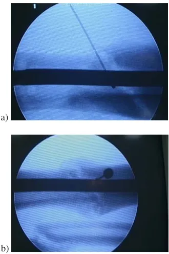
Fig. 1.Patient T.Z., age 30, disease history no. 7647/05, tibial bone. Percutaneous bone marrow graft− ing: a) injection into delayed union, b) injection around delayed union
Ryc. 1.Chory T.Z., lat 30, nr historii choroby 7647/05, kość piszczelowa. Przezskórna iniekcja ko− mórek szpiku kostnego: a) iniekcja w miejsce zrostu opóźnionego, b) iniekcja wokół zrostu opóźnionego
nine months has elapsed since injury and the frac− ture shows no visible progressive signs of healing for three months, and delayed union when it has not healed after three months. These criteria have been modified, as it is difficult to put a fixed time period on the definition. Taylor [4] stated that the criteria of nine months cannot be applied to every fracture and suggested that fracture nonunion of a long bone could be considered when there is a minimum of six months with no progression towards union. Our criteria for defining delayed union were six months and no evidence of union or three months without progression towards union. In our opinion it is very important to treat delayed union, before nonunion, despite the fact that some authors have written about good results also with nonunion [19, 20].
The next essential problem is to define exact− ly which cells work and what amount gives the best effects. The next problem is that there is no agreement on the name to be given to these cells which are injected. Names such as “mesenchymal stem cells” and “bone marrow stromal stem cells” (SSCs) are frequently used. Difficulties in giving a single name to these cells is also a consequence of their nature. They have been shown to be pre− cursors of many specialized cell types, not of only
mesynchemal lineage, such as bone, cartilage, and fat, but they have also been shown to became hepatic and neuronal cells, making the name “mes− enchymal stem cells” inaccurate. Bone marrow is a good source of stem cells, but not unlimited. It is estimated that there are 32 million nucleated cells per milliliter, and only 1 in 18,000 of nucleated cells is a stem cell. Because only a few milliliters of bone marrow is aspirated, there are only a few thousand SSCs that we can obtain from the bone marrow. Muschlera [21] wrote that increasing the amount of bone marrow aspirated is not a solution, because each single aspiration does not coincide with a significant increase in the number of SSCs, suggesting that after a certain amount, what is aspirated is blood and not bone marrow, and aspi− ration of 10 ml gives the best result [22].
Treatment by percutaneous injection with autologous stem cells is one of the methods which can be used in the treatment of delayed union of
Fig. 2.Patient T.Z., age 30, disease history no. 7647/05. Tibial delayed union treated with percuta− neous bone marrow grafting: a) 12 months delayed union, b) progression to union after 3 months
Ryc. 2.Chory T.Z., lat 30, nr historii choroby 7647/05. Kość piszczelowa leczona przezskórnymi in− iekcjami szpiku kostnego: a) zrost opóźniony po 12 miesiącach leczenia, b) zrost kostny po 3 miesiącach od zastosowania iniekcji szpiku kostnego
a)
b)
a)
long bones which give good results [14, 15]. Goel [16] reported achieving union in 75% of patients in 14 weeks, Niedźwiecki [14, 15] achieved union in every case of delayed union in 3.2 months and in every case of nonunion in 3.4 months following the first injection, Connolly et al. [17] used autol− ogous bone marrow injection and reported healing the fractures of 18 of 20 patients in an average time of seven months. In the present study, bone union was achieved in 70% and the average time to union was 16 weeks. The differences in the results are most likely due to the definition of causes of bone union disturbances. This should be remembered. Treatment with mesenchymal pluri− potent marrow cells is a complementary therapy applied after ensuring a correct reduction of frac− tures, immobilization, and a blood supply ade− quate to needs [18]. In the present study, cases of incorrect stabilization of fractures and accompany− ing damage to vessels and nerves were the main cause of treatment failure. Diagonal drilling of a fracture fissure overcoming the cartilage appear− ing on the fragments’ endings and enabling the
migration of cells and restoring common circula− tion between both fragments seems essential [19]. Introducing bone marrow into those fissures com− pletes the new method of Beck’s bone drilling with the new osteogenic material [20].
Percutaneous injection with autologous stem cells is an easy and safe procedure. This method is inexpensive, does not require extra instruments, has regulatory reasons, and can be preformed as a “minimally invasive procedure”. In comparison with an “open” technique, the risk of devascular− ization and infection at the fracture site, where healing is already impaired, is minimized.
Stromal stem cells possess osteogenic proper− ties which can be useful in the treatment of delayed union. Percutaneous injection with autol− ogous stem cells is an effective treatment in induc− ing bone healing and preventing nonunion. Percutaneous injection with autologous stem cells is a safe, time−saving, and economical “minimally invasive” alternative to “open” techniques in treat− ing delayed union.
References
[1] Tylman D, Dziak A: Traumatologia narządu ruchu. PZWL 1996.
[2] Górecki A:Czynniki wzrostu i tkanka kostna. Oficyna Wydawnicza ASPRA−JR; Warszawa 2004. [3] Zgliczyński SzL: Radiologia. PZWL, Warszawa 1989.
[4] Taylor CJ: Delayed union and nonunion of fractures. In: Campbell’s Operative Orthopaedics. 1992, 28 1287–1345.
[5] Russell AT, Taylor CJ, Lavelle DG:Fractures of tibia and fibula. In: Fractures in Adults, Rockwood and Green, 1991, 3, 1915–1982.
[6] Saleh M: Non−union surgery, part 1. Basic principles of management. Int J Orthop Trauma 1992, 2, 4–18. [7] Bruder S et al.: Growth kinetics, self−renewal, and the osteogenic potential of purified human mesenchymal stem
cells during extensive subcultivation and following cryopreservation. J Cell Biochem, 1997, 64, 278–294. [8] Bruder S et al.: The effect of implants loaded with autologous mesenchymal stem cells on the healing of canine
segmental bone defects, J Bone Joint Surg Am 80A, 985–996.
[9] Ohgushi H, et al.:Stem cell technology and bioceramics: from cell to gene engineering, J Biomed Mater Res 1999, 48, 913–927.
[10] Pittenger M, et al.:Multilineage potential of adult human mesenchymal stem cells, Science 1999, 284 (5411), 143–147.
[11] Richards M, et al.:Marrow−derived progenitor cell injections enhance new bone formation during distraction. J Orth Res 1999, 17, 6, 900–908.
[12] Słynarski K:Osteoindukcyjne właściwości wielopotencjalnych komórek szpiku. Ortop Trauma Rehab 2000, 3, 8–10. [13] Hammer R, Hammerby S, Lindholm B:Accuracy of radiologic assessment of tibial shaft fracture union in
humans. Clin Orthop 1985, 199, 233–238.
[14] Niedźwiecki T: Wpływ szpiku kostnego na gojenie złamań, zrostów opóźnionych i stawów rzekomych kości długich. Chir Narz Ruchu Ortop Pol 1993, LVIII, 3, 194–203.
[15] Niedźwiecki T, Niedźwiecki Ł: Wspomaganie gojenia stawów rzekomych kości piszczelowej przeszczepianiem autogennego szpiku kostnego. Nowiny Lekarskie 2001, 70, 4, 322–328.
[16] Goel A, Sangwan S, Siwach R, Ali A: Percutaneous bone marrow grafting for the treatment of tibial non−union. Injury, Int J Care Injured 2005, 36, 203–206.
[17] Connolly J, et al.: Autologous Marrow Injection as a Substitute for Operative Grafting of Tibial Nonunions. Clin Orthop 1991, 266, 259–270.
[18] Lucarelli E, Donati D, Cenacchi A, Fornasari P: Bone reconstruction of large defects using bone marrow derived autologous stem cells. Transfusion and Apheresis Science 2004, 30, 169–174.
[19] Kettunen J, Makela E, Turunen V, Suomalainen O, Partanen K: Percutaneous bone grafting in the treatment of the delayed union and non−union of tibial fractures. Injury Int J Care Injured 2002, 33, 239–245.
[21] Muschler G, Nitto H: Age− and gender−related changes in the cellularity of human bone marrow and the preva− lence of osteoblastic progenitors. J Orthop Res 2001, 19 (1), 117–125.
[22] Buewell R: The Function of Bone Marrow in the Incorporation of a Bone Graft Clin Ortop 1985, 200, 125–141.
Address for correspondence:
Paweł Reichert
Klinika Chirurgii Urazowej i Chirurgii Ręki AM we Wrocławiu ul. R. Traugutta 57/59
50−417 Wrocław Fax: +48 71 7332706
e−mail: [email protected]
Conflict of interest: None declared
Received: 31.10.2006 Revised:
Accepted:
Praca wpłynęła do Redakcji: 31.10.2006 r. Po recenzji: