ABSTRACT
WANG, GONGBO. Sindbis Virus Interaction with Cells. (Under the direction of Dennis T. Brown)
Sindbis Virus Interaction with Cells
by
Gongbo Wang
A dissertation submitted to the Graduate Faculty of
North Carolina State University
in partial fulfillment of the
requirements for the Degree of
Doctor of Philosophy
Biochemistry
Raleigh, North Carolina
2008
APPROVED BY:
Dr. Dennis T. Brown Dr. Keith Weninger
Chair of Advisory CommitteeVice Chair of Advisory Committee
Dr. Paul Wollenzien Dr. E. Stuart Maxwell
Dr. Jason A. Osborne
DEDICATION
谨以此博士论文献给我远在中国的父亲王怀波和母亲公维芝。这篇总结我二十一年受 教育历程的博士论文中,只有在这短短的一页里我可以使用母语来表达我感恩的心 情。我的双亲用他们所能承担的一切来养育我、教育我、激励我、支持我走到今天。 这篇论文以及我即将获得的博士学位不仅仅是我一个人的成就,而是我们全家人的。 我所能在此提及的支言片语并无法示及父母养育大恩的万分之一。
To my parents, Wang, Huaibo and Gong, Weizhi. This is the only page I can express
myself in my native language throughout this Ph.D. thesis that concludes my 21-year
education. My parents, who have raised me, educated me, inspired me and
supported me all my life, contributed everything they could afford to lead me towards
this grand achievement. This thesis and the degree I will receive is not only my own
achievement, but my family’s. All I can describe in this short page is not even
BIOGRAPHY
I was born Wang, Gongbo in Shenyang, China on November 11th, 1980 to Wang, Huaibo and Gong, Weizhi. As the only offspring of my parents, I have no
siblings but seven cousins. My father is the Chief Accountant of the biggest
securities company in Shenyang. My mother works for an international company
where she is the Chairman of the Employees’ Union.
My education life started in 1987 when I went to Qinggonger Elementary
School in the Tiexi District of Shenyang. After passing ridiculous numbers of exams
in 1993, I was admitted into Northeast Yucai Middle School where I finished both
middle school and high school education. In the year of 1999, I was admitted into
School of Life Sciences, Fudan University located in Shanghai, China. Without many
surprises, I graduated in four years from college with a bachelor’s degree. Then I
came to North Carolina State University located in the City of Raleigh, State of North
Carolina in U.S.A. seeking a higher degree. Things went really well. And I will get my
Ph.D. in the August of 2008 at the age of 27.
Being the first Chinese generation born after the “Culture Revolution” and
“Mao era”, my personal life verifies the prosperity of China and her economic growth.
At the time I was born, both my parents were labor workers, who did not get the
chance of higher education because of the Culture Revolution. They earned only
enough money not to starve. Fortunately, through their vigorous personal endeavors
economy policy. By the time I finished elementary school, my family started having
savings. My parents paid to get me started on taking advanced courses in math and
English outside regular school. All of this helped me in passing the exams required
to go to the best middle school in Shenyang. We also moved several times to bigger
and bigger apartments, which are common accommodations for most people in big
Chinese cities. Middle school and high school were fun. I was with the same
classmates for six years and made a lot of life-time friends. After high school, I left
my hometown. College life was hard and competitive in Fudan. In the first two years
of my college life, I worked so hard that I suffered from depression. This also
partially led to my breakup with a girl I grew up with. That chaotic period became the
turning point of my life, as I realized for the first time in my life that there were always
more important things than academic achievement and career. I also learned to
cherish what I already have. For the rest of my college life, I found a perfect balance
among academic achievement, health and pursuit of happiness. Other than that, I
need to mention I made a lot of very good friends in college, most of whom are
Shanghainese.
helped me in research, but also treated me as their family member. Besides in the
lab, I made a lot of friends by taking courses. It is impossible to mention all of their
names here. My American friends helped me get furniture, buy a car and do course
work when I first arrived. They also took me into their homes to meet their families,
drove me to the capitol of the country and hung out with me. It was quite an
experience to make so many friends in a foreign country.
Also in North Carolina State University, I met Qiqi Wang, who shares a very
common Chinese family name with me that sometimes confuses Americans. She
has been my companion, best friend and family in the past four years.
I will go to the University of Texas Medical Branch at Galveston in the
September of 2008 to start a post-doctoral position. I am switching gears to study
pathology and immunology.
ACKNOWLEGEMENTS
Committee members: Dennis T. Brown (NC State, Biochemistry), Keith Weninger
(NC State, Physics), E. Stuart Maxwell (NC State, Biochemistry), Paul Wollenzien
(NC State, Biochemistry), Jason A. Osborne (NC State, Statistics).
Lab mentors: Dennis Brown (NC State, Biochemistry), Raquel Hernandez (NC State,
TABLE OF CONTENTS
LIST OF FIGURES………..………..………….viii
LIST OF TABLES……….…………..………..xi
CHAPTER 1: INTRODUCTION……..………..……...1
CHAPTER 2: SINDBIS VIRUS INFECTION AT LOW TEMPERATURE
………..………...………...18
CHAPTER 3: INTRODUCTION OF THE GREEN FLUORESCENT PROTEIN GENE
INTO THE SINDBIS VIRUS GENOME AS A REPORTER OF SUCCESSFUL
INFECTION………..…….34
CHAPTER 4: IMPACT OF DELETIONS IN THE SINDBIS VIRUS E1
TRANSMEMBRANE DOMAIN ON VIRUS FUNCTION …..……….52
CHAPTER 5: METHODS………75
LIST OF FIGURES
CHAPTER 1
Figure 1.1: Sindbis virus outer surface from a cryo-electron microscopy
reconstruction………..4
Figure 1.2: Cut-away image of Sindbis virus from cryo-electron microscopy
reconstruction………..5
Figure 1.3: Organization, replication and expression of Sindbis virus genome
……….16
Figure 1.4: A schematic representation of Sindbis virus structural polyprotein in the
ER membrane………...17
CHAPTER 2
Figure 2.1: : Sindbis virions lose their RNA content at the cell surface at neutral pH
……….23
Figure 2.2: Formation of infectious centers by Sindbis virus at low temperature
CHAPTER 3
Figure 3.1: Expression of GFP gene in bacterial cells at 37 oC………...……38 Figure 3.2: Expression of GFP gene in bacteria cells at 15 oC………39 Figure 3.3: Genome organization of wild type Sindbis virus and modified virus with
the GFP gene………40
Figure 3.4: The normal inverted microscope mode………...42
Figure 3.5: The laser fluorescent mode of the microscope……….43
Figure 3.6: Cells infected by “green virus” express green fluorescent protein at 37
oC within 24 hours post-infection
………46
Figure 3.7: Cells infected by “green virus” express green fluorescent protein at 15
oC at 72 hours post-infection
………..47
CHAPTER 4
Figure 4.1: The E1 transmembrane domain amino acid sequence of wild type
Sindbis virus and deletion mutants………57
Figure 4.2: Virus infectivity determined by plaque assay. Viral RNA was transfected
into BHK cells………58
Figure 4.3: Particle to PFU (plaque forming unit) ratio of the mutants with short
deletions in E1. ………61
Figure 4.4: Estimated concentration of assembled particles………...62
Figure 4.5: Viral protein synthesis in mutants with large deletions in the E1
Figure 4.6: Wild type virus assembly and budding in BHK cells. ………..…66
Figure 4.7: BHK cells transfected with non-viral RNA………...67
Figure 4.8: BHK cells transfected with E1Δ420-433 RNA………68
Figure 4.9: BHK cells transfected with E1Δ411-428 RNA………69
Figure 4.10: BHK cells transfected with E1Δ416-433 RNA………..70
LIST OF TABLES
CHAPTER 5
Chapter 1
Alphavirus and its transmission pathway
Sindbis virus was first isolated in 1952 from a pool of mosquitoes in Egypt. It
is the prototype of Togaviridae family, Alphavirus genus. 25 other members of this
genus have been identified. Viruses in this genus are called alphaviruses. Eastern
Equine Encephalitis (EEE), Venezuelan Equine Encephalitis (VEE), Semliki Forest
(SF), Chikungunya (CHIK), Ross River (RR) and Western Equine Encephalitis (WEE)
are commonly seen alphaviruses (Strauss and Strauss, 1994). Alphaviruses are
transmitted in nature among birds, rodents and domestic animals by blood sucking
insects such as mosquitoes, ticks, midges and sand flies. Therefore they are also
categorized as arthropod borne viruses (arboviruses), which include alphaviruses,
flaviviruses and (-)-strand RNA bunyaviruses.
The life cycle of alphavirus involves insects, birds, mammals and humans.
Rodents and birds are natural reservoirs of these viruses. Blood sucking insects
carry viruses they obtain from a blood meal and infect other host animals during later
bites. The transmission can be enzootic within the same animal population.
Alphaviruses can also be transmitted epizootically to domestic animals and humans.
Some alphaviruses may cause severe diseases in infected birds and animals
leading to their deaths. After being bitten by an infected mosquito, host humans
develop a febrile disease. Most alphaviruses induce potent immune response in
human body which results in the elimination of virus and no severe symptoms. While
humans. In this case, viremic humans produce sufficient virus for the mosquito
vectors to transmit the disease onto other humans or animals after a blood meal.
The structure of Sindbis virion
Sindbis virus has stable structure and high yield from lab cell cultures, making
it the ideal subject for studying alphavirus structure. Additionally, it is non-pathogenic
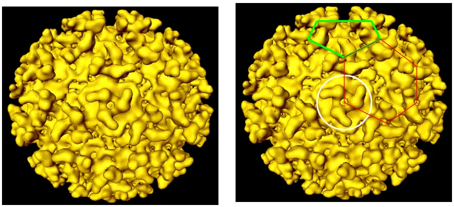
in humans rendering it easier and cheaper to handle. Figure 1.1 shows the
cryo-electron microscopy (cryo-EM) reconstruction image of Sindbis virion (Paredes et al.,
1993). The Sindbis virion is about 70 nm in diameter. The outer surface is covered
exclusively by E1 and E2 proteins, noted in yellow. E1 and E2 have 439 and 423
amino acids, respectively. Both proteins have a molecular mass of about 50 kDa.
The ectodomains are glycosylated and palmitoylated post-translationally. The two
glycoproteins form heterodimers, every three of which form a trimer. The white circle
in Figure 1.1 outlines one trimer composed of three E1 and three E2 proteins. Each
Sindbis virus has 80 of these trimers. If we draw lines between the centers of
adjacent trimers, we can find pentagons (shown in green lines) and hexagons
Figure 1.1: Sindbis virus outer surface from a cryo-electron microscopy
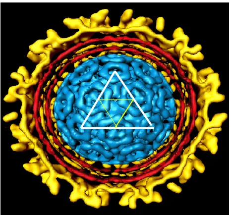
Figure 1.2: Cut-away image of Sindbis virus from cryo-electron microscopy
inner protein shell is composed of 240 copies of the same capsid protein, 30-kDa in
size. The triangles drawn on the inner protein shell signify the traditional way of
identifying T=4 icosahedral symmetry structure. The red lipid bilayer is derived from
the host membrane. E1 and E2 glycoproteins have transmembrane domains that
anchor them in the host derived membrane bilayer. E2 has a 33-residue cytoplasmic
domain, while E1 has a short cytoplasmic domain of only 2 residues (Strauss and
Strauss, 1994). The cytoplasmic domains are involved in an interaction with the
capsid protein, which is important for the stability of Sindbis virus. As shown in
Figures 1.1 and 1.2, the inner nucleocapsid shell has the same T=4 icosahedral
symmetry as the outer glycoprotein shell. In the center of the whole structure is one
plus sense, single stranded genomic RNA, which is not shown in Figure 1.2.
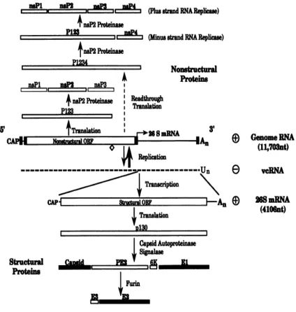
Sindbis viral genome
The Sindbis viral genome is composed of one RNA molecule with a 5’ cap
and 3’ poly-A tail. The molecule is packed in the core of nucleocapsid with high
density, filling the provided space. The Sindbis viral genome can be seen as two
parts: the 5’-terminal two-thirds and the 3’-terminal one-third of the RNA. The 5’
portion contains the genetic information of four non-structural proteins, nsP1, nsP2,
nsP3 and nsP4. The non-structural proteins have methyltransferase, helicase,
protease and RNA polymerase activities. The 3’ part of the genome encodes a
polyprotein that is processed into capsid, E2, E1 structural proteins and two small
virion. Trace amount of E3 and 6K protein has been reported to be present in mature
Sindbis virions. (Gaedigk-Nitschko and Schlesinger, 1990; Liljestrom et al., 1991;
Lusa, Garoff, and Liljestrom, 1991)
Alphavirus infection and life cycle: attachment
It is generally believed that alphavirus infection is started by the attachment
of a virion to a cell receptor, like all other viruses. The receptor of alphavirus
infection is not known. Since Sindbis virus can reproduce in many laboratory cell
cultures derived from different organs of both insects and mammals, the cell
receptor/s must be a universal class of molecules that are presented in all of these
cells. It is also possible that alphaviruses can utilize different receptors, as insect
and mammalian cells differ in many aspects.
Alphavirus infection and life cycle: penetration
Following receptor attachment, alphavirus penetrates into the cell and
releases its genome into the cytosol. There are two proposed mechanisms on how
particle and release of the viral genome into the cytosol (Helenius, 1984; Helenius
and Marsh, 1982; Marsh, 2006). This infection pathway was first found in influenza
viruse and later proposed to be the entry mechanism for all enveloped viruses and
some non-enveloped viruses (Marsh, 2006).
Evidence supporting the role of endocytosis in alphavirus penetration
includes inhibition of virus production by cellular mutations (DeTulleo and
Kirchhausen, 1998), lysosomotropic weak bases (Cassell, Edwards, and Brown,
1984; Helenius, Marsh, and White, 1982) and other drugs that raise the pH of
endosomes (Marsh, Bolzau, and Helenius, 1983). The assays employed in these
experiments measure viral RNA or structural protein synthesis; processes which
occur after expression of the non-structural proteins, negative strand synthesis and
subgenomic RNA production must have taken place. Viral RNA and structural
protein synthesis are comparatively late events that require the correct functioning of
the infected cell. However, drugs and mutations may have multiple effects. In other
words, they may directly or indirectly impair other functions related to viral RNA and
structural protein synthesis. These indirect effects were demonstrated in mosquito
cells in which, it was shown that some weak bases such as chloroquine can not
inhibit virus production and others interfere with virus production by blocking
proteolytic processing of the non-structural precursor protein (Hernandez, Luo, and
Brown, 2001).
Evidence supporting alphavirus entry by low-pH dependent fusion comes
can induce cell-cell fusion (Bron et al., 1993; Kielian, 1995; Kielian and Jungerwirth,
1990; Lu and Kielian, 2000; White, Kartenbeck, and Helenius, 1980). The ability of
alphaviruses to induce fusion between cells in a pH dependent manner was
developed to support the notion that viral and cellular membranes can fuse at low
pH. This fusion event was proposed to be the same as what happens between the
viral and endosomal membranes when endosome acidification occurs. However, it
was later shown that this fusion property is actually a two-step event (Edwards and
Brown, 1986). Fusion does not occur until the pH is switched to acidic and brought
back to neutral. Maximum fusion was achieved when pH went to 5.3 and came back
to 7.4. No fusion occurs at pH 5.3. This is a different scheme as the one-time pH
switch to acidity which occurs in endosomes. Another line of evidence supporting the
role of fusion in alphavirus infection comes from observing the interaction between
virus and liposomes, which are the substitute of cells in the system (Kielian, 1995;
Kielian et al., 2000; Kielian and Jungerwirth, 1990). But there are several issues with
this artificial system concerning the validity of replacing cells with liposomes. First of
all, the liposomes used did not have cell receptors or any other protein contents.
Rietveld, Koorengevel, and de Kruijff, 1995). Additionally, depending on the
composition of the liposomes, fusion can occur at different pH values (Bron et al.,
1993; Haywood and Boyer, 1985). By changing the proportion of lipids and
cholesterol, virus can fuse with liposomes at neutral pH. Finally, when fusion
between virus particles and cells in the context of pH switches was examined by
electron microscopy, the two steps of the pH switch are still required (Paredes et al.,
2004).
The crystal structure of alphavirus E1 protein has been deemed to support
the fusion model (Gibbons et al., 2004; Jardetzky and Lamb, 2004). The crystal
structure of E1 was achieved at both neutral and acidic pH. An analogy of the fusion
protein in influenza virus to E1 has been proposed. However, the crystal structure of
E1 was achieved using treatment with detergent, which suggests that the obtained
structure may not be in its native configuration (Mulvey and Brown, 1994; Whitehurst
et al., 2007). According to the crystal structures, the fusion protein of influenza virus,
hemmaglutinin, undergoes a reorganization of the domains with a pH change to
acidic. Although E1 protein completes a similar change according to crystal
structures and molecular modeling, this change requires refolding of the protein in a
manner which is hard to achieve in the native environment of an intact virus. Plus,
E1 and E2 form heterodimers instead of functioning entirely independently. There is
no evidence that E1 can undergo the same conformational changes found in the
A novel proposed mechanism is the direct penetration of virus at the cell
surface in a pH independent manner (Paredes et al., 2004). In this scheme, the
binding of virus to the cell receptor is followed by a pore formation between virus and
the infected cell. This pore (Koschinski et al., 2005) is hypothesized to provide a
channel through which the viral RNA is injected into the cytosol. This mechanism
does not require endocytosis or fusion. Chapters 2 and 3 of this thesis will focus on
this aspect of the alphavirus infection cycle. More details on data supporting this
mechanism and how the whole process is proposed to occur will be provided.
Alphavirus infection and life cycle: genome replication
After the viral genome is introduced into the cells, part of the cellular
machinery for translation is hijacked. Alphavirus has four non-structural proteins on
the 5’ end of viral genome. They are named after nsP1, nsP2, nsP3 and nsP4,
according to their order in the RNA. The non-structural proteins are translated from
the 5’ end of full length genomic RNA as two polyproteins, nsP123 and nsP1234.
There is an opal codon at nucleotide 1897 of the non-structural open reading frame
during infection. Some of them may form a complex with host proteins to carry out
replicase activity (Strauss and Strauss, 1994). Non-structural proteins are not as
extensively studied as the structural proteins, but there is much information available.
nsP1 appears to be necessary for minus-strand RNA synthesis. nsP2 has
proteinase activity and is required for 26S subgenomic RNA synthesis. nsP2 may
have the RNA helicase activity. The functions of nsP3 are not well defined. nsP4
seems to possess RNA polymerase activity to help with (-)-strand synthesis
(Keranen and Kaariainen 1979; Sawicki, Sawicki et al. 1981; Strauss and Strauss
1994).
Following expression of these essential non-structural proteins,
complementary minus strand RNA is produced. In the presence of both viral and
cellular proteins that have RNA dependent RNA polymerase activity, two plus strand
RNAs are made from the minus strand, the full length genomic RNA and a 26S
subgenomic RNA. The subgenomic mRNA, one third of the full genome in size, is
also 5’ capped and 3’ polyadenylated. This RNA is simply a copy of the 3’ end
nucleotide sequence in the full length genomic RNA. Only the full length genomic
RNA is packaged into a mature virion.
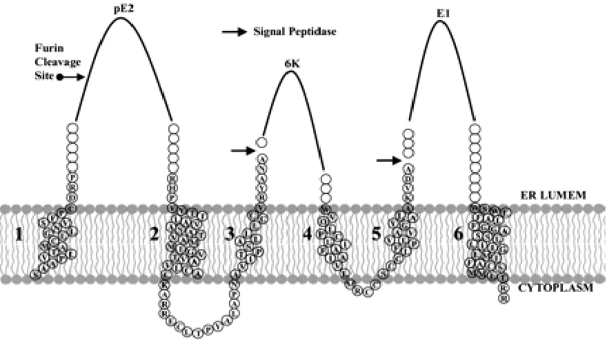
Alphavirus infection and life cycle: assembly of nucleocapsid
The structural proteins are first translated as a polyprotein from the 26S
subgenomic RNA, which is posttranslationally cleaved into capsid, E1, E2, E3 and
ribosomes, the capsid protein is removed from the polyprotein by an autoproteolytic
activity. The signal sequence exposed by the removal of capsid directs the insertion
of a polyprotein with six transmembrane domains (Fig. 1.4) into the endoplasmic
reticulum membrane (Hernandez et al., 2003) and then processed into pE2, 6K and
E1 by signalase (Liljestrom and Garoff, 1991). pE2, the precursor of E2, is then
cleaved into E2 and E3 proteins by furin during the transport of pE2-E1 complex.
The transmembrane domain near the C-terminus of pE2 reorients into the cytoplasm
during maturation (Liu and Brown 1993; Liu and Brown 1993). E1 and E2 proteins
also undergo posttranslational decorations such as glycosylation and palmitoylation.
After the expression of structural polyprotein and autoproteolytic removal of
capsid, assembly starts as genomic RNA is associated with a capsid protein. This
complex serves as a base for nucleocapsid assembly. The encapsidation process is
specific, as normally only full length RNA is packaged (Weiss et al., 1989).
Nucleocapsids that are empty or contain other RNAs are not assembled. Only a
single RNA molecule is packaged in the nucleocapsid, and presumably the size of
interaction between the endodomain of E2 glycoprotein and capsid directs budding
and maturation of virus particles (Lopez et al., 1994). Each capsid protein finds and
binds to an E1-E2 heterodimer. While this pairing process occurs, virus particles bud
through the plasma membrane. At the end of the process, nucleocapsid is sealed in
a protein shell of E1-E2 glycoproteins. During this process, the lipids associated with
the transmembrane domains of glycoproteins become part of the virus structure. The
lipid bilayers finally merge and form a membrane bilayer sandwiched between the
capsid and glycoprotein shells. Upon completion of this process, the mature virus
can proceed to infect the next host cell and reproduce.
Transmembrane Domain Interaction between E1 and E2
The Alphavirus genome is protected by two protein shells. Between the
protein shells is a host derived membrane bilayer. E1 and E2 proteins have
transmembrane domains so that they can pass through the bilayer and contact the
inner shell. There is evidence that E1 and E2 transmembrane domains interact with
each other at the molecular level (Strauss, Lenches, and Strauss, 2002). Sequential
deletions were made in the E2 transmembrane domain (Hernandez et al., 2003;
Whitehurst et al., 2006). Deleting a single methionine (M379) dropped the titer by
four orders of magnitude. Interestingly, the titer is restored gradually when more
amino acids were deleted from the domain. Deletions in the E2 transmembrane
domain do not affect the assembly process but have a dramatic impact on virus
domain, serial deletions were made that cover all faces of the helix throughout the
length of the domain. The data indicate that deletions closer to the cytoplasmic face
reduced virus infectivity much more than deletions closer to the luminal side of the
membrane (Whitehurst et al., 2006).
In looking for E1-E2 transmembrane domain interactions, amino acids in E1
transmembrane domain were also deleted. These results including virus titer, viral
Figure 1.3: Organization, replication and expression of Sindbis virus genome.
Figure 1.4: A schematic representation of Sindbis virus structural polyprotein in the
ER membrane. The polyprotein is processed by signal peptidase and furin activity
after integration so that 6K protein and transmembrane domain 1 are cleaved.
Transmembrane domain 3 flips during the maturation process and becomes the
CHAPTER 2
Sindbis virus infection at low temperatures
Gongbo Wang, Raquel Hernandez, Keith Weninger and
Dennis Brown
Published in:
It is widely held that membrane containing viruses employ those membranes
as tools of infection (Eckert and Kim 2001; Marsh and Helenius 2006). These
enveloped viruses gain entry to potential host cells by fusing the virus membrane
with the host cell membrane. This fusion event is mediated by proteins integrated
into the virus membrane and serve as motors to drive the fusion process (Jardetzky
and Lamb, 2004). Virus fusion proteins can mediate the process of fusion with a
host cell membrane in a low pH dependent or independent manner (Marsh and
Helenius, 2006). In the low pH independent pathway, it is proposed that virus
glycoproteins interact with a cell receptor and that this interaction induces protein
conformational changes which drive the fusion of the virus membrane with the cell
membrane. An example of virus entering cells by fusion at the cell surface is HIV
(Cardoso et al., 2005; Eckert and Kim, 2001). In the low pH dependent pathway it is
proposed that upon interaction with a receptor, virus particles are internalized into
endosomes. In the late endosome and upon acidification, pH induced conformational
changes in the virus proteins lead to membrane fusion (Kielian and Jungerwirth,
1990). This latter pathway has been proposed for many families of viruses and is
(Gibbons et al., 2004). A significant difference in the putative fusion inducing
proteins of influenza virus and the alphaviruses is a dramatic dissimilarity in structure
and sequence (Gibbons, Vaney et al. 2004; Modis, Ogata et al. 2004; Zaitseva,
Mittal et al. 2005). Because of these differences, the proteins are referred to as
Type I (Flu) and Type II (alphavirus) fusion proteins. Type I fusion proteins are
found in a wide variety of unrelated virus families (Eckert and Kim, 2001). Type II
fusion proteins are found only in the insect vectored Alpha and Flaviviruses. Their
ability to fuse with liposomes and the observations that inhibitors of endosome
acidification or mutations which block endosome formation can prevent subsequent
virus RNA or protein synthesis have been presented as evidence that Alpha and
Flaviviruses also penetrate cells from the acidic environment of endosomes
(Helenius, Marsh et al. 1982; DeTulleo and Kirchhausen 1998). Each of these
experiments has presented problems in their interpretation. Fusion with protein free
liposomes has been demonstrated to “absolutely” require the presence of
cholesterol in the liposome membrane (Kielian and Helenius, 1984). Liposomes
employed in these studies typically contain 25-50 mole% cholesterol. Insects are the
alternate host for these viruses and their membranes have less than 3% cholesterol
(Rietveld et al., 1999). Insect cell lines, such as SF21 cells (Cha et al., 1997) (Gibco,
Invitrogen) have been adapted to synthetic growth medium (Weiss et al., 1993)
(SF900IISFM, Gibco, Invitrogen) that contains < 5% cholesterol. It has been shown
that Japanese Encephalitis Virus can infect and replicate in these cells (Kim et al.,
(to be published elsewhere). Studies using endosome acidification inhibitors or cells
encoding genetic defects in the endocytic pathway measure the products of late
events such as RNA or protein synthesis to determine that penetration has not taken
place. These events occur several steps after penetration. These experiments raise
the possibility that the defect in virus production may occur after penetration. In one
instance it has been shown that a reagent blocking endosome acidification does not
block penetration but rather blocks processing of virus non-structural proteins to
form the RNA replicating complex (Hernandez, Luo, and Brown, 2001).
The structures of Alpha and Flaviviruses present an innate barrier to the
fusion process. The Alphavirus particle is composed of two symmetrically identical
T=4 icosahedral protein shells nested one inside the other (Paredes et al., 1993).
The virus membrane is sandwiched between the two shells and protected by the
outer shell. Intramolecular disulfide bridges are involved in the lateral associations
which stabilize the outer protein shell (Anthony, Paredes et al. 1992; Mulvey and
Brown 1994). The E1 protein is assembled in a lateral matrix with fenestrations at
the 5 and 6 fold axes above the membrane bilayer. Disassembly of this protein
liposomes which does not require a return to neutral pH and suggests that
interactions with living cells are more complex in nature. In electron microscope
studies we have found that when infectious Sindbis virus particles interact with the
surface of living cells they lose their electron dense core. This suggests that the
virus RNA has been transferred to the cell (Figure 2.1) (Paredes, Ferreira et al.
2004). These data led to the hypothesis that interaction of the virus spike
glycoproteins with the cell surface resulted in the formation of a protein pore in the
cell plasma membrane through which the virus RNA passed to initiate infection.
Evidence supporting the formation of such a pore during virus infection has
subsequently been provided (Koschinski et al., 2005). Events related to the process
of penetration occurred at the cell surface in the absence of endocytosis and at
neutral pH (Paredes, Ferreira et al. 2004). In these experiments we found that about
25% of particles attached to the cell surface became empty at 37 oC. To our surprise 3% of particles attached became empty if the cell monolayers were maintained at 4
oC. This number is significant because Sindbis does not assemble empty particles,
the virus employed was purified 2X by density gradient centrifugation and no empty
particles were seen if virus was fixed prior to addition to cells. These observations
suggested that some events related to the infection process could occur at
temperatures which prevent endocytosis and indeed prevent all vesicular transport
(Lippincott-Schwartz, Roberts, and Hirschberg, 2000).
Figure 2.1: Sindbis virions lose their RNA content at the cell surface at neutral pH.
stopped (Lippincott-Schwartz, Roberts, and Hirschberg, 2000). To further
characterize the interaction of Sindbis virus with cells under conditions which block
endocytosis, we examined the ability of the virus to establish infection at 5 oC and 15
oC. Our approach was to create an antibody escape experiment consisting of
thefollowing steps: 1. A known quantity of virus was bound to identical monolayers of
cells at 5 oC for 15 minutes (attachment). 2. The cells were washed to remove
unbound virus and placed in media at either 5 oC, 15 oC or 22 oC for either 30 or 60 minutes (penetration). 3. The cells were then washed and incubated at 5 oC in media containing 0.4 mg/ml anti-SVHR whole virus IgG for 60 minutes (to inactivate
virus particles which had not infected the cell or undergone a conformational change
leading to infection) or in medium without antiserum. The antiserum used was
polyclonal and the same as what was used in the electron microscopy studies of
Paredes et. al. (2004, see also figure 2.1). In a virus inactivation experiment the
antiserum inactivated 97% and 100% of virus infectivity in suspension in 15 and 30
minutes respectively at 5 oC in suspension. 4. The cells were then overlaid with agarose in media, incubated at 37 oC for 24 hrs. and stained with neutral red as in a standard plaque assay (Renz and Brown, 1976). In order to interpret the result of
this experiment, it was necessary to demonstrate that the antiserum employed could
inactivate virus particles after the virus had attached to the cell surface. This was
done by following the protocol presented above but eliminating step 2, the
penetration step. In this control experiment, we found that 13.9% of the virus
experiment are presented in figure 2.2. We found that significant numbers of virus
particles could establish infection at low temperature. The overall levels of virus
infection increased as temperature was increased from 5 oC to 15 oC and time was increased from 30 to 60 minutes. The majority of viral particles (97.7%) were
capable of initiating the infection process at room temperature (22 oC) in 60 minutes, while this number falls to 78.4% if the incubation period is reduced to 30 minutes. A
significant fraction of virions are also capable of initiating infection activity at 15 oC in 30 minutes (60.4%) and 60 minutes (72.4%). Even when the incubation is done at 5
oC we can detect viral infection in 28.3% and 35.7% of the virions, at 30 and 60
minutes respectively. These results indicate that events leading to the infection of
cells can occur at temperatures which do not permit endocytosis and that this
process occurred in a time and temperature dependent fashion yielding measurable
kinetics of the infection reaction.
The results presented in figure 2.2 could alternatively be explained by the
possibility that endocytosis is not blocked but rather slowed as temperature is
lowered. While the effects of temperature on endocytosis have been extensively
Figure 2.2: Formation of infectious centers by Sindbis virus at low temperature.
Cells were infected with Sindbis, washed and exposed to antiserum as described in
the text. The amount of virus which was able to infect cells under the conditions of
time and temperature indicated was determined as the percentage of the total
number of plaque forming units applied to the cell monolayers. The 0 time point at 5
oC is the control for the ability of the antiserum to inactivated virus after attachment
to cells. Checkered: control. Squares: 5 oC. Chevron: 15 oC. White: 22 oC. Each entry is the average of three independent experiments. The error bars represent
standard deviation of the data.
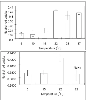
red was extracted from the cells with organic solvent (1:1 acetic acid/ethanol). The
amount of cell associated neutral red was determined by measuring absorbance of
the extract at 550nm. The results of these experiments are presented in figure 2.3.
We found that at low temperatures (5oC, 10 oC and 15 oC), a low amount of neutral red was cell associated. This amount remained constant in all three of the lower
temperatures tested. At temperatures of 22 oC and higher, a much greater amount of
cell associated neutral red was recovered. A statistical analysis of the data produced
gave a p-value of less than 0.0001 of the data from all six temperatures, which
affirms a 99.99% confidence that the shift of neutral red uptake was not due to
variance in the experiment. The same statistical procedure was applied to the
subgroups of neutral red uptake data. Data from 5 oC, 10 oC and 15 oC experiments
resulted in a p-value of 0.3664, while 22 oC, 28 oC and 37 oC gave a p-value of 0.2027. Thus, there is no significant difference among the three groups of data either
below or above 15 oC. These data are in agreement with the observation that
endocytosis is efficient at temperature of 22 oC and higher but raised the possibility that lower temperatures slowed but did not arrest the process of endocytosis. To
due to background association and not to endocytosis. These data support the
previous investigation demonstrating that endocytosis in mammalian cells does not
take place at 15 oC or below.
The data presented above suggest that events leading to the infection of
living cells by Sindbis virus can occur in the absence of endocytosis. We have
proposed that these events also occur without the need for membrane fusion. To
further examine this possibility we have determined if the phenomenon of Sindbis
virus mediated cell-cell fusion can take place at low temperatures. We have
previously shown that in contrast to fusion of virus with artificial liposomes, fusion of
wild type virus with living cells does not occur at acidic pH but requires a return to
neutral pH (Edwards and Brown, 1986; Paredes et al., 2004). We examined the
ability of Sindbis virus to fuse BHK cells at 15 oC and 37 oC using standard procedures described previously (Edwards and Brown, 1986; Paredes et al., 2004).
The result of this experiment is presented in figure 2.4. We found that at 15 oC, no
evidence of cell fusion could be detected. At 37 oC, significant fusion of the cell monolayer was seen.
The use of low temperatures to block endocytosis and other events related to
vesicular transport has been widely employed by cell biologists to study these
processes (Lippincott-Schwartz, Roberts, and Hirschberg, 2000). We have used the
benign mechanism of lowering the incubation temperature to establish conditions for
studying the roles of vesicular transport/endocytosis in the penetration of cells by
Figure 2.3: Uptake of neutral red by BHK cells at different temperatures. Cells were
preincubated at the temperature and treated with neutral red in PBS-D for one 0.3 0.32 0.34 0.36 0.38 0.4 0.42 0.44
5 10 15 22 28 37
Temperature (oC)
Neutral red uptake
NaN3 0.3400 0.3600 0.3800 0.4000 0.4200 0.4400
5 15 22 22
Temperature (oC)
in a plaque assay) and other researchers also employed the assay of alternate virus
replication events (RNA or protein synthesis as indicators that penetration had taken
place). Our result differs from previous reports in that the data is positive indicating
penetration must have taken place rather than a negative result (indicating that an
event not related to penetration did not occur). In the case of a negative result,
many known factors and unknown host cell mediated responses may have been
involved.
The data presented above suggest that interaction of virus with living cells at
low temperature allows for conformational changes in the virion which place the
virus beyond inactivation by antivirus serum. The serum employed in this study was
polyclonal and rapidly neutralized virus in solution. The antiserum was the same
reagent used in the immuno gold-labeling experiments examining the process of
Sindbis virus infection by electron microscopy (Paredes et al., 2004). In that EM
study, we found that this antiserum could bind to virus which had lost its electron
dense core and presumably had delivered its RNA into the host cell (see also figure
2.1). That observation suggested that the antiserum could bind to virus after RNA
penetration and after protein conformational changes related to that process had
taken place. We expect that the ability to bind virus in various states is retained in
this study and that the escape from inactivation by antibody seen in figure 2.2 is
likely not due to an inability of the antibody to bind a virion which has attached to
cells and undergone structural rearrangements. The data presented in this paper
37
oC, pH5.3 pH5.3
37
oC, pH5.3 pH7.4
Figure 2.4: Virus mediated cell-cell fusion at different temperatures induced by pH
cells by a mechanism that does not require endocytosis, exposure to acidic pH or
high concentrations of cholesterol in the target membrane (Koschinski et al., 2005;
Paredes et al., 2004; Rietveld et al., 1999). The observation that events leading to
infection can be initiated at low temperature, conditions which also prevent
membrane fusion, support the notion that penetration occurs by production of a
protein pore in the cell plasma membrane (Koschinski et al., 2005; Paredes et al.,
2004). Evidence for the existence of such Sindbis virus induced pores produced in
the plasma membranes of infected cells has existed for some time established by
the observation that the newly infected cell becomes leaky and ion permeable (Ulug
et al., 1984). This observation has been confirmed recently in experiments
elaborating differences in pores formed as a result of penetration and pores formed
as a result of membrane modification by virus proteins (Koschinski et al., 2005).
Electron microscopy has also indicated the presence of a hollow connector
developing between the virus and the cell surface during infection (Paredes et al.,
2004). The early cytopathic effect in living cells exemplified by changes in ion
permeability is in direct contrast to the fusion between virus and liposomes induced
by low pH which is found to be non-leaky (Smit et al., 2002).
The difference in the structure of the envelope glycoproteins which make up
the Type I and Type II “fusion” proteins suggest that while both classes of proteins
are capable, under very specific conditions, of producing fusion of the virus
membrane with a target membrane, they may function differently during the process
Type I fusion proteins which are capable of inducing a fusion event may differ from
Type II fusion proteins. Type II fusion proteins may, in concert with the virus
receptor, produce a protein pore in the host cell membrane. Infection of host cells by
“Type II” proteins which are found in membrane containing viruses that must infect
both vertebrates and invertebrates may require that they employ structural proteins
in a very different ways to breach the barrier presented by these very different
membranes. That the alternate, insect host, is poikilothermic and functions at
ambient temperature may explain the ability of the Alphaviruses to infect cells at low
CHAPTER 3
Introduction of the green fluorescent protein
gene into the Sindbis virus genome as a
In the antibody escape experiment discussed in chapter 2, we carried out
most of the treatments at low temperatures and added antibody at the last step to
stop infection. After antibody treatment, virus particles should settle into two
categories: those that have started the infection process before antibody addition
and the others that have not. The virus particles that escaped antibody treatment
should reproduce themselves in the infected cells and form plaques by spreading
out and killing the peripheral cells. Those virions that did not get a chance to
complete the initial steps of infection should be bound by antibody molecules and
lose the ability of infection. By comparing the number of plaques formed in the
sample treated with antibody to that of no-antibody treatment, we could calculate the
percentage of infection that had occurred before addition of antibody. In that
experiment however, we had to bring the cells to 37 oC after antibody treatment and overlay the cell monolayers with agarose in media so that we could count plaques.
The assay part of the experiment was actually finished after antibody treatment.
Raising the temperature to 37 oC was only for counting plaques and should not affect the assay result.
15 oC. We introduced ZsGreen gene, a derivative of
Zoanthussp. green fluorescent
protein gene, into the wild type Sindbis viral genome (Figure 3.1). Then we infected
Baby Hamster Kidney (BHK) cells with this newly constructed virus. When we did
both the infection and incubation at temperatures no higher than 15 oC, we detected with the help of laser microscopy the presence of green fluorescent protein.
Expression and maturation of green fluorescent protein at 15 oC
The green fluorescent protein (GFP) gene was bought from Clonetech. It
came in the DNA form, vectored in a pUC19 plasmid backbone. In this plasmid,
named pZsGreen, the GFP gene is under the control of the LacZ operon. The gene
itself encodes a protein that is the derivative and brighter version of Zoanthus sp.
green fluorescent protein. Zoanthussp. is a species of corals that can grow on reefs
and express green fluorescent protein at 20 oC in the sea. The protein needs a maturation step after expression to form tetramers before becoming fluorescent. The
reported “maturation time” between transfection and seeing obvious green
fluorescence provided by the vendor is 8-12 hours at 37 oC (Clontech, product information sheet).
To evaluate the expression and maturation process of GFP, we transformed
the commercial vector that GFP gene resides into E.coli cells, grew the culture and
measured bulk green fluorescence versus time. A 10-ml culture of bacterial cells
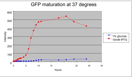
containing pZsGreen was grown overnight in LB media containing 1% glucose.
expressed. Then the culture was split into two equal volumes. One of them was kept
in 1% glucose as the control. IPTG was added to the other one to induce gene
expression controlled by the LacZ operon. We did this experiment at both 15 oC and 37 oC. In 5 hours at 37 oC, massive maturation of GFP began (Fig. 3.1). After 10 hours, the production and degradation of GFP tetramers reached equilibrium. The
amount of GFP plateaus from this time on, until the last time point recorded.
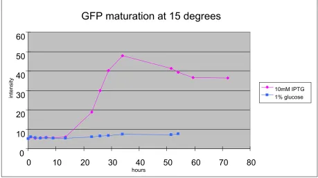
While at 15 oC, the maturation process takes much longer time (Fig. 3.2). No GFP was detected until 20 hours post-induction and the concentration of GFP fades
quickly. The highest concentration of GFP reached in bacteria at 15 oC is only one tenth that seen at 37 oC (Fig. 3.1, Fig. 3.2). Part of this difference is due to the growth of bacteria at 37 oC and absence of bacterial self-replication at 15 oC. A
general approximation we can get from the graphs is that metabolism slows at 15 oC to require triple the time at 15 oC compared to 37 oC. Although metabolism is much
slower at 15 oC, we have confirmed that green fluorescent protein can be
synthesized and maturate at 15 oC (Fig. 3.2).
Figure 3.1: Expression of GFP gene in bacterial cells at 37 oC. The same source of bacterial cells containing GFP gene in the plasmid were split into two at the 0
time point. In the control, lacZ operon was inhibited by 1% glucose. Expression of
GFP was induced by the addition of IPTG. Samples were kept shaking in a 37oC incubator. Expression of GFP was quantified by measuring absorbance at 505nm
with excitation at 493nm in a fluorometer.
GFP maturation at 37 degrees
0 100 200 300 400 500 600
0 5 10 15 25 30 35
hours
Intensity
GFP maturation at 15 degrees
0 10 20 30 40 50 60
0 10 20 30 40 50 60 70 80
hours
intensit
y
10mM IPTG 1% glucose
Figure 3.2: Expression of GFP gene in bacteria cells at 15 oC. Same source of bacteria cells containing the GFP gene in the plasmid were split into two aliquots
after the log phase was reached. In the control, lacZ operon was inhibited by 1%
glucose. Expression of GFP was induced by addition of IPTG. Samples were kept
Figure 3.3: Genome organization of wild type Sindbis virus and modified virus
with the GFP gene. The wild type viral genome has 11703 nucleotides. The
two XbaI sites. pTE3’2J is a plasmid DNA that contains all the genomic information
of Sindbis virus. At the 5’ end of the viral genome between the structural protein
genes and poly-adenylated tail, there is a repeated subgenomic promoter. pTE3’2J
also has a unique XbaI cutting site, which is located between this extra subgenomic
promoter and poly-adenylated tail. GFP gene was introduced into pTE3’2J plasmid
by a ligation reaction. The product DNA was then transformed into E.coli cells. The
colonies containing product DNA with GFP gene at the correct orientation were
screened out by PCR.
Production of “green virus” from plasmid DNA clone
As the first step in making virus from plasmid DNA, DNA was linearized using
the unique XhoI restriction site. Full length viral genomic RNA was produced by in
vitro transcription using SP6 RNA polymerase, followed by transfection into BHK
cells.
The “green virus” from the transfection has an infectious virus titer of
Figure 3.4: The normal inverted microscope mode. The differential interference
contrast (DIC) lamp must be turned on for this mode. Sample images can be
Figure 3.5: The laser fluorescent mode of the microscope. The laser mode is
controlled by a shutter. Laser activated fluorescence from the sample can be
Real-time laser microscopy observational system
To look at virus infection at the single-cell level, we exploited a real-time laser
microscopy observational system. A schematic representation of how the system
works and how it switches between the two modes is shown in Figures 3.4 and 3.5.
The microscope can be used as an inverted microscope to look at cells by
Differential Interference Contrast (DIC) mode (Fig. 3.4). In this mode, the laser beam
is blocked before entering the system. The DIC lamp is the light source. Image of
cells can be seen through the eyepiece lens. There is also a camera connected as
the alternative observation pathway (not shown in the figure). The camera can
receive the signal and pass it onto a computer. Software on the computer can show
the images and collect data as images or movies.
By controlling the on and off of the shutter, we can shoot the laser beam
perpendicularly through the center of the lens. With the DIC lamp off, the system
switches to the laser-fluorescence mode. In this mode, the laser is the light source
that activates fluorescence energy in the sample. The fluorescent signal is then
transmitted through the lens into the observational path. Before the signal reaches
the eyepiece lens or camera, a filter in the light path makes sure only light at the
correct wave length can penetrate. This setup is to get rid of the image of
unnecessary background fluorescence from the cells. In our experiment, a blue laser
is used to activate fluorescence from mature GFP. Data in image or movie form
Infection of BHK cells by “green virus” at 37 oC
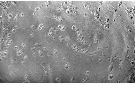
With the virus and observational system ready, we infected cells with the
modified “green virus” and incubated the samples at 37 oC. BHK cells infected by this modified virus can express green fluorescent protein at 37 oC as observed by laser microscopy (Fig. 3.6). After the cells were infected by wild type virus, we could
see a normal cell monolayer in DIC mode (Fig. 3.6 A). When we looked at this same
area under the laser mode, we could only see the blue background (Fig. 3.6 B). The
cells infected by “green virus” do not look different under DIC mode (Fig. 3.6 C).
However, these cells have potent green fluorescence under a blue laser (Fig. 3.6 D).
The camera in this system can only measure the strength of signal at each pixel, but
not the colors. The colors in the image generated by computer software represent
different strengths of the signal. The dark red area has the strongest green
fluorescence. The yellow area has less green fluorescence than the dark red area,
but more than the bright blue area. The blue area is background.
The titer data and laser microscope observation results confirmed that the
virus we constructed with an extra gene can infect cells with almost wild type virus
The question that naturally follows this observation is, if endocytosis is not the
(A) (B)
(C)
(D)
Figure 3.6: Cells infected by “green virus” express green fluorescent protein at 37
oC within 24 hours post-infection. (A) DIC image of cells infected by wild type virus.
(A)
(B)
infect cells, replicate and spread into the media to infect more cells. At 15 oC,
however, all vesicular transport is inhibited. Virus infection of one cell may not lead
to infection of neighboring cells. Additionally, the virus life cycle at lower
temperatures is longer than normal. All these aspects of the low temperature
experiment make it harder to detect GFP-positive cells than at 37 oC. Virus harvested from a transfection is concentrated enough to do an infection and allow us
to see the majority of cells turn green within 18 hours at 37 oC. A small portion of infected cells may reproduce and circulate virus so that most of the cells become
green in 18 hours at this temperature. When the same concentration of virus was
added to cells and incubated at 15 oC, only a few green cells from a field of 100,000 cells were detected.
Multiplicity of Infection (MOI) is an index to control the average infectious
virus particles added per cell. This number is very important when we do
low-temperature infection, as the successful infection rate is very low. Several protocols
for purifying and concentrating virus to maintain the required Multiplicity of Infection
(MOI) were tested. We transfected green virus RNA into BHK cells and used the
virus produced to do a large scale infection. Then we used iodixanol as the gradient
medium (Ford T., 1994) to concentrate virus from the large scale infection. Iodixanol
is iso-osmotic to both virus and cells. We used iodixanol in purification and infection
does not harm the virus or cells. These properties were helpful in the
incubated infected cells at 15 oC in fresh media. After 72 hours, we detected cells
that expressed GFP (Fig. 3.6).
Discussion
The result from this experiment shows that Sindbis virus can release its viral
RNA into the host cell at 15 oC and the viral genome can be expressed in an
infection at this temperature. This confirmed our previous finding that at 15 oC Sindbis virus can infect BHK cells. This protocol is a better experiment because the
cells and virus were never exposed to warm temperatures after infection. This
experiment completely rules out the possibility of infection initiation by endocytosis.
Paredes and colleagues sought to answer this question with the help of electron
microscopy (Paredes et al., 2004). In that study virus was added to cells at neutral
pH followed by the addition of polyclonal antibody against Sindbis. The antibody
molecules were conjugated to gold beads so that the antibody molecules could be
visualized as dark spots. In the images produced by their experiment, viruses
surrounded by antibodies can be located on the cell surface. After attaching to the
In addition to pore structures found in both empty and RNA containing
particles, Paredes and colleagues observed the flow of density presumably from the
inside of virus into the pore portion. The observed particle is a Sindbis virion as it is
surrounded by specific antibody molecules. The resolution seen in an EM
thin-section can define the cell boundaries, which confirmed that the virions were at the
cell surface. The image obtained may be of an intermediate stage in the process of
viral RNA injection into the cell.
Alphaviruses and flaviviruses are close relatives. Both virus groups belong
to the arboviruses. There are over 700 known members of this group including
Yellow Fever Virus, Dengue Fever Virus, West Nile Virus and many encephalitic
viruses. There are about 100 million cases of diseases annually caused by
arboviruses. Understanding the precise entry mechanism is critical for the prevention
of viral infection and disease.
Our data suggest an entry mechanism that does not require endocytosis or
fusion. We propose a direct penetration process at the cell plasma membrane at
neutral pH by Sindbis virus. This scenario starts when virus binds to a cell receptor.
The binding may promote a local or overall conformational change in the outer
surface protein shell of virus. A pore structure is then formed in the presence of cell
receptor, forming a channel between the virus and host cell. The conformational
changes required for pore formation may be related to a localized acidic
released into the cell cytosol and disassembled as this would be energetically
Chapter 4
Impact of deletions in the Sindbis virus E1
Introduction
Sindbis virus is the prototype of Togaviridae family, Alphavirus genus. It is
propagated in nature by blood sucking insect vectors such as mosquitoes, ticks and
midges. The virus can be transmitted from natural reservoirs such as rodents and
birds to domestic animals and humans. Insects that take blood meals from animals
can transmit the virus to other mammalian individuals. But Sindbis virus cannot grow
to high concentration during a viremia in humans. Thus enough virus can not be
provided in a blood meal from humans for insects to transmit the virus to other
targets. This is due to the potent immune response possessed by humans against
Sindbis virus infection and does not cause any human disease.
The mature Sindbis virion has two protein shells with a host derived
membrane bilayer sandwiched in between. The inner nucleocapsid shell, containing
viral genomic RNA, is built from 240 copies of capsid protein. The outer shell is
composed of two glycoproteins, E1 and E2. E1 and E2 form heterodimers which
trimerize. There are 80 trimers in each Sindbis virion. E1 and E2 have
transmembrane domains which traverse the membrane bilayer. E2 contains a
The structural proteins including capsid, E1 and E2 are synthesized as one
polyprotein from the subgenomic mRNA on cytoplasmic ribosomes,
NH-C-E3-E2-6K-E1-COOH. After capsid protein is removed by autoproteolysis, the
RNA-ribosome is associated with the endoplasmic reticulum where the rest of the
polyprotein is made and incorporated into the membrane with multiple
transmembrane domains. En route to the plasma membrane, the polyprotein is
processed by signal peptidase and furin proteinase activity to form a trimeric
complex. Nucleocapsids are assembled in the cytoplasm and package a single
strand of genomic RNA. During maturation, nucleocapsid associates with the
endodomain of E1-E2 complexes which are found in the plasma membrane. This
interaction directs the budding process of virus particles.
E1 and E2 proteins have 436 and 423 amino acids, respectively. The
predicted transmembrane domains are 24 to 28 amino acids in length. The
transmembrane domains are important not only for correct protein expression and
dimerization, but also for the transportation from endoplasmic reticulum to the
plasma membrane during which time appropriate cleavage and post-translational
decorations are completed. To determine the residues involved in the endodomain
binding reaction, chimeric alphavirus with structural proteins from Sindbis and Ross
River viruses were constructed. The chimera virus is composed of Ross River E1,
Sindbis E2 and Sindbis capsid protein produced low titers of infectious virus.
Passage of the chimeric virus until wild type titers were achieved were selected for
structural proteins in the revertants were sequenced to elucidate amino acid
substitutions. This research revealed several positions in the transmembrane
domains of E1 and E2 that are critical for viral infectivity. Amino acid substitutions at
these positions restored viral infectivity at least two orders of magnitude. This
observation suggests that the hydrophobic anchors may establish critical interaction
between E1 and E2 for virus assembly and function.
To characterize the role of E2 in tansmembrane domain interactions,
sequential deletions in the E2 transmembrane domain were first made (Hernandez
et al., 2003; Whitehurst et al., 2006). Most of these deletions do not impair
assembly, but infectivity. It was also concluded from this research that the length of
E2 transmembrane domain is essential for infectivity rather than the specific amino
acid sequence.
Incremental deletions in the E2 transmembrane domain (26 amino acids long)
show that virus infectivity is not positively correlated to its length (Hernandez et al.,
2003). Instead, with more amino acids deleted from the region, the titer follows a
cyclic pattern of increasing virus titer in both mammalian and mosquito cells. One
virus titer when 12 or 16 amino acids were deleted. Surprisingly, when big deletions
such as 14 and 18 amino acids were made from E2 transmembrane domain, there
was still virus production, although the titers were low.
To investigate the mechanism by which one amino acid deletion dropped
virus titer by four orders of magnitude, single deletions in different regions of the E2
transmembrane domain were produced. The position of single deletion on different
angles of the helix does not have different effects on virus titer. However, deletions
closer to the carboxyl terminus are more detrimental than the ones near the amino
terminus.
E1 transmembrane domain deletion mutants (Fig. 4.1 & Fig. 4.2)
To complete the characterization of the E1-E2 transmembrane domain
interaction, a series of deletions in E1 transmembrane domain were constructed
(Fig. 4.1). The infectivity of these mutants determined by plaque assay is shown in
Figure 4.2.
Deleting one methionine in the E2 transmembrane domain dropped virus titer
by four orders of magnitude (Hernandez et al., 2003). Single deletions near the
carboxyl terminus of E2 transmembrane domain have a dramatic effect on virus
infectivity (Whitehurst et al., 2006). Therefore we deleted one methionine at position
433 from the carboxyl region of E1 transmembrane domain. Surprisingly, there was
E1
Δ
433: WSWLFALFGGASSLLIIGLMIFACSM
M
L
E1
Δ
432-433: WSWLFALFGGASSLLIIGLMIFACS
MM
L
E1
Δ
422: WSWLFALFGGASSLL
I
IGLMIFACSMML
E1
Δ
422-423: WSWLFALFGGASSLL
II
GLMIFACSMML
E1
Δ
413-414: WSWLFA
LF
GGASSLLIIGLMIFACSMML
E1
Δ
420-433: WSWLFALFGGASS
LLIIGLMIFACSMM
L
E1
Δ
411-428: WSWL
FALFGGASSLLIIGLMIF
ACSMML
E1
Δ
416-433: WSWLFALFG
GASSLLIIGLMIFACSMM
L
Wild type (Y420): WSWLFALFGGASSLLIIGLMIFACSMML
Figure 4.1: The E1 transmembrane domain amino acid sequence of wild
type Sindbis virus and deletion mutants. Underlined and colored are the
Titer of E1 TM domain deletion mutants
0 1 2 3 4 5 6 7 8 9
E1Δ433 E1Δ432-433 E1Δ422 E1Δ422-423 E1Δ413-414 Y420
log(pfu/ml)
Figure 4.2: Virus infectivity determined by plaque assay. Viral RNA was
transfected into BHK cells. Virus was harvested by collecting the media and
were deleted. The mutant with both methionines deleted produced the same titer as
wild type.
There are two isoleucines in E1 transmembrane domain. They are the 422nd and 423rd moieties in the E1 protein amino acid sequence (Strauss, Lenches, and Strauss, 2002). The position occupied by these isoleucines had been shown to be
important for E1-E2 interaction. The isoleucine at position 422 was deleted. The
virus titer dropped by three orders of magnitude. Deleting the second isoleucine did
not drop the virus titer further, compared to the single isoleucine deletion.
Deleting amino acids at the distal and proximal regions of the E2
transmembrane domain resulted in differential virus infectivity (Whitehurst et al.,
2006). Thus two amino acids were deleted from the E1 amino terminus (E1Δ
413-414). A titer drop of three orders of magnitude is observed.
Large deletions of 14 and 18 amino acids from the E1 TMD were also
constructed. Deletions were made near both the amino (E1Δ411-428) and carboxyl
termini (E1Δ420-433, Δ416-433). Unlike the E2 transmembrane domain deletion
mutants, mutants with large deletions in E1 did not produce any infectious virus