ISSN(Online): 2320-9801 ISSN (Print): 2320-9798
I
nternational
J
ournal of
I
nnovative
R
esearch in
C
omputer
and
C
ommunication
E
ngineering
(A High Impact Factor, Monthly, Peer Reviewed Journal) Website: www.ijircce.com
Vol. 7, Issue 10, October 2019
Review on “Computer-Aided Image
Processing Techniques for Lung Tumor
Detection Using CT scan Images.”
Prof. Jahnavi Nagaraj Timalapur
1Assistant Professor, JSPM’S Imperial college of Engg and Research, Pune, India
ABSTRACT: Lung cancer is one of the growing problems in world and moreover lung cancer detection in the early stage has become a problem. To save the lives of people early diagnosis should be done. Lung cancer detection can be done with CT scan imaging but it is difficult for doctors toidentifies the cancer cells from CT scan images. So with the increase in demand of computers some computer aided techniques in the medical science, image processing and machine learning has been implemented. The methods help doctor to detect cancer accurately on Time. The aim is to study the different computer-aided techniques, and compare it with current best technique to find out limitation and finally propose the new model. The lung cancer detection technique are sorted and listed on the basis of their detection accuracy. The methods are analyzed and compared to find changes in accuracy. Therefore, target is to increase the accuracy towards 100%. The comparison of different system is done to find out best method for Lung cancer detection
KEYWORDS: CT scan image, Lung nodule, computed tomography, computer-aided detection, support vector machine (SVM)
I. INTRODUCTION
Cancer is diseases that people are particularly worried and afraid now a day. in 2016, 8.9 million people are estimated to have died from the various forms of cancer. Every sixth death in the world is due to cancer and lung cancer is one of the most deadly diseases. The main cause is the formation of cancerous nodules around the lobe or lung. Therefore, early detection of nodules is very importantnowadays lung cancer has rising due to the change in environment and lifestyle. Lung cancer can become the causes of deaths. In some cases early symptoms can detect cancer in early stage but it become difficult to detect cancer in final stage as symptoms are observed late. Cancer spreads very rapidly in our body. However in some cases diagnosis fails to detect cancer in early stages due to poor detection techniques. best practice used in medical science is the CT scanning technique as it can identify every suspected and unsuspected lung cancer nodules [3]. But due to the change in the intensity of CT scan images and anatomical structure misjudgment by doctors and radiologists it becomes difficult to find the cancer cell [4]. Recently, computer aided diagnosis has helped doctors to reduce the rate of failure in cancer detection [5]. Many systems are proposed to detect cancer and more research is undergone. it is found that, some systems don’t give 100 percent accuracy. Image processing techniques and machine learning techniques has been implemented to detect and classify the lung cancer and analysis is done to propose a best system for cancer detection.
II. LITERATURE REVIEW
A survey shows different methods for lung cancer detection.
ISSN(Online): 2320-9801 ISSN (Print): 2320-9798
I
nternational
J
ournal of
I
nnovative
R
esearch in
C
omputer
and
C
ommunication
E
ngineering
(A High Impact Factor, Monthly, Peer Reviewed Journal) Website: www.ijircce.com
Vol. 7, Issue 10, October 2019
improvement.
2. Jin, Zhang and Jin [5] used convolution neural network as classifier in his CAD system to detect the lung cancer. The system has 84.6% of accuracy, 82.5% of sensitivity and 86.7% of specificity. It uses circular filter in Region of interest (ROI) extraction phase which reduces the cost of training and recognition steps. Still it gives unsatisfactory results.
3) Sangamithraa and Govindaraju [6] uses K mean unsupervised learning algorithm for clustering or segmentation. It groups the pixel dataset according to certain characteristics. Features like entropy, correlation, homogeneity, PSNR, SSIM are extracted using gray-level co-occurrence matrix (GLCM) method. The system has accuracy of about 90.7%. Median filter is used for noise removal and to increase accuracy
4) Roy, Sirohi, and Patle [7] developed system to detect lung cancer nodule using fuzzy interference system and active contour model. This system uses gray transformation for image contrast enhancement. Image binarization is performed before segmentation and resulted image is segmented using active contour model. Cancer classification is performed using fuzzy inference method. Features like area, mean, entropy, correlation, major axis length, minor axis length are extracted to train the classifier. Overall, accuracy of the system is 94.12%. Counting its limitation it does not classify the cancer as benign or malignant which is future scope of this proposedmodel.
5) Ignatious and Joseph [8] developed a system using watershed segmentation. In preprocessing it uses Gabor filter to enhance the image quality. It compares accuracy with neural fuzzy model and region growing method. Accuracy of the proposed is 90.1% which is higher than model with segmentation using neural fuzzy model and region growing method. The model solves the over segmentation problem using marker controlled watershedas a limitation it does not classify the cancer as benign or malignant and accuracy is high but still not satisfactory. Some change has increases the accuracy.
The Analyzing the literature reviews, on the basis of accuracy and advantages of the steps used, the system proposed by Ignatious and Joseph [8] is good.
III. DIFFERENT METHODOLOGY
3.1 Lung cancer detection using Gabor filter.
Fig 3.1: Current Method proposed by Ignatious and Joseph [8]
3.1.1 Method uses,
• Gabor filters to enhance the image
• Marker controlled watershed method for segmentation and detects the cancer nodule.
ISSN(Online): 2320-9801 ISSN (Print): 2320-9798
I
nternational
J
ournal of
I
nnovative
R
esearch in
C
omputer
and
C
ommunication
E
ngineering
(A High Impact Factor, Monthly, Peer Reviewed Journal) Website: www.ijircce.com
Vol. 7, Issue 10, October 2019
3.1.2 The system has some limitation
• Only few features has been extracted for cancernodules
• No preprocessing like noise removal, image smoothing available To overcome the limitation of current system a new method is proposed
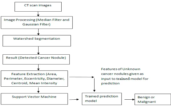
3.2 Lung cancer detection using Median and Gaussian filter
3.2.1 Additional features in proposed system
• Use of Median filter and Gaussian filter Instead of Gabor Filter has been implemented in pre- processing stage.
• In addition to features like area, perimeter and eccentricity, features like Centroid, Diameter and pixel Mean Intensity have been extracted in feature extraction stage for the detected cancer nodules.
• The feature extraction and calculation of accuracy is also done.
• But, its classification as benign or malignant has not been implemented. An additional stage of classification of cancer nodule is performed using Support Vector Machine.
Fig 3.2: proposed model using Median and Gaussian filter
3.2.2 Function of each block in proposed system
• Image Preprocessing
Median filter is used on gray scale image of CT scan images. Noise gets added at the time of image acquisition and that noise can be detected as cancer. Median filter helps to remove these noises. Median filter removes salt and pepper noise from the CT images [11].Gaussian filter smoothes the image and remove speckle noise.
• Segmentation
Segmentation helps to get area of interest by location object or boundaries. This process locates objects or boundaries which help in acquiring the region of interest in the image. It divides the image into parts. In lung cancer detection it segments the cancer nodule from the CT scan image. Watershed segmentation is implemented. Its main feature is that it can separate and identify the touching objects in the image. This feature helps in proper segmentation of cancer nodules if it is touching to other false nodules.
• Features extraction
In this stage, features like area, perimeter, centroid, diameter, and eccentricity and Mean intensity. These features later on are used as training features to developclassifier.
• Classification
ISSN(Online): 2320-9801 ISSN (Print): 2320-9798
I
nternational
J
ournal of
I
nnovative
R
esearch in
C
omputer
and
C
ommunication
E
ngineering
(A High Impact Factor, Monthly, Peer Reviewed Journal) Website: www.ijircce.com
Vol. 7, Issue 10, October 2019
that classifies data into two classes [9].The function is defined as D(x) =wTxi+ b where xi is training inputs, w T
is m dimensional vector, and b is bias term. Here, i=1….M.
D(x) =wTxi+ b ≥ 1 for yi=1 D(x) =wTxi+ b ≤-1 for yi=-1
3.2.3 Advantages and Limitations
Advantages of proposed model. Increase in accuracy
Classifies the detected lung cancer as malignant orbenign.
Removes salt-pepper noises and speckle noise that creates false detection of cancer
Limitation of proposed model.
Accuracy has increased but not reached 100 percent
It classifies cancer as just malignant or benign but does not classify into different stages like stage I, II, III,and IV.
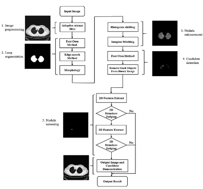
3.3 Lung detection using image processing techniques in computed tomography
This method includes all type of nodule detection thus increasing the accuracy of system. This is done by an image processing method for detecting ground glass opacity(GGO), partsolid, and solid nodules in chest computed tomography.
3.3.1 The method includes
ISSN(Online): 2320-9801 ISSN (Print): 2320-9798
I
nternational
J
ournal of
I
nnovative
R
esearch in
C
omputer
and
C
ommunication
E
ngineering
(A High Impact Factor, Monthly, Peer Reviewed Journal) Website: www.ijircce.com
Vol. 7, Issue 10, October 2019
Fig 3.3 flow chart of proposed system
3.3.2 Function of each block in proposed system
• Preprocessing -The preprocessing uses an adaptive Wiener filter to optimize the images.
• Lung segmentation-The lung segmentation uses Fast Otsu method and the edge search method for rapidsegmentationanduseshistogramshiftingtocreateimageswiththesame reference.The edge search is replaced by computing-intensive iterative hole-filling method. The advantage is to extract extensively distributed grey levels.
• Nodule enhancement–The image accumulation and cumulative method is used to enhance grey level of nodules
• Candidate detection-Uses fast Otsumethod to get preliminary objects and reduces false positives the system uses quadratic SVM to obtain the nodules To remove false positive SVM is used twice , in first run nodules are obtained and results are detected in second run
3.3.3Advantages
ISSN(Online): 2320-9801 ISSN (Print): 2320-9798
I
nternational
J
ournal of
I
nnovative
R
esearch in
C
omputer
and
C
ommunication
E
ngineering
(A High Impact Factor, Monthly, Peer Reviewed Journal) Website: www.ijircce.com
Vol. 7, Issue 10, October 2019
• A new 3D feature is proposed for lung nodule detection
IV. DISCUSSION AND CONCLUSION
1. Lung cancer detection using Gabor filter has no satisfactory result of accuracy with 82 %. The system does not classify degree of cancer nodules.
2. Therefore the modified method of Lung cancer detection using Median and Gaussian filter uses SVM for detection of nodule as malignant and benign with 92 percent accuracy which is higher than previous method. But the new system does not classify cancer in stages of I, II, III, and IV. So in future a system can be implemented to classify cancer in different stages.
3. The third method Lung detection using image processing techniques in computed tomography uses an innovative edge detection which save 3 times the computing time. Based on thecharacteristics of repeated nodules system can effectively and quickly find GGOs with low brightness. The fast Otsu method helped to obtain binarized threshold quickly
4. To reduce number of object after binarization a two dimensional classification is implemented, which saved time and accelerated the acquisition of real nodule
5. The total system sensitivity is increased to 92.05% and system accuracy o find out smaller nodules also to 93.73%
REFERENCES
1.Suren Makajua, P.W.C. Prasad, Abeer Alsadoona, A. K. Singhb, A. Elchouemi” Lung Cancer Detection using CT Scan Images .”6th International Conference on Smart Computing and Communications, ICSCC 2017, 7-8 December 2017, Kurukshetra, India, 107-114.
2.Chung-Feng Jeffrey Kuo, Chang-Chiun Huang, Jing-Jhong Siao, Chia-Wen Hsieh,VuQuangHuy ,Kai-HsiungKo ,Hsian-HeHsu ” Automatic lung nodule detection system using image processing techniques in computed tomography.”,Elsevier journal 17th
august 2019, 1-20.
3. Xiuhua, G., Tao, S., & Zhigang, L. (2011) “Prediction models for malignant pulmonary nodules based –on texture features of CT images.” In theory and application of CT Imaging and Analysis. DOI:10.5772/14766.
4. Aggarwal, T., Furqan, A., & Kalra, K. (2015) “Feature extraction and LDA based classification of lung nodules in chest of CT.” 2015 international conferences on Computing, Communications and Informatics (ICACCI),
DOI:10.1109/ICACCI.2015.7275773.
5.Jin, X, Zhang, Y& Jin, Q. (2016) “Pulmonary nodule detection based on the CT Images using convolution neutral network.” 2016 9Th International Symposium on Computational Intelligence and Design (ISCID). DOI: 10.1109/ISCID.2016.1053.
6. Sangamithraa, P., & Govindaraju, S. (2016) “Lung tumour detection and its classification by using EK-MEAN EK-Mean clustering.” 2016 International Conference on Wireless Communications, signal processing and networking (Wispnet). DOI: 10.1109/WiSPNET.2016.7566533. 7. Roy, T., Sirohi, N., & Patle, A. (2015) “Classification of lung image and nodule detection using fuzzy inference system.” International conference on computing, communication and automation.DOI:10.1109/CCAA.2015.7148560.
8. Ignatious, S., & Joseph, R. (2015) “computer aided lung cancer detection system.” 2015 Global Conference on Communication Technologies (GCCT), DOI:10.1109/GCCT.2015.7342723.
9. Rendon-Gonzalez, E., & Ponomaryov, V. (2016) “Automatic lung nodule segmentation and classification in CT images based on SVM.” 2016 9Th International Kharkiv Symposium on Physics and engineering on microwave, Millimeter andSub millimeter Waves (MSMW). DOI:10.1109/MSMW.2016.7537995.
10. Miah, M.B.A, & Yousuf M.A. (2015) “Detection of lung cancer from CT image using image processing and neural network.” 2015 conference on electrical engineering and Information communication technology (ICEEICT):1-6.
11. Khobragade, S., Tiwari, A., Patil, C., & Narke, V. (2016)“Automatic detection of major lung diseases using Chest Radiographs and classification by feed-forward artificial neural network.” IEEE International Conference on Power Electronics, Intelligent Control and Energy Systems (ICPEICES):1-5.
12. Armato, I., Samuel McLennan, G., McNitt-Gray, F. R., Michael, Charles, Reeves, Anthony Clarke, Laurenc, (2015) “Data from LIDC-IDRI. The Cancer Imaging”Archive.http://doi.org/10.7937/K9/TCIA.2015.LO9QL9SX.
13. Gindi ,A.M , Al Attiatalla, T. A., & Sami, M.M. (2014) “ Comparative Study of Comparing 2 Feature Extraction methods are 2classifiers in classification of the early stage lung cancer diagnosis of chest X-rays images.” Journal of America science, 10(6):13-22.
![Fig 3.1: Current Method proposed by Ignatious and Joseph [8]](https://thumb-us.123doks.com/thumbv2/123dok_us/1371631.1169898/2.595.191.412.527.709/fig-current-method-proposed-ignatious-joseph.webp)