Type of the Paper (Article)
1
Affinity Purification and Comparative Biosensor
2
Analysis of Citrulline-Peptide Specific Antibodies in
3
Rheumatoid Arthritis
4
Eszter Szarka1, Petra Aradi1, Krisztina Huber1, Judit Pozsgay1, Lili Végh1, Anna Magyar2,
5
Gergő Gyulai3, György Nagy4, 5, Bernadette Rojkovich4, Éva Kiss3, Ferenc Hudecz2 and Gabriella
6
Sármay 1,*
7
1Department of Immunology, Eötvös Loránd University, Budapest, Hungary, [email protected]
8
[email protected], [email protected], [email protected], [email protected],
9
2MTA-ELTE Research Group of Peptide Chemistry, Budapest, Hungary, [email protected]
10
11
3Laboratory of Interfaces and Nanostructures, Institute of Chemistry, Eötvös Loránd University, Budapest,
12
Hungary, [email protected], [email protected]
13
4Buda Hospital of Hospitaller Brothers of St. John, [email protected],
14
5Semmelweis University, Department of Rheumatology, Budapest, Hungary, [email protected]
15
16
* Correspondence: [email protected]; Tel.: +36-30 2116018
17
Abstract:
18
Background: In rheumatoid arthritis (RA), anti-citrullinated protein/peptide antibodies (ACPAs)
19
are responsible for disease onset and progression, however, our knowledge is limited on ligand
20
binding affinities of autoantibodies with different citrulline-peptide specificity.
21
Methods: Citrulline-peptide specific ACPA IgGs were affinity purified and tested by ELISA.
22
Binding affinities of ACPA IgGs and serum antibodies were compared by surface plasmon
23
resonance (SPR) analysis. Bifunctional nanoparticles harboring a multi-epitope citrulline-peptide
24
and a complement activating peptide were used to induce selective depletion of ACPA producing
25
B cells.
26
Results: KD values of affinity purified ACPA IgGs varies between 10-6-10-8 M and inversely correlate
27
with disease activity. Based on their cross-reaction with citrulline-peptides we designed a novel
28
multi-epitope peptide, containing Cit-Gly and Ala-Cit motifs in two-two copies, separated with a
29
short, neutral spacer. This peptide detects antibodies in RA sera with 66 % sensitivity and 98 %
30
specificity in ELISA and is recognized by 90% of RA sera, while none of the healthy samples in SPR.
31
When coupled to nanoparticles, the multi-epitope peptide specifically targets and depletes ACPA
32
producing B cells ex vivo.
33
Conclusions: The unique multi-epitope peptide designed on the basis of ACPA cross-reactivity
34
might be suitable to develop better diagnostics and novel therapies for RA.
35
Keywords: rheumatoid arthritis; Citrulline-peptides; autoantibody; affinity; cross-reaction;
36
targeting
37
38
39
1. Introduction
40
Anti-citrullinated protein/ peptide antibodies (ACPA) are highly specific and sensitive clinical
41
marker of rheumatoid arthritis (RA), a chronic inflammatory autoimmune disease [1]. Approximately
42
70-80 % of RA patients are ACPA positive, and high level of ACPA has been associated with bad
43
prognosis of the disease [2]. ACPA is produced against citrullinated self-proteins, emerging as a
44
result of the activation of peptidyl arginine deiminase (PAD) enzymes [3, 4]. PADs are activated at
45
inflammatory sites and in the presence of high level of intracellular free calcium in activated cells.
46
PADs deiminate arginine to citrulline and this post-transcriptional modulation drastically modify the
47
structure of the protein, initiating unfolding [5]. The newly appearing citrullinated epitopes will then
48
be recognized by the immune system, resulting in the production of ACPA [6].
49
Most of the citrullinated target molecules of ACPA are localized in the joints, such as collagen II
50
[7], fibrinogen/fibrin [8, 9], and the intracellular protein, vimentin [10, 11], which is released from
51
macrophages in the inflamed synovial [12]. Binding of ACPA to synovial target proteins in vitro and
52
in vivo has been demonstrated [13]. Deposition of ACPA and ACPA containing immune complexes
53
induce the release of inflammatory cytokines, such as TNFα, eventually leading to the escalation of
54
inflammation [8, 14, 15]. ACPA appears many years before the disease onset in the sera of patients,
55
and its detection is extremely important for the early diagnosis. ACPA is not only a specific and
56
sensitive disease marker, but also play key role in the pathogenicity of RA [16].
57
A wide variety of citrulline containing peptides (Cit-peptides) are recognized by ACPA, these
58
include peptides representing citrullinated sequences in the major proteins of the joints, such as
59
fibrinogen [8] and type II collagen [7], the intracellular structural protein, vimentin, which is also
60
detected in the synovium [11, 17, 18], as well as peptides derived from bacterial (α-enolase) [19] and
61
viral (Epstein-Barr virus nuclear antigen (EBNA)) origin [20]. An ACPA cross-reacting, citrullinated
62
peptide, derived from filaggrin produced by epithelial cells has also been described [9], [21] and used
63
in the first diagnostic tests of RA.
64
Citrullination in general is a physiological process, it may occur for example upon gene
65
regulation [22], apoptosis [23], inflammation [24] and NET formation [25] without inducing an
66
autoimmune response. In RA, however, probably due to the abnormal humoral immune response,
67
citrullinated proteins induce ACPA production in the vast majority of patients. Since ACPA is a
68
crucial player and has a pathological role in RA, detailed characterization of this autoantibody would
69
be of great importance both for diagnostic accuracy and prognostic evaluation.
70
Many groups described the multiple specificity and cross-reactivity of ACPA [26-30], and
71
several citrullinated peptide epitopes have been identified, however, only limited data are available
72
so far on the binding affinity of individual purified ACPA IgGs. Therefore our aim was to analyze in
73
parallel by Surface Plasmon Resonance (SPR) and ELISA both the affinity and the specificity of
74
affinity purified autoantibodies from RA patients.
75
In accordance with previous data, we detected a high degree of cross-reactivity between affinity
76
purified ACPA IgGs, with apparent dissociation constants (KD) between 10-6 -10-8 M. Based on the
77
pattern of cross-reaction we designed a novel multi-epitope peptide that was specifically recognized
78
by RA sera and was able to deplete specific B cells from in vitro culture. Thus we suppose that this
79
multi-epitope peptide might be further developed for RA diagnosis and therapy.
80
2. Results
81
2.1. Affinity purification of ACPA
82
We have synthesized a panel Cit-peptides corresponding to known ACPA epitopes, listed in
83
Table 1., and prepared insoluble matrixes for the immune-purification of ACPA from the sera of
84
selected RA patients. Patients’ selection was based on the positivity of the sera in an in house
Cit-85
peptide-based ELISA [31, 32].
86
Table 1. List of Citrulline containing peptides used in the assays
91
92
Peptides Sequences References
Filaggrin (306-326) Ac-SHQESTX1GXSXGRSGRSGSK-NH2 [33]
Collagen II (359-369) Ac-AXGLTGXPGDA-NH2 [7]
Fibrin β (60-74) H-XPAPPPISGGGYXAX-NH2 [8, 34]
Vimentin (65-77) Ac-SAVRAXSSVPGVRK-NH2 [10]
α-enolase (5-21) Ac-KIHAXEIFDSXGNPTVE-NH2 [35]
EBNA-2 (341-361) Ac-GQSXGQSXGXGXGXGXGXGKG-NH2 [36]
multi-epitope H- Ttds2-AXAXGSGSGXGXG-NH2
1X stands for citrulline. 2Ttds - linker 1,13-diamino-4,7,10-trioxatridecan-succinamic acid. For screening the
93
serum samples by ELISA both citrulline and arginine containing variants of the same peptides were used.
94
95
The citrulline containing filaggrin19, collagen II, fibrin β60-74 and vimentin peptides were
96
already thoroughly tested with our cohort of serum samples [31, 32, 37]. Therefore we only show here
97
the screening of sera of RA patients (n=177) and healthy volunteers (n=104) with arginine and
98
citrulline containing EBNA-2 and α-enolase peptides, respectively (Fig.1).
99
100
a)
b)
c) d)
101
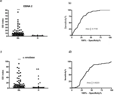
Figure 1. Recognition of citrulline-containing EBNA-2 and α-enolase peptide epitopes by sera of patients
102
(RA, n =177) and healthy volunteers (H, n= 104). (a), (c), OD index: OD with Cit-peptide/OD with Arg peptide.
103
(b), (d), Receiver operating (ROC) curves. *** p≤0.001.
104
105
EBNA-2 and α-enolase peptides detected autoantibodies in RA serum samples by 48 % and 27
106
% sensitivity, respectively. For comparison, the sensitivity values for the previously tested
Cit-107
filaggrin, fibrin, collagen II and vimentin peptides were 62 %, 56 %, 44 % and 41 %, respectively [31,
108
32]. Based on these data we have selected the sera, which had a high OD index at least with one of
109
EBNA 2
RA H
0 10 20 30 40 50 60 70 80
90 ***
O
D
in
d
ex
0 25 50 75 100
0 25 50 75 100
Area 0.7730
100% - Specificity%
S
e
n
siti
vity
%
enolase
RA H
0 20 40 60 80
100 ***
OD
in
d
ex
0 25 50 75 100
0 25 50 75 100
Area 0.6223
100% - Specificity%
S
e
ns
itiv
ity
the peptides and purified the anti-peptide antibodies by affinity chromatography. ACPAs were
110
purified in two steps [38]. First the IgG fractions were obtained on a protein G column and this was
111
followed by the affinity purification of anti-peptide antibodies on the corresponding citrulline
112
containing filaggrin19-, collagen II-, fibrin-, vimentin- and α-enolase-peptide-coted matrixes,
113
respectively.
114
115
2.2 Cross-reaction of affinity purified ACPA IgG fractions
116
To see if the affinity purified anti-peptide antibodies are able to recognize a different peptide,
117
ELISA plates were coated with the individual peptides directly (fibrin, enolase, EBNA-2) or
118
indirectly, using neutravidin coated plates and biotinylated peptides (filaggrin, collagen II, or
119
vimentin) and the binding of the affinity purified IgG fractions was monitored (Fig.2).
120
121
122
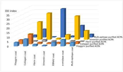
Figure 2. Reactivity of affinity purified IgG fractions with the relevant and irrelevant citrulline containing
123
peptides. OD ratios that are OD with Cit /OD with arg containing peptides are shown. Cut-off values for each
124
Cit and Arg containing peptide pair were calculated from OD indexes of 120 healthy samples (the means of OD
125
indexes + 2*SD). These were below 1.5 for all peptides. Results of a typical experiment.
126
127
Interestingly, the affinity purified IgG prepared on Cit-filaggrin19 peptide recognized citrulline
128
containing collagen II, fibrin β, EBNA-2 and α-enolase as well, beside the filaggrin19; IgG prepared
129
on Cit-collagen II peptide bound to citrulline containing filaggrin, fibrin β, and EBNA-2 peptides,
130
while IgG purified on Cit-vimentin peptide recognized all Cit-peptides tested, although at different
131
level. ACPA purified on the Cit-fibrine β peptide has shown the lowest degree of cross-reactivity in
132
ELISA.
133
134
We compared the Cit-peptide sequences and observed that certain short motifs containing
Ala-135
Cit and/or Cit-Gly residues are present in all peptides. We supposed that both of these short motifs
136
might be important for the recognition. Thus we designed and synthesized a novel “multi-epitope”
137
peptide consisting of two-two copies of Ala-Cit and Cit-Gly motifs separated with a neutral spacer,
138
SGSG. As expected, IgG purified on citrulline-containing multi-epitope peptide coated matrix
139
recognized all other peptides, but with different intensity (Fig. 2., dark blue columns). Vice versa, the
140
multi-epitope peptide bound to almost all other ACPA IgG, fibrin β was the exception. These data
141
verify earlier data suggesting that ACPAs are highly cross-reactive and indicate that Ala-Cit as well
142
2.3. Analysis of the multi-epitope peptide by ELISA screening of RA sera
144
Next, we screened 210 RA sera and 90 healthy control samples using the biotinylated, citrulline
145
or arginine containing multi-epitope peptide as a coat in ELISA.
146
147
a) b)
148
Figure 3. The Cit-multi-epitope peptide identifies RA sera with the highest specificity and 66 % sensitivity.
149
(a), ELISA, OD indexes (OD with Cit-peptide/OD with Arg-peptid) of RA (n=210) and healthy (n=90) samples,
150
(b) ROC curve of ELISA. Area under the curve (AUC): 0.7843.
151
152
The designed multi-epitope peptide identified ACPA in RA serum samples with 66 %
153
sensitivity, while none of the healthy control sera showed binding (Fig. 3). This sensitivity value is
154
somewhat higher than that of fibrin β peptide, and the shape of the ROC curve (AUC 0.7843) suggest
155
that a diagnostic test based on the multi-epitope peptide would be more accurate.
156
157
2.4. SPR analysis of affinity purified antibodies and sera of RA patients
158
For affinity measurements of IgGs purified from RA sera on insolubilized citrulline containing
159
filaggrin19, collagen II, fibrin β, vimentin and multi-epitope peptides first we had to test the ability
160
of the individual peptides to immobilize on GLH sensor chip. Amine coupling immobilization
161
strategy was used. Immobilization buffers were selected separately for each peptide, according to
162
their isoelectric point. From these five peptides only filaggrin19, vimentin and the multi-epitope
163
peptide could couple covalently to the surface of the GLH chip. Therefore fibrin β, and collagen II
164
peptides were biotinylated and immobilized using the neutravidin coated NLC chip.
165
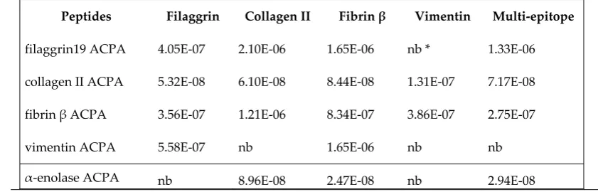
First, apparent KD of affinity purified IgG fractions of anti-peptide antibodies were tested on
166
various immobilized Cit-peptides. The dissociation constants are in the micromolar - 10 nanomolar
167
range, and as well as in the ELISAs, a high level of cross-reactivity can be observed (Table 2.).
168
169
Table 2. KD of affinity purified IgG of anti-peptide antibodies as measured by SPR
170
171
Peptides Filaggrin Collagen II Fibrin β Vimentin Multi-epitope
filaggrin19 ACPA 4.05E-07 2.10E-06 1.65E-06 nb * 1.33E-06
collagen II ACPA 5.32E-08 6.10E-08 8.44E-08 1.31E-07 7.17E-08
fibrin β ACPA 3.56E-07 1.21E-06 8.34E-07 3.86E-07 2.75E-07
vimentin ACPA 5.58E-07 nb 1.65E-06 nb nb
α-enolase ACPA nb 8.96E-08 2.47E-08 nb 2.94E-08
*nb: no binding
172
multi-epitope peptide
RA H
0.1 1 10
100 ***
OD
in
d
ex
0 20 40 60 80 100
0 20 40 60 80 100
Area 0.7843
100% - Specificity%
S
en
sit
iv
it
2.4.1. KD determination of serum IgG on GLH chip
173
Fig. 4. shows representative sensograms of IgG from an RA patient purified by affinity
174
chromatography on immobilized Cit-filaggrin19 peptide. As a negative control, intravenous
175
immunoglobulin (IVIG) was used, pooled from the plasma of more than a thousand healthy blood
176
donors, which clearly did not show any specific binding.
177
To calculate the KD values for serum samples, the concentration of an individual peptide specific
178
IgG was estimated in all sera by ELISA, using a standard concentration series from the given affinity
179
purified IgG fraction. Typical binding curves of two serum samples are shown on Fig. 4. c) and d).
180
181
a) b)
c) d)
182
Figure 4. Representative sensograms of (a) Cit-filaggrin19-affinity purified IgG; (b) negative control IVIG
183
and two typical sera with (c) higher and (d) lower affinity on Cit-filaggrin19 coupled GLH chip.
184
185
The distribution of the apparent KD values of serum antibodies measured on vimentin,
Cit-186
filaggrin19 and multi-epitope peptides immobilized by covalent coupling is shown on Fig. 5. The
187
majority of KD values were in the range between 104 - 107 M/L. All sera selected for this assay (n=68)
188
were highly positive for the given Cit-peptide in ELISA. SPR analysis has shown that 92 % of sera
189
bound to Cit-vimentin and all samples bound to the multi-epitope peptide on the chip. The
190
distribution of KD values were similar, however, only 41 % of the Cit-filaggrin19 peptide positive sera
191
bound to this peptide under the flow condition on the chip. The Ka / Kd plots shown at the lower
192
panels of the figure represent the data of individual sera. In general, more sera have shown a lower
193
dissociation rate from the multi-epitope peptide as compared to the other two.
194
196
Figure 5. Distribution of KD values of serum antibodies as measured from RA samples on GLH chip, upper
197
panels; isoaffinity diagrams, lower panels
198
199
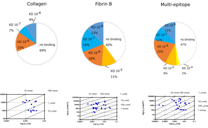
2.4.2. Binding of serum IgG to the biotinylated peptides immobilized on neutravidin coated chip (NLC)
200
Fibrin and collagen II peptides that did not immobilize on GLH chip, were biotinylated and
201
immobilized on a neutravidin coated surface. Additionally, multi-epitope peptide was also
202
biotinylated and re-tested on NLC chip (Fig. 6.)
203
204
205
Figure 6. Distribution of KD values measured from RA sera on biotinylated Cit-collagen II, Cit-fibrin β
206
and multiepitope peptides immobilized on NLC chip.
207
208
When the biotinylated peptides were immobilized on the NLC chip, 31 % of selected RA sera
209
bound to Cit-collagen peptide with apparent 10-6 – 10-8 KD, while 60 % of sera bound to the Cit-fibrin
β peptide with 10-5 - 10-8 KD. Interestingly, although a lower proportion of sera (53 %) showed binding
211
to the multi-epitope peptide immobilized on the NLC chip and among those who bound, a larger
212
proportion showed higher KD (10-8 M/L).
213
214
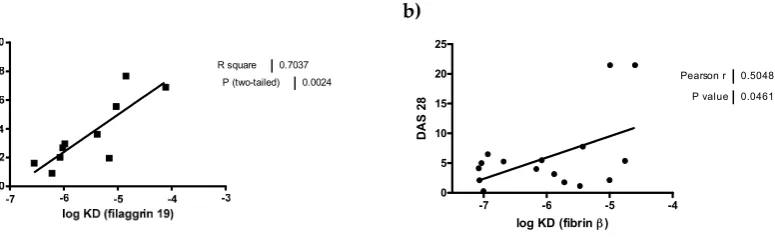
Low avidity ACPA was previously shown to associate with a higher rate of joint destruction
215
[39]. Therefore we tested if the disease activity score, DAS28, correlates with the apparent KD (Fig. 7.).
216
We found an inverse correlation between DAS and KD of IgG purified on Cit-filaggrin19 peptide, and
217
on Cit-fibrin β peptide, indicating that low KD auto-antibodies are more pathogenic.
218
219
a) b)
220
Fig. 7. Correlation between the disease severity (DAS28) and the apparent binding affinity (KD) of serum
221
antibodies from RA patients.
222
223
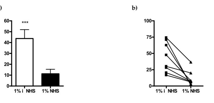
2.5. Selective depletion of autoantibody producing B cells ex vivo by the multi-epitope peptide covalently
224
coupled to poly (lactic-co-glycolic acid) (PLGA) nanoparticles (NP)
225
We have reported earlier that bifunctional nanoparticles covalently coupled to Cit-fibrinβ60-74
226
peptide and a peptide inducing cell lysis deplete the peptide specific B cells in ex vivo cultures of
227
PBMC from RA patients. Here we tested if the newly designed multi-epitope peptide would have a
228
similar effect. Therefore, the multi-epitope peptide and a complement activating peptide, a modified
229
form of the peptide derived from HIV1 GP120 [32, 40]
(Ac-230
233C(Acm)NNQTFNGTGPC(Acm)TNV247-K-NH2, (Acm: acetamido-methyl)) were simultaneously
231
coupled to PLGA nanoparticles, and added to pre-activated PBMC from RA patients in the presence
232
or absence of complement source (1% human sera, NHS). The Cit-peptide specific antibody
233
production was monitored by ELISpot assay. The bifuctional PLGA nanoparticles targeted to B cells
234
recognizing the multi-epitope peptide significantly suppressed the peptide specific IgG secretion in
235
the presence of complement (Fig. 8.), while total IgG production was not affected (data not shown).
236
These data indicate that the newly designed multi-epitope peptide might be suitable for the
237
development of autoantigen-specific immunotherapy.
238
239
R square 0.7037 P (two-tailed) 0.0024
-7 -6 -5 -4
0 5 10 15 20 25
Pearson r 0.5048 P value 0.0461
log KD (fibrin)
DAS
a) b)
240
Figure 8. Ex vivo inhibition of the Cit-peptide specific antibody production by bifunctional PLGA
241
nanoparticles covalently coupled to multi-epitope peptide and a complement activating peptide. Antibody
242
synthesis was tested by ELISpot assay according to a modified procedure of Manufacturer (Mabtech, Stockholm,
243
Sweden). As complement source 1 % normal human sera (NHS) was used, for the negative control the sera were
244
heat-inactivated (iNHS). a) Average number of peptide specific antibody secreting cells (ELISpots) from 106
245
PBMC of eight RA patients, b) ELISpot data of individual RA patients.
246
247
3. Discussion
248
In the present study we have analyzed the specificity and the apparent affinity of anti-peptide
249
antibodies isolated from the sera of RA patients by affinity chromatography. The citrulline containing
250
peptides we used for the isolation and analysis represent the immunodominant epitopes of fibrin β
251
chain, filaggrin, collagen II, vimentin, α-enolase and EBNA-2. Most of the affinity purified IgG
252
fractions recognized other cit-peptides, beside the ones used for the isolation, and their apparent KD
253
were comparable. Additionally, in some cases (e.g. anti-cit-vimentin) anti-peptide antibodies bound
254
to a different peptide with even higher affinity, indicating a high degree of cross-reactivity.
255
Comparing the individual peptide sequences we have observed that all peptides contain Cit-Gly
256
and/or Ala-Cit motifs, which are seemingly essential for the recognition. Thus we designed an
257
artificial peptide, containing two copies of each small motifs separated with a neutral spacer. This
258
“multi-epitope” peptide was recognized by sera from RA patients with 66 % sensitivity and 98 %
259
specificity, and the binding affinities were comparable with that of the natural citrulline containing
260
peptides. Antibodies purified on the multi-epitope peptide cross-reacted with most other peptides
261
tested, although at different degree. The functional activity of the multi-epitope peptide was
262
equivalent to that of Cit-fibrin β peptide [32], it was able to specifically deplete Cit-peptide specific B
263
cells, thus significantly inhibit ACPA production.
264
265
Cross-reactivity of ACPA was reported by several groups, and the concept that Cit-Gly motif
266
has importance in the recognition was also studied earlier [26, 29, 30, 36, 41]. Several combinations of
267
Cit-peptides and ACPAs have been tested for cross-reactivity; antibodies to Cit-EBNA(35-58)
268
strongly cross-reacted with the immunodominant epitope of citrullinated fibrin [42] and vice versa,
269
two proteins encoded by Epstein-Barr virus (EBV), EBNA-1 and EBNA-2 were specifically recognized
270
by antibodies of the ACPA family [43]. Additionally, monoclonal antibodies obtained from an RA
271
patient and were specifically directed against a citrullinated fibrin peptide also recognized
Cit-272
peptides derived from enolase and vimentin [28]. Recently Trier et al. concluded that ACPAs are a
273
collection of largely cross-reacting but occasionally non-cross-reactive antibodies. Cross-reactivity is
274
largely depends on Cit-Gly motif in combination with the peptide backbone, while non-cross-reactive
275
ACPAs depend on a specific citrullinated epitopes, rather than a citrulline containing motif [41, 44].
276
277
The data presented here expand these earlier findings, first, since here a panel of affinity purified
278
anti-peptide antibodies were screened with a panel of Cit-peptides representing immunodominant
279
1% i NHS 1% NHS
0 10 20 30 40 50
60 ***
1% i NHS 1% NHS 0
epitopes of in vivo ACPA target proteins, and second, in addition to binding studies by ELISA,
280
affinities of the interaction were also analyzed by SPR. Both the specificity and the affinity play a key
281
role in the pathological effect of an antibody, therefore we have monitored these two parameters by
282
analyzing their interactions with seven Cit-peptides derived from major target proteins of ACPA.
283
The suggested central role of Cit-Gly [41, 44] in the recognition of autoantigens by ACPA does
284
not explain how vimentin and fibrin β peptides are recognized. Cit-vimentin and fibrinβ peptide do
285
not contain Cit-Gly, instead has Ala-Cit motif. Similarly, collagen II and enolase peptides also have
286
Ala-Cit motif. Although vimentin peptide had restricted reactivity with affinity purified IgG
287
fractions, IgG purified on Cit-vimentin recognized all other peptides, even without Ala-Cit, such as
288
EBNA-2 and filaggrin, indicating that antibodies specific for the Ala-Cit motif cross-react with
Cit-289
Gly and thus Ala-Cit motif is also important in the recognition. Based on these, we designed an
290
artificial peptide containing two Cit-Gly and two Ala-Cit motifs. This artificial peptide has a higher
291
sensitivity to detect serum ACPA than fibrin β and filaggrin peptides, has a similar binding affinity,
292
and is able to target in vitro the peptide specific B cells of RA patients.
293
294
We determined the apparent binding affinities of Cit-peptides to affinity purified antibodies by
295
SPR biosensor analysis. The IgG fractions purified by affinity chromatography show a comparable
296
KD and a similar distribution of dissociation constants as RA serum antibodies. Most of the KD values
297
were between 1 micromolar - 10 nanomolar range and showed high heterogeneity. While almost all
298
serum samples bound to Cit-vimentin and to the multi-epitope peptide, only 30-60 % of RA sera
299
reacted with citrulline containing collagen II, fibrin β and filaggrin19 peptides on the chip, although
300
all samples were positive in ELISA. This may be explained by the difference between the static nature
301
of the binding to ELISA plate in contrast to the dynamic flow rate over the surface of the chip, which
302
induce a faster dissociation of the analyte from the peptide ligand coated surfaces. Thus antibodies
303
with low affinity might be lost.
304
305
In concert with previous findings [39] we detected an inverse correlation between KD values and
306
the disease severity (DAS 28), in case of Cit-filaggrin19 and Cit-fibrinβ peptides, the lowest KD was
307
associated with the most severe disease. A possible explanation could be that low binding affinity
308
might allow higher degree of cross-reaction, leading to more extensive tissue damage.
309
310
ACPA play a pathological role in the development of RA, in the perpetuation of inflammation
311
and the pathophysiology of the disease [45]. ACPA positive patients have a faster progress of the
312
disease and a worst radiological outcome [27, 46] thus the detailed characterization of ACPA has high
313
importance. These results presented here contribute to the understanding of the interactions between
314
citrullinated epitopes and ACPA in RA patients thus may support the development of better
315
diagnostics and precision therapy for RA.
316
317
4. Materials and Methods
318
4.1. Patients
319
Blood samples were collected from 210 RA patients (165 women/45 men; median age 63 years,
320
interquartile range 51–70; median disease duration 6 years, interquartile range 3–13) at the Buda
321
Hospital, Budapest, Hungary of Hospitaller Brothers of St John. The diagnosis of the disease was
322
established on the basis of the revised classification criteria of the American College of Rheumatology
323
(ACR)/European League against Rheumatism (EULAR) [47].
324
Blood samples were taken after the patients signed a written consent, and with ethical
325
permission provided by the National Public Health and Medical Officer Service (49468-/2013/EKU
326
(576/2013)). Selected patients with a high level of anti-CCP antibodies were repeatedly recruited for
327
the functional assays. A total of 120 age-matched healthy control sera were obtained from healthy
328
volunteers.
329
4.2. ACPA affinity purification
331
We purified IgG from RA sera via Protein G agarose (Thermo Fischer Scientific, MA, USA)
332
affinity chromatography. To isolate ACPAs, Citrulline containing peptides were covalently bound (5
333
mg/ml) to GE NHS HiTrap 1 ml column, according to manufacturer’s instructions. Purified IgG was
334
diluted 2x in 0.1 M NaH2PO4, 0.15 M NaCl, pH=7 buffer and circulated through the column for 3
335
days. After washing, ACPA-containing fractions were eluted with 0.1M glycine buffer (pH 2.5). The
336
pH was immediately restored by adding 10 µl 2M TRIS pH 9 to 1 ml eluted ACPA. The eluate
337
fractions were dialyzed against PBS and after determining the IgG concentration via Nanodrop
338
Spectrophotometer (Thermo Scientific, Waltham, MA, USA) the samples were stored at -20oC.
339
4.3. ELISA
340
ELISA was used to determine serum IgG ACPA concentrations and to test ACPA
cross-341
reactions. The biotinylated, citrulline or arginine containing filaggrin19, vimentin, collagen II and the
342
multi-epitope peptides were added at 1 µg/ml to the Medisorp plates (Greiner GmbH) pre-coated
343
with 5 µg/ml Neutravidin (Pierce Biotechnology, Rockford, IL, USA) overnight, at 4 °C, and then the
344
plates were incubated for 1h at 37 °C. EBNA2, α-enolase and fibrin β peptides were coated directly
345
to Maxisorp (Nunc) plates at 2.5 µg/ml. After washing, the plates were blocked with 150 mM NaCl
346
and 2 % BSA containing PBS for 1h, at 37 °C.
347
To determine the specific ACPA concentrations in sera a calibration curve was prepared from
348
the affinity purified ACPA as standard, in a two-fold dilution series, starting from 0.5 mg/ml. Sera
349
samples were diluted in 1:100. Dilution buffer was 2 M NaCl, 2 % BSA in PBS. In cross-reaction assays,
350
ACPAs were diluted to 0.1 mg/ml. Sera/ACPA samples were incubated overnight at 4 °C, shaking.
351
After washing, the plates were incubated with 1:15000 dilution of rabbit anti-human IgG HRP (H+L)
352
(Southern biotech) for 1h at 37°C. Signal was developed with TMB substrate (Sigma) and reaction
353
was stopped with 2 N H2SO4. Plates were read at 450 nm with THERMO Multiscan EX ELISA-reader.
354
4.4. SPR analysis
355
All experiments were conducted using a ProteOn™ XPR36 Protein Interaction Array System
356
from Bio-Rad. All solutions and sensor chips were purchased from Bio-Rad. Citrulline containing
357
peptides were immobilized on GLH sensor chip, with amine coupling strategy. Immobilization
358
buffers were selected separately for each peptide, according to the isoelectric point of the peptide.
359
Running buffer was 0.005% PBST pH 7.4.
360
For immobilization, sensor chip surface was activated with 1:1 mixture of 400 mM EDAC and
361
100mM Sulfo-NHS for 300 sec at a flow rate 30 µl/min. Immobilization buffers were selected
362
separately for each peptide using the pH scouting procedure, using the following buffers: sodium
363
acetate buffer (10 mmol/L, pH 4, 4.5, 5.0), phosphate buffer (10 mmol/L, pH 6, 6.5, 7, 7.5 and 8) and
364
borate buffer (10 mmol/L, pH 9 and 9.5). Peptides were solubilized in each buffer at a final
365
concentration of 10 µg/ml. The selected buffer and immobilized peptide quantities are reported in
366
Table 2. Each peptide, solubilized in the previously selected immobilization buffer (10 µg/ml), was
367
injected for 300 s at a flow rate of 30 µl/min. To deactivate free reactive sites, ethanolamine–HCl (1
368
mol/L, pH 8.5) was injected for 300 s at a flow rate of 30 µl/min. Reference channel was activated
369
injecting NHS/EDC (1:1) and PBST and deactivated with ethanolamine–HCl.
370
Peptides that were not able to bind to the surface of the GLH chip were biotinylated and then
371
coupled to neutravidin functionalized NLC sensor chips (Bio-Rad). After the immobilization of the
372
biotinylated peptides (2.5 µg/ml, 30 µl/min, 300 s) the excess of neutravidin binding sites were
373
neutralized by biocytin (Sigma) in concentration of10 µg/ml for 200 s at a flow rate of 30 µl/min.
374
Affinity purified anti-citrullinated peptide antibodies were diluted in running buffer to final
375
concentrations of 2000, 1000, 500, 250, 125 nmol/L. Sera samples were used in two-fold dilutions,
376
starting dilution was 10x in running buffer. Diluted samples were injected over each immobilized
377
peptide for 120 s contact time at a flow rate of 50 µl/min. Dissociation was performed over a 600 s
378
(10 mmol/L, pH 2) for 30 s, 100 µl/min and washed with running buffer at 90 µl/min flow rate for 150
380
s. Binding kinetics and data analysis were calculated with Bio-Rad ProteOn Manager software,
381
Langmuir kinetic model.
382
The chip surface was regenerated by adding Glycine-HCl (10 mM, pH 2.5), and then PBST
383
solution at a flow rate of 30 µl/min. The stability of the chip surface was controlled throughout the
384
experiments.
385
386
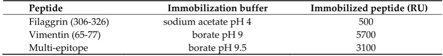
Table 3. Characteristics of peptide immobilization on GLH chip
387
388
Peptide Immobilization buffer Immobilized peptide (RU)
Filaggrin (306-326) sodium acetate pH 4 500
Vimentin (65-77) borate pH 9 5700
Multi-epitope borate pH 9.5 3100
4.5. Peptide synthesis
389
All peptides were synthesized by solid-phase peptide synthesis as described previously [31, 37,
390
42]. The N-terminus of peptides was free or acetylated. Some of the peptides (filaggrin 19, vimentin,
391
collagen II) were C-terminally labeled and some (fibrinβ and multi-epitope peptide) were N
392
terminally labeled by biotinyl-aminohexanoic acid [37]. The –COOH group on the C-terminus was
393
always amidated (Table 1). Peptides were purified by semipreparative reversed-phase
high-394
performance liquid chromatography (HPLC) and were characterized by analytical reversed-phase
395
HPLC and electrospray ionization mass spectrometry (ESI-MS).
396
4.6. Preparation and characterization of bifunctional PLGA NPs
397
Carboxylate-functionalized PLGA NPs were prepared by the nanoprecipitation method as
398
described [32]. The NPs were dispersed in doubly distilled water, finally containing 1.6 × 10¹¹ NPs/ml.
399
The average diameter of NPs was 160–180 nm, each NP containing approximately 4–5000 carboxyl
400
groups available for covalent binding of peptides. NPs were converted to PLGA-active ester
401
derivatives with high excess N-hydroxysuccinimide and
1-ethyl-3-(3-dimethylaminopropyl)-402
carbodiimide. The activated particles were covalently linked to a 1:1 (mol:mol) mixture of
403
complement activating and targeting peptide. This strategy resulted in uniformly oriented peptides
404
of the two types.
405
4.7. Cells
406
Peripheral blood mononuclear cells (PBMCs) were isolated from RA patients and healthy
407
volunteers as described previously [38]. B cells were purified by negative selection using magnetic
408
bead-activated cell sorting (MACS) according to the manufacturer’s protocol (Miltenyi Biotec,
409
Auburn, CA, USA). The purity of the resulting B-cell population was 85–98 %. B cells (106/ml) were
410
cultured with 1 µg/ml CD40L, 1 µg/ml R848 (Mabtech, Stockholm, Sweden) 10 ng/ml interleukin
411
(IL)-2, and 50 ng/ml IL-21 (ImmunoTools GmbH) for 48 hours to increase the frequency of activated
412
memory B cells.
413
4.8. Detection of IgG secreting cells by enzyme-linked immunospot assay (ELISpot)
414
PBMCs were cultured in RPMI-1640 containing 10 % FCS in the presence of 10 ng/ml
415
recombinant IL-2 and 1 µg/ml R848 polyclonal activator provided with the enzyme-linked
416
immunospot assay (ELISpot) kit (Mabtech, Stockholm, Sweden). The cells were harvested at day
417
three, and incubated with the bifunctional PLGA NPs at 1000-fold excess for 1 hour. After removing
418
the unbound NPs, the cells were incubated at 37 °C for 30 minutes in the presence of 1 % pooled
419
normal human serum (NHS) as a complement source or in heat-inactivated serum. After washing, 4
420
×105 PBMCs were transferred into the wells of ELISpot plates (Millipore) pre-coated with the
421
developed after18 hours according to the manufacturer’s instruction (Mabtech, Stockholm, Sweden).
423
The frequency of Cit-containing multiepitope peptide-specific IgG and non-specific IgG producing
424
cells was determined using a C.T.L. Immunospot analyzer (CTL-Europe GmbH, Bonn, Germany).
425
4.9. Statistical analysis
426
For the statistical analysis of data the two-tailed t test and Pearson correlation were used and the
427
results were analyzed with GRAPHPAD PRISM 4 software (GraphPad Software, La Jolla, CA, USA).
428
In all tests, p <0.05 was considered significant.
429
430
Acknowledgments: National Research Development and Innovation Office, OTKA NK 104846, OTKA NK
431
105898 and OTKA 119459, and ELTE KMOP-4.2.1/B-10-2011-0002.
432
The open access publication cost was covered by OTKA NK 105898 fund.
433
Author Contributions: G.S. conceived and designed the experiments; E.S., P.A. and L.V. purified IgG fractions
434
and performed affinity purification and ELISA; E.S. and P.A. performed SPR measurements and analyzed the
435
data, K.H. contributed to ELISA and SPR measurements and analysis of data, J.P. isolated PBMC and performed
436
ELISpot assays, B.R. and G.N. provided blood samples and clinical data of RA patients, G.G. and E.K. designed
437
and prepared nanoparticles, A.M. and F.H. contributed to peptide design and performed the synthesis, A.M
438
coupled the peptides to nanoparticles, G.S. and E.S. wrote the paper.
439
Conflicts of Interest: The authors declare no conflict of interest. The founding sponsors had no role in the design
440
of the study; in the collection, analyses, or interpretation of data; in the writing of the manuscript, and in the
441
decision to publish the results.
442
Abbreviations
443
ACPA Anti-Citrullinated Protein/peptide Antibodies Cit Citrulline
DAS Disease Activity Score
EBNA Epstein-Barr virus Nuclear Antigen EBV Epstein-Barr virus
IVIG Intravenous Immunoglobulin NHS Normal Human Sera
NP Nanoparticles
PBMC Peripheral Blood Mononuclear Cells PLGA Poly (lactic-co-glycolic acid) RA Rheumatoid arthritis SPR Surface Plasmon Resonance
444
References