Structure-function analysis of NF-
k
BI pl05
Soren Beinke
A thesis submitted in partial fulfilment of the requirements of the University o f London
for the degree of Doctor of Philosophy
March 2003
Division of Immune Cell Biology
All rights reserved
INFORMATION TO ALL USERS
The quality of this reproduction is dependent upon the quality of the copy submitted.
In the unlikely event that the author did not send a complete manuscript and there are missing pages, these will be noted. Also, if material had to be removed,
a note will indicate the deletion.
uest.
ProQuest U642858
Published by ProQuest LLC(2016). Copyright of the Dissertation is held by the Author.
All rights reserved.
This work is protected against unauthorized copying under Title 17, United States Code. Microform Edition © ProQuest LLC.
ProQuest LLC
789 East Eisenhower Parkway P.O. Box 1346
NF-kB transcription factors regulate the expression o f genes that promote
immunity, inflammation and cell survival. The p i 05 precursor protein of NF-kB1 p50
acts as an NF-kB inhibitory protein (IkB), retaining associated NF-xB/Rel subunits
inactive in the cytoplasm o f cells. The pro-inflammatory cytokine tumor necrosis factor-
a (TNFa) and bacterial lipopolysacharide (LPS) stimulate p i 05 proteolysis via IkB
kinase (IKK) mediated phoshorylation o f p i 05. Subsequent ubiquitination o f p i 05
precedes its degradation by the proteasome, releasing associated NF-KB/Rel subunits to
translocate to the nucleus.
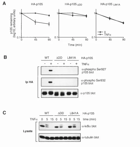
In this study, it is demonstrated that p i 05 binds to IKK via the death domain (DD)
m otif in its C-terminus. Furthermore, the p i 05 DD is required for the efficient
phosphorylation and proteolysis o f p i 05 induced by IKK or in response to TN Fa
stimulation. These data suggest that the p i 05 DD is a docking domain that couples p i 05 to signal-induced proteolysis.
The MAP 3-kinase tumor progression locus-2 (TPL-2), which is required for the
activation of M EKl/2 MAP 2-kinases in response to LPS in macrophages, associates
stoichiometrically with p i 05. Here, it is demonstrated that TPL-2 stability relies on its
high affinity association with p i 05 through two interaction sites. While the TPL-2 C-
terminus binds to residues 497-538 of p i 05, the p i 05 DD interacts with the TPL-2 kinase
domain. Binding to the pi 05 DD inhibits TPL-2 MEK kinase activity, but concomitant
interaction of the TPL-2 C-terminus with residues 497-538 of p i 05 is required for
efficient TPL-2 inhibition by p i 05. Consequently, the C-terminally truncated form of
TPL-2 is insensitive to p i 05 regulation m vivo, which may explain why such a mutation
is oncogenic. These data indicate that in addition to its role as a precursor for p50 and a
Publications arising from this thesis
Beinke, S., Belich, M. P., and Ley, S. C. (2002). The death domain of NF-kappa B1 pl05
is essential for signal-induced pl05 proteolysis. J Biol Chem 277, 24162-24168.
Beinke, S., Deka, J., Lang, V., Belich, M., Walker, P. A., Howell, S., Smerdon, S. J.,
Gamblin, S. J., and Ley, S. C. (2003). NF-kappaBl pl05 negatively regulates TPL-2
A B S T R A C T ... 3
P U B L IC A T IO N S A R IS IN G F R O M T H IS T H E S I S ... 4
T A B L E O F C O N T E N T S ... 5
T A B L E O F D IA G R A M S ... 7
T A B L E O F F IG U R E S ...7
A B B R E V IA T IO N S ...8
1. I N T R O D U C T IO N ... 13
1.1. Re c ept o r sinim m u n it ya n din f l a m m a t io n... 13
1.2. NF-KB SIGNALLING PATHWAYS...17
1.2.1 Structure and function o f NF- kB...17
1.2.2 NF-KB/Rel proteins...20
1.2.2.1 NF-KB/Rel dimérisation and DNA binding... 20
1.2.2.2 Generation of p50 and p 52... 21
1.2.2.3 Transactivation potential of NF-KB/Rel dimers... 24
1.2.2.4 Genetic analysis of NF-KB/Rel protein function... 25
1.2.3 N F-kB regulation by IkB proteins... 29
1.2.3.1 Function and specificity of IkB proteins... 29
1.2.3.2 Targeting IkB « for the proteasome...34
1.2.3.3 The IKK complex - structure and function... 36
1.2.3.4 Upstream of IK K ...40
1.2.3.5 Signal-induced plOO processing...41
1.2.3.6 Signal-induced pIOS proteolysis...42
1.3. M A P KINASE SIGNALLING PATHWAYS...45
1.4. T N F RECEPTOR SIGNALLING... 49
1.5. IL-1 /To l l-l ik er e c e p t o rs ig n a l l in g... 52
1.6. TUMOR PROGRESSION LOCUS-2 (T P L -2 )... 53
1.7. Spec ifica im so ft h iss t u d y...55
2. R E S U L T S ... 59
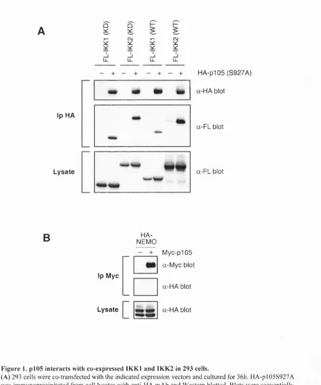
2.1. THE P l0 5 D D IS A DOCKING DOMAIN REQUIRED FOR SIGNAL-INDUCED P i05 PROTEOLYSIS 59 2.1.1 IK K l and 1KK2 interact with N F -kB 1 p i 05 in transiently transfected 293 cells...59
2.1.2 p i 05 interacts with the endogenous IK K com plex...59
2.1.3 The p i 05 DD is required fo r the interaction o fp l0 5 with the IK K complex...61
2.1.4 A functional p l0 5 DD is required fo r efficient serine 927/932 phosphorylation in vitro by IK K l or 1KK2...66
2.1.5 Efficient p i 05 proteolysis induced by over-expressed 1KK2 requires a functional p l0 5 D D...68
2.1.6 The DD is not required fo r H A -pl05 proteolysis induced by high concentrations o f FL-1KK2...71
2.1.7 p i 05 DD is essential fo r TNFa-induced degradation o f p i 0 5...74
2.2. Ch a r a c t e r is a t io no f TPL -2 b in d in gtop105 ... 76
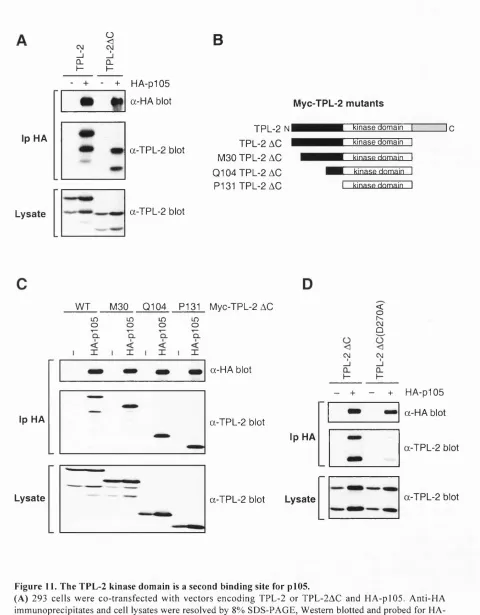
2.1.8 The TPL-2 C-terminus binds to residues 497-539 o f p i 0 5...76
2.1.9 The TPL-2 kinase domain interacts with the p l0 5 D D...78
2.1.10 The p l0 5 DD and residues 497-538 o f p 105 contribute equally to the interaction with TPL-2...82
Table of contents
2.4. TPL-2-INDUCED P l0 5 PROTEOLYSIS DEPENDS ON ITS BINDING TO P l 0 5 ... 89
2.5. P i05 INHIBITS T PL -2 M E K KINASE ACTIVITY... 92
3. DISCUSSION... 101
3.1. H o w DOES THE P105 D D MEDIATE P105 BINDING TO I K K ? ... 101
3.2. Sig n if ic a n c eo f IK K b in d in gt op105 f o rs ig n a l-in d u c e dp105 p r o t e o l y s is...103
3.3. Is TPL-2 UPSTREAM OF P i 0 5 ? ... 106
3.4. Re g u l a t io no f T PL -2 - an o v e lf u n c t io no fp105... 107
3.5. A RATIONALE FOR THE ONCOGENIC POTENTIAL OF C-TERMINALLY TRUNCATED T P L -2 ...109
3.6. Do e sp105 r e g u l a t e TPL-2 a c t iv it yinr e sp o n s et o L P S ? ... 110
3.7. Wh a tist h em e c h a n ismo f TPL -2 a c t iv a t io n? ... 111
3.8. Im p l ic a t io n so f T PL -2 s t a b il is a t io nb yp1 0 5 ... 113
3.9. Im p l ic a t io n sf o rp 105 a n d T PL -2 a st h e r a p e u t ict a r g e t s...114
4. MATERIALS AND METHODS... 118
4.1. CDNA CONSTRUCTS, RECOMBINANT PROTEINS AND ANTIBODIES...118
4.2. Cellsa n dt r a n s f e c t io n...120
4.3. IMMUNOPRECIPITATION AND WESTERN BLOTTING ANALYSIS...121
4.4. An a l y se so fp r o t e ind e g r a d a t io n... 122
4.5. St a t is t ic s...123
4.6. An a l y se so fp r o t e inin t e r a c t io n... 123
4.7. Ce l lf r a c t io n a t io n... 124
4.8. An a l y siso fp r o t e inp h o s p h o r y l a t io n... 125
4.9. R N A ISOLATION AND R T -PC R ANALYSIS... 126
BIBLIOGRAPHY... 128
DIAGRAM 2. NF-KB ACTIVATION...18
Dia g r a m 3. N F-kB a n d IkB f a m il ym e m b e r s... 19
Dia g r a m 4. IkB d e s t r u c t io nb o x e s... 35
Dia g r a m 5. He t e r o g e n ic it yo f N F -kB s ig n a l l in gp a t h w a y s... 44
Dia g r a m 6. M A P k in a s es ig n a l l in gp a t h w a y s... 48
Dia g r a m 7. T L R /IL -IR a n d T N F R s ig n a l l in g... 50
Dia g r a m 8. St r u c t u r eo fd e a t hd o m a in s... 57
Table of Figures
Fig u r e 1. P l0 5 in tera ctsw it hc o-e x p r e ss e d IK K in 293 c e l l s...60Fig u r e 2. En d o g e n o u sp105 in t e r a c t sw it h IK K l a n d IK K 2 ...62
Fig u r e 3. Th epI05 d ea thd o m a inisr e q u ir e df o rpI0 5 b in d in gto I K K l a n d IK K 2 ...64
Fig u r e 4. p I0 5 d ea thd o m a inm u t a n t sb in dto Re lA a n da r er e t a in e dint h ec y t o p l a s m...65
F i g u r e 5. P h o s p h o r y l a t i o n o f p105 s e r i n e 927/932 b y IK K l a n d IK K 2 invitro r e q u i r e s a FUNCTIONAL P105 DEATH DOMAIN... 67
Fig u r e 6. In d u c t io no fap5 0/pI0 5 r a t ios h if tby IK K 2 r e q u ir e st h epI0 5 d ea thd o m a in... 69
Fig u r e 7. Th epI05 d ea thd o m a inisr e q u ir e df o r IK K 2-in d u c e dpI0 5 p r o t e o l y s isa n dse r in e 927/932 PHOSPHORYLATION O F P I0 5 ... 70
Fig u r e 8. Hig hlev elso fo v e r-e x p r e ss e d IK K 2 in d u c epI05 p r o t e o l y s is in d e pe n d e n to ft h epI05 DEATH DOMAIN... 73
Fig u r e 9. Th epI05 d ea th d o m a inises s e n t ia lfo r T N F a -in d u c e dse r in e 927/932 p h o s ph o r y l a t io n AND PROTEOLYSIS OF P i 0 5 ... 75
Fig u r e 10. Th e TPL -2 C -t e r m in u sb in d st or e sid u e s 4 9 7-539... 77
Fig u r e 11. Th e TPL-2 k in a s ed o m a inisase c o n db in d in gs it efo rpI0 5 ...80
Fig u r e 12. Th ek in a s ed o m a ino f T PL -2 d ir e c t l yin ter a ctsw it ht h epI0 5 d ea thd o m a in...81
Fig u r e 13. Th epI05 d ea thd o m a ina n dr e sid u e s 497-538 o f p105 e q u a l l yc o n t r ib u t et o TPL-2 BINDING TO P i0 5 ...83
Fig u r e 14. TPL-2 m e t a b o l ics t a b il it yisr e g u l a t e db ypI 0 5 ...87
Fig u r e 15. Bin d in gtop105 is r e q u ir e dbu tt h epI05 d ea thd o m a inisin h ib it o r yf o r T PL -2-INDUCEDPI05 PROTEOLYSIS...90
Fig u r e 16. Th epI05 d ea thd o m a inin h ib its TPL-2 M E K k in a s ea c t iv it y... 93
Fig u r e 17. Th e TPL-2 C -t e r m in u sisr e q u ir e dfo rt h ee f f ic ie n tin h ib it io no f TPL -2 M E K k in a s e ACTIVITY BY P l0 5 ... 96
Abbreviations
AP-1 activator protein 1
APC antigen presenting cell
ATP adenosine tri-phosphate
P-TrCP p-transducin repeat containing protein
BAFF B cell activating factor
BCR B cell antigen receptor
BMDM bone marrow-derived macrophages
BSA bovine serum albumin
bZIP basic region leucine zipper protein
cAMP cyclic adenosine mono-phosphate
CARD caspase recruitment domain
CD cluster of differentiation
CKII casein kinase II
Cot cancer osaka tyroid
CRD cysteine-rich domain
DD death domain
DED death effector domain
DNA desoxyribonucleic acid
E gestation day
ERK extracellular signal-related kinase
EV empty vector
FADD Fas-associated death domain
FL FLAG
GRR glycine-rich region
GST glutathione-iS-transferase
h hours
HA haemaglutinine
HAT histone acetyl transferase
HDAC histone deacetylase
HRP horse raddish peroxidase
HTLV-1 human T-cell leukemia virus-1
ICAM-1 intercellular adhesion molecule-1
Ig immunoglobin
IKK IkB kinase
IL interleukin
IL-IR IL-1 receptor
iNOS inducible nitric oxide synthetase
IP incontinentia pigmenti
IRAK IL-1 receptor associated kinase
IV? in vitro precipitation
JNK Jun amino-terminal kinase
KD kinase-inactive
LFA-1 leukocyte function-associated antigen-1
LPS lipopolysacharide
LT lymphotoxin
MAP mitogen activated protein
MAP 2-kinases MAP kinase kinase
MAP 3-kinases MAP kinase kinase kinases
MAPK MAP kinase
MAPKAPK MAPK-activated protein kinase
Abbreviations
MCP-1 macrophage chemoattractant protein-1
M-CSF macrophage-colony stimulating factor
MEK MAPK ERK kinase
MEKK MEK kinase
MHC major histocompatibility complex
min minutes
M IP -la macrophage inflammatory protein-la
MKK MAP kinase kinases
MKKK MAP kinase kinase kinases
mRNA messenger RNA
MUK MAPK upstream kinase
MyD88 myeloid differentiation factor 88
NF-AT nuclear factor of activated T cells
NF-kB nuclear factor-KB
NIK NF-KB-inducing kinase
NIMR National Institute for Medical Research
NLS nuclear localisation signal
NO nitric oxide
p90 ribosome S6 kinase
PAGE polyacrylamid gel electrophoresis
PAMP pathogen-associated molecular pattern
PBS phosphate-buffered saline
PCR polymerase chain reaction
PEST proline/glutamate/serine/threonine-rich
PKA protein kinase A
PKB protein kinase B
PP2A protein phosphatase 2A
PRR pattern recognition receptor
PVDF polyvinylidene difluoride
RHD Rel-homology domain
RIP receptor-interacting protein
RKIP RAF kinase inhibitory protein
RNA ribonucleic acid
SCF SKpl-Cullin-F-box
SCID severe combined immunodeficiency
SDS sodium dodecyl sulfate
SODD silencer o f death domains
STAT signal transducer and activator of transcription
TAB TAKl binding protein
TAKl transforming growth factor-p activated kinase 1
TCR T cell antigen receptor
ThI T helper type 1
Th2 T helper type 2
TIR Toll-like/lL-1 receptor homology
TLR Toll-like receptor
TNF tumor necrosis factor
TNFR TNF receptor
TPL-2 tumor progression locus-2
TRADD TNF receptor-associated death domain
TRAF TNF receptor-associated factor
VCAM-1 vascular cell adhesion molecule-1
^ max maximal velocity
1. Introduction
1.1. Receptors in immunity and inflammation
The immune system protects vertebrates from invading pathogens by both innate
and adaptive immune responses. Both achieve the recognition of “non se lf’ molecules by
specialised receptors, but use different effector mechanisms to finally eliminate
pathogens that carry these molecules.
Innate immune responses can be activated very rapidly in response to infection
and provide the first line of defence (Janeway and Medzhitov, 2002). The innate immune
system relies on germ line encoded pattern recognition receptors (PRRs), which
recognise general pathogen-associated molecular patterns (PAMPs). PAMPs are essential
components of pathogens with little variability and are absent in mammals (e.g.
lipopolysacharide (LPS), peptidylglycans, lipoproteins, unmethylated bacterial DNA or double-stranded RNA).
Ten members of the Toll-like receptor (TLR) family have been identified, which
specifically mediate responses to particular PAMPs in mammals (Medzhitov, 2001).
TLRs are expressed by a broad range of cells (e.g. monocytes/macrophages, dendritic
cells, neutrophils or endothelial cells), which come in close contact with the pathogen at
the site of antigen encounter. They are transmembrane receptors and mediate responses to extracellular PAMPs. Ligation of TLRs activates nuclear factor (NF) -kB and mitogen
activated protein (MAP) kinase signalling pathways. The nucleotide-binding
domain/leucine-rich repeat proteins, NODI and N 0D 2, have been suggested to be
involved in the recognition of intracellular PAMPs and also activate NF-kB and MAP
Introduction_________________________________________________ Receptors in immunity and inflammation
Pathogen recognition induces the up-regulation of the pro-inflammatory cytokines
tumor necrosis factor-a (TNFa) and interleukin (IL) -1. TN Fa and IL-1 affect most cell
types and their biological functions are remarkably similar (Dinarello, 1996; Vassalli,
1992). However, the two cytokines and their receptors are not related in sequence.
TN Fa can bind to both the p55 TNF receptor (TNFRl) and the p75 TNF receptor
(TNFR2). However, the majority of biological functions of TN Fa is mediated through
the activation of NF-kB and MAP kinase signalling pathways by TNFRl (Wallach et al.,
1999). Upon ligation TNFRl also activates a caspase-cascade leading to apoptosis, but
this is usually suppressed by the simultaneous induction o f anti-apoptotic proteins via the induction of NF-kB. IL-1 binds to the IL-1 type 1 receptor (IL -lR l), which also triggers
NF-kB and MAP kinase signalling pathways (O'Neill, 2000).
The major source of TN Fa is cells o f the monocyte/macrophage lineage, with T
lymphocytes, neutrophiles, mast cells and endothelial cells also contributing under different circumstances (Vassalli, 1992). The production and release of TN Fa can be
induced by all potentially noxious stimuli. In vivo, TN Fa is the most rapidly produced
pro-inflammatory cytokine and acts as a key inductor and central coordinator of the
innate immune response (see Diagram 1). TN Fa up-regulates the production o f pro-
inflammatory cytokines, including TN Fa itself. This initiates an auto-regulatory positive
feedback loop, which plays an important role for the amplification o f inflammatory
immune responses. Furthermore, TN Fa induces the expression of adhesion molecules
(e.g. vascular cell adhesion molecule-1 (VCAM-1), intercellular adhesion molecule-1
(ICAM-1), leukocyte function-associated antigen-1 (LFA-1), selectins) and the secretion
of chemokines (e.g. IL-8, macrophage inflammatory protein-la (M IP -la), macrophage
chemoattractant protein-1 (MCP-1), RANTES, eotaxin), which attract mononuclear
phagocytes and granulocytes to the site of infection. In macrophages, TN Fa induces the
and enhances the killing ability o f macrophages. Activation of macrophages by TN Fa
also induces the up-regulation of the major histocompatibility complex (MHC) and co
stimulatory molecules (e.g. CD80, CD86), as well as the secretion of effector cytokines
(e.g. IL-6, IL-12, Lymphotoxin (LT) a and p), thereby facilitating the activation of
adaptive immune responses.
Adaptive immune responses involve a highly diverse repertoire o f somatically
rearranged antigen receptors expressed on B and T cells (B cell antigen receptors or
BCRs and T cell antigen receptors or TCRs), which recognise virtually every possible
structure of foreign protein antigens. Each B or T cell produces receptors o f a single
specificity. Recognition by TCRs requires phagocytosis and proteolysis of the antigen by
antigen presenting cells (APCs; e.g. macrophages, dendritic cells or B cells), which
present the antigenic polypeptide bound to the MHC on their surface. BCR and TCR ligation leads to the Ca^^-dependent activation of nuclear factor of activated T cells (NF-
AT), as well as the induction of NF-kB and MAP kinase signalling pathways. In concert,
activation of these signalling pathways induces the expression of proteins that support
lymphocyte proliferation and differentiation. The expansion and differentiation of antigen
specific T or B cell clones in response to primary antigen encounter requires 1-2 days.
The main effector function of mature B cells in adaptive immunity is the secretion
of antibodies (also referred to as immunoglobin (Ig)), which have a similar structure and
the same specificity as the respective BCR. Antibodies bind to pathogens or their toxic
products, which elicited the immune response, in the extra cellular spaces of the body,
thereby neutralising the pathogen or recruiting phagocytic cells and molecules of the
complement system to eliminate them. T cells differentiate into either CD8^ effector T
cells, which specifically eliminate cells that host pathogens, or CD4^ helper T cells,
TNFa
TNFR
MAPK MARK
iNOS
TNFR
TNFa
Chemokines TLR
Adhesion Molecules
M acroph age
T-cell
Endothelial cell
1.2. NF-kB signalling pathways
1.2.1 Structure and function of NF-kB
Nuclear factor (NF) -kB is an eukaryotic transcription factor that is ubiquitously
expressed. It was first described in 1986 as a nuclear protein required for the B cell
specific transcription of the immunoglobin kappa light chain (Sen and Baltimore, 1986).
However, subsequent research revealed that NF-kB is an inducible factor, which is
involved in the regulation o f an exceptionally large number of genes, most importantly
during immune responses, inflammation or lymphoid organ development (Baeuerle and
Henkel, 1994; Baldwin, 1996; Ghosh et al., 1998; Siebenlist et al., 1994). NF-kB is also
important for the prevention o f apoptosis, which is vital for its function in immune
responses (Karin and Lin, 2002).
The term NF-kB describes a family o f transcriptional regulators composed of
hetero- and homodimeric complexes of NF-KB/Rel proteins (Baldwin, 1996; Siebenlist et
al., 1994). All NF-KB/Rel proteins contain a conserved Rel-homology domain (RHD)
that contributes to dimérisation and DNA binding. In mammalian cells, this family
comprises RelA (p65), RelB, c-Rel, NF-kB 1 p50 and NF-kB2 p52 (see Diagram 3). NF-
kB 1 p50 and NF-KB2 p52 are produced as larger inactive precursor molecules o f lOSkDa (p i05) and lOOkDa (p i00), respectively (Mercurio et al., 1993; Rice et al., 1992). p50 or
p52 are derived from the N-terminal domain of their precursors via processing o f the C-
terminal half by the 26S proteasome (Betts and Nabel, 1996; Heusch et al., 1999; Lin et
al., 1998a; Palombella et al., 1994).
In unstimulated cells, NF-kB is retained inactive in the cytoplasm via the
interaction with inhibitory proteins termed IkBs (see Diagram 2) (Baldwin, 1996;
Siebenlist et al., 1994). IkBs contain multiple ankyrin repeats that bind to RHDs of NF-
Stimulus
Proteasome
G ene transcription
D i a g r a m 2. N F -kB activation.
N F -K B / R e l
l_Z
RelB >
c-R el >
N F-K B1(p 105/p 50) K
N F-K B2(p 100/p 52) = ( ' X
TD 550
TD 587
Ankyrin repeats GRR
^ „ V '
> Q ^ OOŒ]=CEM3:
PEST
k B - a
IkB-P IkB-£
Bcl-3
DD
969
=nofTT==rTT==n=
DD 940
401
IkB
D i a g r a m 3. N F -kB a n d IkB f ami ly m e mb e r s.
Introduction NF-kB signalling: structure and function
nuclear uptake. This family of structurally related proteins includes I KB a , iKBp, IkBe,
together with the precursors NF-KB 1 pl05 and NF-kB2 plOO and the iKB-like protein
BCL-3 (see Diagram 3). In response to appropriate stimulation with agonists, IkBs are
phosphorylated by the high molecular weight IkB kinase (IKK) complex (Ghosh et a l,
1998; Karin and Ben-Neriah, 2000). Subsequent ubiquitination leads to their degradation
by the proteasome, which allows NF-kB dimers to translocates into the nucleus and
regulate gene expression. In addition, the modification of transcriptional activity of
nuclear NF-kB dimers by association with other regulatory proteins is regulated by NF-
KB/Rel subunit phosphorylation.
1.2.2 NF-KB/Rel proteins
1.2.2.1 NF-KB/Rel dimérisation and DNA binding
With the exception o f RelB, all members of the NF-KB/Rel family can homo- or
heterodimerise with each other (Baeuerle and Henkel, 1994; Siebenlist et al., 1994). RelB
forms hetero-dimers with p50 and p52, but can not form homodimers. Crystal structures
o f the p50 homo-dimer and RelA/p50 hetero-dimer reveal that the RHDs of these
proteins are comprised of two immunoglobin-like domains connected by a flexible linker
(Baltimore and Beg, 1995; Chen et al., 1998; Ghosh et al., 1995; Muller et al., 1995).
Dimérisation occurs via inter-digitating hydrophobic site chains exclusively o f the C-
terminal domain, while loops between p-strands o f both N- and C-terminal domains
contribute to DNA binding. The overall structure of NF-kB bound to DNA resembles the
shape o f a butterfly that grips DNA by enveloping it. Different NF-kB dimers exhibit
different affinities for the 10 base pair KB-sites with the consensus sequence 5'-
GGGRNN Y Y CC-3 ' in promotors of NF-kB responsive genes (R is purine, Y is pyrimidin
KB-sites contributes to specificity to one or the other NF-kB dimer, which is presumably
the basis for different NF-kB dimers regulating different genes.
1.2.2.2 Generation of p50 and p52
p50 and p52 are generated as the transcriptionally inactive longer precursor
proteins p i 05 and p i 00, respectively (Bours et al., 1990; Ghosh et al., 1990; Kieran et al.,
1990; Meyer et al., 1991; Neri et al., 1991; Schmid et al., 1991). The N-terminal half of
the precursors is identical to the active NF-KB/Rel subunits. Processing of the precursor proteins results in the complete degradation of the C-termini leaving the N-terminal
transcription factor domain intact. p50 and p52 production has been proposed to occur
co-translationally on polysomes (Heusch et ah, 1999; Lin et al., 1998a; Lin et al., 2000; Mordmuller et al., 2003). However, the physiological relevance of this mechanism is not
clear since most other studies find that the majority of cellular p50 and p52 is produced
post-translational by processing of p i 05 or p i 00, respectively (Belich et al., 1999;
Claudio et al., 2002; Coope et al., 2002; Dejardin et al., 2002; Donald et al., 1995; Mellits
et al., 1993; Orian et al., 2000; Xiao et al., 2001b).
A number of evidence indicate that proteolysis of p i 05 is mediated by the
proteasome. It was demonstrated that p i 05 proteolysis is adenosine triphosphate (ATP)
dependent, requires ubiquitination and can be blocked by proteasome inhibitors in vitro
and in vivo as well as by mutations of the proteasome in yeast (Fan and Maniatis, 1991;
Palombella et al., 1994; Sears et al., 1998). Furthermore, proteasome enriched cytosolic
fractions can facilitate p i 05 processing in vitro (Orian et al., 1995). Similarly, the
proteasome was found to mediate processing o f p i 00 to p52 (Betts and Nabel, 1996;
Introduction NF-kB signalling: p50/p52 generation
p i 05 processing to p50 occurs constitutively, but p i 05 proteolysis is also induced
in response to cellular stimulation. However, it is not entirely clear whether signal-
induced p i 05 proteolysis results in increased processing to p50 or in the complete
degradation o f p i 05. Some studies report increased processing of p i 05 in response to
TN Fa (Mellits et al., 1993; Naumann and Scheidereit, 1994), LPS (Donald et al., 1995)
or phorbol 12-myristate 13-acetate (PMA)/ionomycin (MacKichan et al., 1996), while
others fail to detect an increase in p50 levels equivalent to the decrease in p i 05 levels
(Belich et al., 1999; Harhaj et al., 1996; Zheng et al., 1993). Most stimuli tested induce
p i 05 proteolysis with slower kinetics than IkBœ degradation (0.5-4 hours) (Donald et al.,
1995; Lang et al., 2003). p i 00 is not constitutively processed to p52 and p i 00 processing
is completely unaffected by most stimuli that induce degradation of IkB a . However, p i 00 processing occurs in response to LXP, CD40 and B cell activating factor (BAFF)
within 3-6 hours (Claudio et al., 2002; Coope et al., 2002; Dejardin et al., 2002; Kayagaki
et al., 2002).
Partial proteolysis of proteins by the proteasome is a rare mechanism that only
applies to p i 05, p i 00 and a few other proteins, including the distant yeast homologues of
the NF-kB precursor proteins SPT23p and Mga2p (Hoppe et al., 2000; Rape et al., 2001).
Flexible unstructured regions composed of amino acids with short side chains seem to be
necessary for partial degradation of proteins by the proteasome (Rape and Jentsch, 2002).
A glycine-rich region (GRR) is required for 105 and 100 processing (Betts and Nabel,
1996; Heusch et al., 1999; Lin and Ghosh, 1996; Orian et al., 1999). The GRR could act
as a stop signal that halts proteasomal degradation proceeding from the C-terminus.
Two lysines 441 and 442 and a potential ubiquitin ligase recognition motif C-
terminal to the p i 05 GRR are also required for constitutive ubiquitination and processing
of p i 05 (Orian et al., 1999). Matouschek and collègues proposed a model for proteasomal
signal (Lee et al., 2001). Accordingly, an alternative model for pl05 processing bases on
a proteolytic cleavage o f the precursor protein before the C-terminus is degraded.
Therefore, the GRR could function as a cleavage site. However, a potential
endoproteolytic activity needs to be associated with the 26S proteasome, because the 26S
proteasome clearly can provide all proteolytic activities necessary for p50 processing in
vitro (Coux and Goldberg, 1998). Indeed, the proteasome was recently demonstrated to
catalyse endoproteolytic cleavage o f polypeptide bonds (Liu et al., 2003). Processing
substrates may enter the proteasome by hairpin loop formation to be proteolytically
cleaved by the active site o f the proteasome (Lee et al., 2001; Liu et al., 2003). Unfolding
of the GRRs of p i 05 and p i 00, as well as regions of low sequence complexity in related
domains o f SPT23p and Mga2p, probably does not require much energy and the lack of
bulky side chains may facilitate the passage into the 20S proteasome barrel. Interestingly,
the yeast proteins SPT23p and Mga2p are cleaved before their C-termini are degraded
(Rape et al., 2001).
This model implies uni-directional proteolysis from the cleavage site into the C-
terminus. Accordingly, tightly folded domains or protein interaction can prevent
proteasom al degradation (Lee et al., 2001). Indeed, processing o f p i 05 and
SPT23p/Mga2p is dependent on homo-dimerisation of the N-terminal Ig-like domains of
the transcription factors (Lee et al., 2001; Lin et al., 2000; Rape et al., 2001). Therefore,
the globular structure of the dimerised transcription factors probably rescues them from
proteasomal degradation. The generation of the p50 and p52 NF-xB/Rel subunits from
inactive precursor proteins provides an additional regulatory step in the activation of NF-
Introduction NF-kB signalling: NF-KB/Rel transactivation
1.2.2.3 Transactivation potential of NF-KB/Rel dimers
RelA and c-Rel function as potent transcriptional activators, while RelB can
activate transcription only in certain cell types. p50 and p52 lack intrinsic transactivation
capacity. This implies that homodimers of p50/p50 or p52/p52 without any co-factors are
transcriptionally repressive, while dimers including RelA, RelB or c-Rel are
transcriptionally activating. Once liberated from the inhibitory IkB proteins the capacity
o f NF-KB/Rel dimers to induce transcription is also a regulated process. Binding affinity
to both DNA and nuclear cofactors is often regulated by direct phosphorylation of NF-
KB/Rel dimers.
Protein kinase A (PKA) (Zhong et al., 1997; Zhong et al., 1998), Akt/protein
kinase B (PKB) (Madrid et al., 2001; Madrid et al., 2000), casein kinase II (CKII) (Bird
et al., 1997; Wang et al., 2000) and phosphatidylinositol 3-kinase (Sizemore et al., 1999)
bave been implicated in NF-KB/Rel subunit phosphoryalation. Knockout studies revealed
that glycogen synthetase kinase 3P (GSK3p) (Hoeflich et al., 2000), protein kinase C (PKC) Ç (Leitges et al., 2001), IKK (Sizemore et al., 2002) and Tbk/t2k/NAK (Bonnard
et al., 2000) are required for in the induction of transactivation capacity in response to a
wide variety o f stimuli, while NF-KB-inducing kinase (NIK) regulates NF-kB
transcriptional competence specifically in response to LXP (Yin et al., 2001).
RelA It was first shown that RelA is phosphorylated after cellular
stimulation (Naumann and Scheidereit, 1994). RelA phosphorylation regulates the
recruitment of histone acetyl transferases (HATs), such as CBP/300 and p/CAF, to
nuclear RelA containing NF-kB dimers, which modify the secondary structure of target
DNA sites to facilitate NF-kB binding (Gerritsen et al., 1997; Merika et al., 1998; Perkins
et al., 1997; Sheppard et al., 1999; Zhong et al., 1998). Nuclear RelA can also interact
expression (Ashbumer et al., 2001; Chen et al., 2001; Ito et al., 2000; Lee et al., 2000;
Zhong et al., 2002).
p50/p52 p50 and p52 lack a transactivation domain and can not directly
recruit HATs (Sheppard et al., 1999). Consistent with this, over-expression o f p50 causes
a decrease in the expression o f NF-kB dependent genes, such as TN Fa (Baer et al.,
1998). Analysis o f naturally occurring promotor mutants has confirmed that binding of
p50 homodimers to the TNFot promotor negatively regulates TN Fa expression (Udalova
et al., 2000). Most nuclear NF-kB complexes in unstimulated cells were shown to consist
of p50 homodimers associated with HDAC-1. These (p50)2/HDAC complexes repress the transcription o f a subset of NF-xB-dependent genes, but can be replaced within certain
target gene promotors by complexes containing p50 and RelA, which is phosphorylated
by PKA (Zhong et al., 2002). However, p50 and p52 homodimers can promote
transcription in vivo via the interaction with the nuclear, IxB-like protein Bcl-3, which recruits associated HATs, such as Tip60 (Dechend et al., 1999). The transcriptional
activation potential of p50 and p52 homodimers, therefore, seems to be dependent on the
availability of Bcl-3 and is likely to be cell type and stimulus specific.
1.2.2.4 Genetic analysis of NF-KB/Rel protein function
RelA The p50/RelA heterodimer was the first form o f N F-kB to be
identified and is the most abundant NF-kB dimer in most cell types. The deletion of the
relA gene in mice causes embryonic death at gestation day (E) 15.5-16.5 due to massive
apoptosis o f liver parenchymal cells, which especially affects hepatocytes (Beg et al.,
1995a). TN Fa readily kills macrophages and fibroblast from RelA-/-, but not wild type
mice, and RelA-/-TNFa-/- or RelA-/-TNFRl-/- double mutants are born with grossly
Introduction NF-KB signalling: NF-KB/Rel function
the protection from TNFa-induced apoptosis, a function that is not compensated by other
NF-KB dimers.
Adoptive transfer of RelA-/- hematopoietic stem cells into irradiated SCID mice
results in a reduced number of B cells, which exhibit impaired proliferative responses and
reduced immunoglobin secretion (Doi et ah, 1997). More severe lymphopoietic defects
with complete absence o f B cells is found in lethally irradiated mice reconstituted with
double deficient RelA-/-NF-KB 1 -/- fetal liver stem cells (Horwitz et al., 1999). This
illustrates that RelA also has specfic functions in B cell development and function.
NF-kB 1 p50 The involvement of p50/RelA in the transcription of a wide variety
of genes and the importance of the RelA subunit in the prevention of apotptosis during
embryonic development would suggest severe histopathological defects in mice deficient
for NF-KB 1. However, these mice, which do not produce either the p i 05 precursor or p50, develop normally, suggesting a great degree o f functional compensation by other
NF-KB family members for p50 in the p50/RelA dimer (Sha et al., 1995).
Nevertheless, NF-KB 1 -deficient mice are unable to clear the intracellular
bacterium Listeria monocytogenes and are more susceptible to infection with the extra
cellular gram-positive bacterium Streptococcus pneumoniae. However, they respond
normally to challenges with extra-cellular gram-negative bacteria Haemophilus influenza
and Escherichia coli K l. NF-KB 1-/- B cells display impaired proliferation in response to
LPS and soluble CD40 ligand, but not in response to membrane-bound CD40 ligand,
anti-IgM or anti-IgD-dextran. They also display defective IgG3, IgE and IgA class
switching (Snapper et al., 1996b). These observations suggest an important role for p50
in B cell proliferation and maturation as well as in cellular immune responses.
Interestingly, NF-KB 1-/- mice are more resistant to murine encephalomycarditis virus,
negative regulatory function of p50 homodimers in regulation of the expression of the 13-
interferon gene. Furthermore, NF-kB 1-/- mice are completely devoid o f allergic airway
inflammation in a murine model for asthma (Yang et al., 1998). This results from a lack
o f CD4^ T helper type 2 ( Th2 ) differentiation o f NF-kB 1-/- T cells, which is due to
impaired IL-4, IL-5 and IL-13 production, correlating with the failure to induce the
expression of the transcription factor GATA-3 (Das et al., 2001). NF-kB 1-/- mice are
also refractory to the induction o f both a chronic (collagen-induced) and acute
(methylated BSA/IL-1 induced) arthritis model, suggesting p50 is important for the
pathology of this inflammatory disease (Campbell et al., 2000).
NF-KB2 p52 Analyses of NF-kB2 knockout mice indicates a particular role for p52 in the maintenance o f the peripheral B cell pool and organisation o f B cell
compartments in secondary lymphoid organs. Deletion o f NF-kB2 in mice results in
disrupted splenic architecture characterised by diffuse B cell areas, indiscreet follicles
and the absence of the perifollicular marginal zone (Caamano et al., 1998). The number
o f B cells in the spleen and other lymphoid organs is reduced and NF-kB2-/- B cells
show mild proliferative defects in responses to CD40 ligand, LPS and anti-IgD-dextran.
NF-kB2-/- mice are also deficient in the formation of germinal centres and Peyer’s
patches, and their T cell-dependent and independent responses are reduced (Yilmaz et al.,
2003). These mice also display increased susceptibility to infection by L. monocytogenes
(Caamano et al., 1998).
NF-KB 1/2 p50/p52 The phenotype of NF-kB 1 and NF-kB2 double knockout
mice suggest these proteins are partially redundant (lotsova et al., 1997). Impaired bone
remodelling leading to osteopetrosis and incomplete odontosis in these mice correlates
with a decrease in the total number of osteoclasts. This indicates an additional role of
Introduction NF-kB signalling: NF-KB/Rel function
hyperplasia, early thymus regression, lack of CD4^ and CD8^ T cells in the periphery and
progressive loss of B cells.
RelB RelB-/- mice are impaired in the formation of germinal centers, the
marginal zone o f the spleen and Peyer’s patches (Weih et ah, 2001; Yilmaz et ah, 2003).
Consistent with this, RelB is required for normal production o f antigen specific IgG in
response to T-dependent and independent stimuli (Snapper et ah, 1996a). This phenotype
is very similar to that of NF-KB2 knockout mice, suggesting that these Rel subunits
functionally cooperate.
RelB knockout mice also display multifocal inflammatory cell infiltration in
several organs accompanied with myeloid hyperplasia and splenomegaly (Weih et ah,
1995). Crossing RelB-deficient mice with mice that lack either T and B cells or only T
cells revealed that this inflammatory phenotype is dependent on T cells (Weih et ah,
1996). Moreover, RelB-/- mice are susceptible to L. monocytogenes and are unable to mount protective immune responses to choriomeningitis virus, indicating defects in T
cell/macrophage interaction and CD8^ cytotoxic T cell responses (Weih et ah, 1997).
RelB is also required for a normal delayed-type hypersensitivity response, an immune function that requires the intact function of epidermal antigen presenting (Langerhans)
and antigen specific CD4^ T helper type 1 (T hI) cells (Weih et ah, 1995). Thus, RelB is
not only essential in the correct development o f the hematopoietic system, but is also
involved in humoral and cellular immune responses.
c-Rel Knockout mice for c-Rel develop normally but are specifically
impaired in humoral and cellular immune responses (Gerondakis et ah, 1996; Kontgen et
ah, 1995). Proliferation o f c-Rel-/- B and T cells in response to most mitogenic stimuli is
impaired, and serum levels of IgG 1, IgG2a, IgG2b and IgG3 are decreased in c-Rel-/-
of inducible nitric oxide synthetase (iNOS), which correlates with diminished nitric oxide
(NO) production. This makes c-Rel-/- mice susceptible to infection with the intracellular
parasite Leichmania major. Mice with an homozygous deletion of the trans-activating C-
terminus of c-Rel (c-Rel"^^^"^^^^) display similar defects in immune functions but with a
more severe phenotype than mice lacking the entire c-Rel protein (Carrasco et al., 1998).
Additionally, c-Rel^^^^^^^ animals present histopathological alterations of hemopoietic
tissues, such as an enlarged spleen due to lymphoid hyperplasia, extramedullary
hematopoiesis and bone marrow hypoplasia. Thus, in c-Rel^^^'"^^^ mice, the lack of c-Rel
activity is less efficiently compensated by other NF-kB proteins, which may result from
c-Rel^^^^^^^ acting as a dominant negative protein.
1.2.3 NF-kB regulation by IkB proteins
1.2.3.1 Function and specificity of IkB proteins
IkB proteins contain six to seven ankyrin repeats of approximately 33 amino
acids, with which they bind to the RHD of NF-KB/Rel proteins (see Diagram 3) (May and
Ghosh, 1998). The small IkB proteins, iKBa, iKBp and IkBe contain six centrally located
ankyrin repeats and an N-terminal domain, which is required for signal-induced
degradation. iKBa and IkBP also contain a C-terminal PEST region, which is involved in
their constitutive protein turnover. NF-kB 1 p i 05 and NF-kB2 p i GO contain seven
ankyrin repeats in their C-terminal half and also function as IkB proteins in addition to
their function as precursor proteins for p50 or p52, respectively (Hatada et al., 1993;
Mercurio et al., 1993; Naumann et al., 1993; Rice et al., 1992). iKBy is produced by
transcriptional initiation from an alternative promotor in the p i 05 gene and is identical in
Introduction NF-kB signalling: IkB function
The association with IkB proteins retains NF-kB dimers inactive in the cytoplasm
of imstimulated ceils. The prevailing view is that this is due to masking of the NTS in the
C-terminus o f the RHD o f NF-KB/Rel proteins (Beg et al., 1992; Ganchi et al., 1992;
Henkel et al., 1992; Zabel et al., 1993). Crystal structures for iKBa bound to p50/RelA
RHD dimers reveal that each repeat consists o f two closely packed helices followed by a
loop and a tight hairpin turn (Huxford et al., 1998; Jacobs and Harrison, 1998). Multiple
ankyrin repeats form a curved cylindrical stack facing the NF-kB RHDs with the loops
forming finger like extensions. In the complex, iKBa directly sequesters the NTS of
RelA, which is not ordered in NF-kB bound to DNA, as an a-helical segment (Huxford
et al., 1998; Jacobs and Harrison, 1998).
iKBa iKBa was the first member of this family to be cloned and is the best
characterised IkB protein (Davis et al., 1991; Haskill et al., 1991). licBa interacts with
heterodimers o f p50 and p52 complexed with RelA and c-Rel, as well as homo- and
heterodimers o f RelA and c-Rel. Most stimuli that have been implicated in NF-kB
activation induce the rapid degradation of Ix B a within minutes, thereby quickly releasing
associated NF-kB dimers to translocate to the nucleus and induce gene transcription
(Henkel et al., 1993). However, Ix B a is efficiently re-synthesised through the auto-
regulatory induction o f iKBa mRNA transcription by NF-kB (Brown et al., 1993; Chiao
et al., 1994; Le Bail et al., 1993; Sun et al., 1993). Newly synthesised iKBa enters the
nucleus and removes NF-kB from DNA (Arenzana-Seisdedos et al., 1995). A non
classical nuclear localisation signal in the second ankyrin repeat of IkB a as well as a
transport mechanism involving the IkB a ankyrin repeats and associated proteins have
been suggested to be responsible for nuclear import of free iKBa (Sachdev et al., 2000;
Turpin et al., 1999).
Recent studies show that lKBa:NF-KB complexes constantly shuttle between
most likely that the NLS o f p50, which is not covered by IkB a in the complex
contributes to its nuclear uptake (Jacobs and Harrison, 1998; Malek et ah, 2001).
However, the strong nuclear export signal of RelA together with nuclear export signals in
the N- and C-terminus o f iKBa removes lKBa:NF-KB complexes immediately from the
nucleus by a CRM 1-dependent mechanism, so that the default position of the complexes
is in the cytoplasm (Harhaj and Sun, 1999b; Huang et ah, 2000; Huang and Miyamoto,
2001; Johnson et ah, 1999; Rodriguez et ah, 1999; Tam et ah, 2000). Therefore, iKBa is
involved in both the steady state inhibition as well as the post-induction attenuation of
NF-kB activation.
iKBa knockout mice develop normally, but after birth exhibit psoriasis-like skin
defects and extensive granulopoiesis, which results in death typically by 8-10 days (Beg
et ah, 1995b; Klement et ah, 1996). Haematopoietic cells from these mice display
elevated basal and sustained induced levels o f NF-kB in the nucleus. mRNAs of some
but not all genes thought to be regulated by NF-kB are induced in these cells, consistent
with iKBa regulating only a subset of NF-kB dimers and the equivalent responsive genes. Signal-induced NF-kB activation in iKBa-/- embyonic fibroblast is minimally effected
and correlates with the degradation of IkBP, suggesting that IkBP can substitute for IkBœ
in this cell type.
IkBP IkBP has a similar binding affinity for RelA and c-Rel containing NF-
kB complexes as iKBa (Chu et ah, 1996; Thompson et ah, 1995). Genetic
complementation studies revealed that IkBP can largely substitute for IkB « (Cheng et ah,
1998). Thus, mice that have the iKBa gene replaced by the IkB ^ gene and express IkB ^
under the iKBa promotor do not show the obvious abnormal phenotype o f the iKBa
knockout mice. However, most defects in iKBa-/- mice are not complemented by
endogenous IkB p. Therefore, IkB a and IkBP acquire their specific functions as a result
Introduction NF-kB signalling: IkB function
LPS, IL-1 and the human T-cell leukemia virus-1 (HTLV-1) Tax protein induce
IkBP degradation with slower kinetics than IkB a degradation (Good and Sun, 1996;
McKinsey et ah, 1996; Thompson et ah, 1995). Therefore, IkBP was proposed to regulate
the persistent activation of NF-kB in a cell type and stimulus dependent manner. KB-Ras
proteins, which display homology to the small GTPase Ras, have been identified to
specifically associate with IkBP and appear to cause the slow kinetics o f IkBP
degradation (Fenwick et al., 2000). In unstimulated cells, IkBP is hyper-phosphorylated
and IkBP:N F-kB complexes reside exclusively in the cytoplasm because the hyper-
phosphorylated IkBP masks both NLS of associated NF-KB/Rel proteins (Malek et al.,
2001). Basal phosphorylation of IkBP involves CKII-mediated phosphorylation of serine
313 and serine 315 in the PEST region (Chu et al., 1996; McKinsey et al., 1997). IkBP,
which is newly synthesised during the post-induction phase, appears unphosphorylated
(Suyang et al., 1996). Unphosphorylated IkBP binds NF-kB in the cytoplasm, but fails to
mask its NLS and DNA binding domains. Therefore, it translocates together with NF-kB
to the nucleus and forms a ternary complex with DNA, which may shield associated NF-
kB from feedback inhibition by IkBœ (Tran et al., 1997).
IkBe Degradation of IkBe is also induced with slow kinetics (Whiteside et
al., 1997). IkBe was found to interact specifically with RelA/RelA and RelA/c-Rel
complexes and is most likely involved in the expression o f specific genes, whose
promotors bind preferentially RelA and c-Rel complexes, e.g. IL-8 (Kunsch and Rosen,
1993; Whiteside et al., 1997). IicBe-null mice develop normally and display only minor
abnormalities including up-regulation of IgM and IgGl Ig-isotypes and constitutively
elevated IL -1, IL -IR antagonist and IL-6 mRNA levels. The failure o f observable
augmentation o f constitutive nuclear NF-KB-binding activity is probably due to
compensatory mechanisms involving iKBa and iKBp, which are up-regulated in several
NF-KB 1 p l0 5 p i 05 has been shown to have a higher affinity for p50 than
other members of the NF-KB/Rel family (Hatada et al., 1993; Lion et al., 1992). Deletion
of the iKB-like C-terminal half o f p i 05 {nf-Kbl^^'^^) in mice results in an increase of
nuclear NF-kB activity in several organs, which consists mainly o f p50 homodimers.
This indicates that p i 05 is required to specifically control p50 homodimers and other
IkBs are not able to compensate for the loss o f the p i 05 (Ishikawa et al., 1998).
Proteolysis of p i 05 in response to TN Fa coincides with the nuclear appearance of p50
homodimers complexed with Bcl-3, which is independent o f iKBa degradation
(Heissmeyer et al., 1999). Therefore, signal-induced proteolysis of p i 05 is an important
mechanism to specifically activate p50 homodimers, presumably inducing the expression
of a specific subset of genes.
mice display a severe hyper-inflammatory phenotype with enlarged
spleens and lymph nodes, perivascular infiltration in lung and liver as well as increased
susceptibility to opportunistic infections. Correspondingly, B cell proliferation in response to a-IgM and LPS is enhanced. However, T cell functions are moderately
reduced, and cytokine production in macrophages is severely impaired. Therefore,
analysis of these mice clearly demonstrates the dual function o f nuclear p50 homodimers
functioning either as transcriptional activators or repressors depending on the cell type.
NF-KB2 plOO Deletion of the C-terminal half of p i 00 {nf-Kb2^^'^^) in mice
results in the enhanced nuclear activity of p52 homodimers, suggesting that p i 00 is
particularly im portant for the control o f p52 (Ishikaw a et al., 1997). The
pathophysiological consequences o f increased nuclear p52 in these mice are gastric
hyperplasia, atrophy o f thymus and spleen, enlarged lymph nodes and increased
granulopoiesis. Splenic T cells of nf-Kb2^^'^^ mice have increased proliferation responses
Introduction NF-kB signalling: IkB function
p i 00 was also shown to be the bone fide inhibitor for RelB (Solan et ah, 2002).
Indeed, anti-CD40 and LT|3 stimulation, which induces p i 00 processing, results the
nuclear translocation of p52/RelB dimers (Coope et al., 2002; Yilmaz et al., 2003).
Bcl-3 Bcl-3 is not involved in the retention o f NF-kB in the cytoplasm of
cells. It is a nuclear protein, which binds specifically to nuclear p50 and p52 homodimers
(Bours et al., 1993; Hatada et al., 1992). There are two proposed mechanisms by which
Bcl-3 modulates their transcriptional effector function. First, Bcl-3 has been shown to be
able to activate transcription by removing inhibitory homodimers from DNA (Franzoso et
al., 1993; Nolan et al., 1993). Second, it recruits proteins, e.g. the HAT Tip60, which
provide transactivation potential to homodimers (Bours et al., 1993; Dechend et al., 1999;
Fujita et al., 1993). Bcl-3 knockout mice fail to clear infection with the intracellular
bacterium L. monocytogenes and succumb upon infection with the gram-positive
bacterium S. pneumoniae (Schwarz et al., 1997). They display defects in humoral
immune responses characterised by a reduction in B cells, disorganised B and T cell areas in the spleen and impaired formation of germinal centres. This phenotype is most similar
to that of nf-Kb2'^' micQ and suggests that Bcl-3 mainly functions together with p52 terms
of gene activation.
1.2.3.2 Targeting iKBa for the proteasome
The rapid degradation of iKBa in response to potent NF-kB stimuli, e.g. TNFa,
IL-1 and LPS, requires phosphorylation of serines 32 and 36 in its N-terminus (Brockman
et al., 1995; Brown et al., 1995; DiDonato et al., 1996; Traenckner et al., 1995; Whiteside
et al., 1995). The two phosphorylated serines lie in a sequence m otif DS*G\|/XS* (*
marks phospho-serines, \j/ is hydrophobic), which represents one o f the best defined
IkBP 11 A D A D E W C D S G L G S L G P D
IkBe 10 E A E E S Q Y D S G I E S L R S L
NF-KB2/p100 858 P S T E V K E D S A Y G S Q S V E
NF-KB1/p105 919 R D S D S V C D S G V E T S F R K I I
927 932/
GRR Ankyrin repeats PEST
pio5
DD
D iag ra m 4. IkB d e s tru c tio n boxes.
Introduction NF-kB signalling: IkB induction
IkBP of all species so far examined (see Diagram 4) (Yaron et al., 1997). The equivalent
serines in IkBP have similarily been shown to be required for its signal-induced
degradation (Weil et al., 1997).
Phospho-lKBa is recognised by P-transducin repeat containing protein (p-TrCP),
a F-box WD repeat protein that is the receptor subunit o f the SKpl-Cullin-F-box (SCF)^‘
E3 ubiquitin ligase complex (Kroll et al., 1999; Spencer et al., 1999; Winston et al.,
199; Yaron et al., 1998). The concerted action of the complex and a specific
E2, UbcH5, results in the attachment of ubiquitin chains to two specific lysines (lysine 21
and 22) N-terminal to the recognition site (Baldi et al., 1996; Chen et al., 1995; Scherer et
al., 1995). This modification then targets IkBœ for rapid degradation by the 26S
proteasome releasing associated NF-KB/Rel dimers to translocate to the nucleus.
Inhibition of the proteasome efficiently blocks NF-kB activation indicating that neither
phosphorylation nor ubiquitination results in the release o f NF-kB from IkBs (Alkalay et
al., 1995; DiDonato et al., 1995; Lin et al., 1995).
1.2.3.3 The IKK complex - structure and function
A cytokine responsive kinase activity specific for the regulatory serines 32 and 36
in iKBa was purified as a 700-900kDa protein complex (DiDonato et al., 1997; Mercurio
et al., 1997; Zandi et al., 1997). Some activity was also found at a size of 300kDa (Zandi
et al., 1997). Three components of the high molecular weight IkB kinase (IKK) complex
were subsequently identified by means of protein purification, micro sequencing and
molecular cloning: IK K l, 1KK2 and NEMO.
IKKl and 1KK2 (also referred to as IK K a and 1KK(3), which are 85 and 87kDa
Regnier et al., 1997; Woronicz et al., 1997; Zandi et al., 1997). They contain an N-
terminal kinase domain as well as leucine zipper and helix-loop-helix motifs. IKKl and
IKK2 serve as the catalytic subunits of the IKK complex.
The third component of the 700-900kDa IKK complex, NEMO (also designated
as IKKy), is a 48kDa regulatory subunit (Mercurio et al., 1999; Rothwarf et al., 1998).
NEMO was also identified by genetic complementation of a cell line unresponsive to NF-
kB activating stimuli (Yamaoka et al., 1998). Furthermore, NEMO is identical to FIP-3, which interacts with an adenovirus protein (Ad E3-14.7K), which had been shown to
prevent TNFa-induced cytolysis (Li et al., 1999d). NEMO does not contain a catalytic
domain but is composed mostly of three a-helical regions containing a leucine zipper and
a zinc finger m otif in its C-terminus. NEMO exist in either a dimeric, trimeric or
tetrameric form (Agou et al., 2002; Rothwarf et al., 1998; Tegethoff et al., 2003). It is possible that the ability o f NEMO to bind to itself and to interact with IKKl and IKK2 is
sufficient to account for the formation o f the 700-900kDa IKK complex (Miller and
Zandi, 2001). However, NEMO may also recruit other components into the complex.
The prevalent form of active IKK in mammalian cells contains both catalytic
subunits (DiDonato et al., 1997; Mercurio et al., 1999; Mercurio et al., 1997; Rothwarf et
al., 1998). However, studies with recombinant IKKl and IKK2 expressed in insect cells
indicate that IKKl and IKK2 can form both hetero- and homodimers (Mercurio et al.,
1999; Zandi et al., 1998). Indeed, in IKKl or IKK2-deficient cell lines, homodimeric
forms of the remaining catalytic subunit are complexed with NEMO (Hu et al., 1999; Li
et al., 1999a; Li et al., 1999c; Li et al., 1999e; Tanaka et al., 1999). In NEMO-deficient
cells, IKKl and IKK2 elute on a gel filtration column with a molecular size o f 300kDa,
suggesting that the low molecular weight IKK complex may consist solely o f IKKl and
Introduction NF-kB signalling: IkB induction
but not IK K l was detected in HeLa cells (Mercurio et al., 1999). Therefore, IKK
complexes presumably display heterogeneity in terms of their subunit composition.
The kinase domains of IKKl and IKK2 are similar to those o f serine threonine
kinases. Mutation of a highly conserved lysine (Lys44) in the ATP binding site generates
catalytically inactive forms o f IKKl and IKK2 (Mercurio et al., 1997; Woronicz et al.,
1997; Zandi et al., 1998; Zandi et al., 1997). Considerable evidence indicates that IKK
activation is dependent on phosphorylation. Purified IKK is inactivated upon incubation
with protein phosphatase 2A (PP2A), whereas treatment o f HeLa cells with the PP2A
inhibitor, okadaic acid, results in the activation of IKK (DiDonato et al., 1997). Like
other kinases, IKKl and IKK2 contain an activation loop in their kinase domains. This
region contains specific sites, whose phosphorylation causes a conformational change
that results in kinase activation (Johnson et al., 1996). Replacement o f serines 177 and
181 in the activation loop o f IKK2 with alanines prevents activation o f IKK2 (Delhase et
al., 1999; Mercurio et al., 1997). Replacement o f these serines with glutamic acid (which
mimics phospho-serine) results in constitutive IKK2 activity (Mercurio et al., 1997).
Similarily, mutation of serine 176 to alanine inactivates IK K l, whereas substitution with
glutamic acids results in its full activation (Ling et al., 1998).
In vitro both catalytic subunits of the IKK complex, IK K l and IKK2, can
phosphorylate the regulatory serines in iKBa (Mercurio et al., 1997; Zandi et al., 1998;
Zandi et al., 1997). However, functional analyses of IKKl and IKK2 by gene targeting in
mice demonstrate that only IKK2 mediates IxBct degradation in response to pro-
inflammatory stimuli (Li et al., 1999a; Li et al., 1999e; Tanaka et al., 1999). IKK2-/- cells
exhibit very little NF-kB activation in response to TN Fa or IL-1 and are more sensitive
to TNFa-induced apoptosis than wild type cells. As a result, IKK2 knockout mice die at
E l 3.5 to 14.5 due to massive apoptosis in the liver. This phenotype is essentially identical
deficient in both RelA and p50 (Horwitz et ah, 1997), which die at E l 3.5 to 14.5. IKKl
knockout mice are bom alive, but die within 4 hours after birth (Hu et al., 1999; Li et al.,
1999b; Takeda et al., 1999). IK K l-deficient mice display a defect in keratinocyte
differentiation, which results in a thickened epidermis that causes fusion of extremities to
the bodies o f these animals. However, the function o f IK K l in keratinocyte
differentiation is not dependent on IKK kinase activity or NF-kB activation (Hu et al.,
2001). TN Fa, IL-1 or LPS-induced IicBa degradation and IKK activation are normal in
IKKl-deficient cells.
The C-terminus of NEMO is required for IKK activation induced by TN Fa and
IL-1, but not for basal IKK activity (Mercurio et al., 1999; Rothwarf et al., 1998),
suggesting it may be required for the recognition by activators of IKK. Consistent with
this hypothesis is the observation that over-expression of full-length NEMO or deletion mutants that contain its C-terminus inhibit activation of IKK or NF-kB in response to
pro-inflammatory stimuli (Li et al., 1999d; Mercurio et al., 1999). NF-kB activation in
response to TNFa, IL-1 or LPS is completely ablated in NEMO-deflcient cells (Makris et al., 2000; Rudolph et al., 2000; Schmidt-Supprian et al., 2000). NEMO is an X-
chromosome linked gene and NEMO-deflcient male knockout mice die at El 1 .5-E l2.5
from enhanced liver apoptosis due to the lack of TNFa-induced NF-kB activation.
However, heterozygous NEMO -/+ female mice develop a unique dermatopathy similar
to the human X-linked disorder incontinentia pigmenti (IP) characterised by keratinocyte
hyper-proliferation, skin inflammation and increased apoptosis (Makris et al., 2000;
Schmidt-Supprian et al., 2000). Human IP is also caused by NEMO mutation (Smahi et
Introduction NF-kB signalling: IkB induction
1.2.3.4 Upstream of IKK
Over-expressed in cells, IKKl or IKK2 can auto-phosphorylate the regulatory
serines in their activation loops and exhibit strong activity in the absence o f additional
stimulation (Li et ah, 1998; Zandi et ah, 1998; Zandi et ah, 1997). Therefore, it is
possible that IKK auto-phosphorylation is sufficient for IKK activation. Physiologically,
auto-phosphorylation may play a central role in response to the Tax protein o f HTLV-1.
Tax, which can stabilise dimer formation o f proteins (Bex et ah, 1998), has been shown
to directly interact with NEMO (Chu et ah, 1999; Harhaj and Sun, 1999a; Jin et ah, 1999)
and may bring two IKK catalytic domains in close contact, thereby inducing their auto activation.
Alternatively, IKK activation may be mediated by upstream kinases. PKC
isoforms (Lallena et ah, 1999) and the MAP 3-kinase family members NIK (Lin et ah,
1998b; Woronicz et ah, 1997), MEK kinase (MEKK) 1 (Lee et ah, 1998; Nemoto et ah, 1998), MEKK2 (Zhao and Lee, 1999), MEKK3 (Zhao and Lee, 1999), TPL-2/Cot (Lin et
ah, 1999), transforming growth factor-|3 activated kinase 1 (TAKl) (Ninomiya-Tsuji et
ah, 1999; Sakurai et ah, 1999) and Akt/PKB (Ozes et ah, 1999; Romashkova and
Makarov, 1999) have been implicated in the activation o f the IKK complex. The
activation loop m otif in IKKl and IKK2 is similar to those found in many MAP 2-
kinases (S-X-X-X-S, see section 1.3). It is, therefore, not surprising that MAP 3-kinases
can activate IKKs when over-expressed in cells. Catalytically inactive forms of MAP 3-
kinases can bind to IKKs upon over-expression and function as potent inhibitors of their
activation by pro-inflammatory stimuli. However, it is unclear whether these activities
reflect their physiological function. Nevertheless, given the multiple and diverse
upstream stimuli that activate NF-kB through IKK, it seems reasonable to predict that
Genetic studies are more definitive in delineating the physiological function of
MAP 3-kinases in specific pathways that lead to NF-kB activation. Deletion o f MEKK3
in mice disrupts TNFa-induced iKBa degradation and NF-kB activation, suggesting that
MEKK3 may be upstream of IKK and function as the IKK kinase in the TNFR signalling
pathway (Yang et al., 2001). Interestingly, PKCÇ and PKCP were demonstrated to be
specifically required for TCR or BCR-induced NF-kB activation, respectively (Saijo et
al., 2002; Su et al., 2002; Sun et al., 2000). However, knockout mice for MEKKl, NIK
and TPL-2/Cot do not display an obvious defect in NF-kB activation in response to pro-
inflammatory cytokines or LPS (Dumitru et al., 2000; Xia et al., 2000; Yin et al., 2001;
Yujiri et al., 2000).
1.2.3.5 Signal-induced plOO processing
Slow processing o f p i 00 is specifically induced by LTP, BAFF and CD40
(Claudio et al., 2002; Coope et al., 2002; Dejardin et al., 2002; Kayagaki et al., 2002; Yilmaz et al., 2003). Interestingly, plOO processing is impaired in mice that carry the
alymphoplasia (aly) mutation, which causes an amino acid substitution in the carboxy-
terminal interaction domain of NIK and effects its interaction with IKKl (Claudio et al.,
2002; Coope et al., 2002; Dejardin et al., 2002; Matsushima et al., 2001; Shinkura et al.,
1999; Yilmaz et al., 2003). Furthermore, NIK interacts with p i 00 and over-expressed
NIK induces p i 00 processing, which requires two serines that reside in a m otif
homologous to the iKBa degradation box in the p i 00 C-terminus (Xiao et al., 2001b).
NIK-induced p i 00 processing is dependent on IKKl but not IKK2, suggesting that NIK
functions upstream o f IKKl (Senftleben et al., 2001). LTp requires IKKl but not IKK2
or NEMO to induce p i 00 processing (Dejardin et al., 2002; Yilmaz et al., 2003) and