P red ictin g S tru ctu ral D om ain s
in P ro tein s
Richard Anthony George
This thesis is submitted in partial fulfilment of the
requirements of the University of London
for the degree of Doctor of Philosophy
November 2001
Division of Mathematical Biology
National Institute for Medical Research
ProQuest Number: 10015668
All rights reserved
INFORMATION TO ALL USERS
The quality of this reproduction is dependent upon the quality of the copy submitted. In the unlikely event that the author did not send a complete manuscript and there are missing pages, these will be noted. Also, if material had to be removed,
a note will indicate the deletion.
uest.
ProQuest 10015668
Published by ProQuest LLC(2016). Copyright of the Dissertation is held by the Author. All rights reserved.
This work is protected against unauthorized copying under Title 17, United States Code. Microform Edition © ProQuest LLC.
ProQuest LLC
789 East Eisenhower Parkway P.O. Box 1346
The copyright of this thesis rests with the author and no quotation from it or derived
A b stract
Domains are the structural and functional units of proteins. N ature often brings
several domains together to form multidomain-multifunctional proteins with the
possibility of an almost infinite number of combinations.
Delineation of a protein sequence into its structural domains is often an essential
pre-requisite in sample preparation for structure determination, for protein engi
neering, site directed mutagenesis experiments and for the optimisation of structure
prediction methods. Individual domains are likely to correspond to recurring
functional and evolutionary units of a protein; for this reason, using individual
domains to search a database for related sequences is often more successful than
using the whole protein sequence.
Until now there have been no accurate methods to delineate the boundaries of
structural domains from sequence information alone. This thesis describes com puta
tional tools to characterise the essential properties of domains and their boundaries.
Several observations render domain prediction feasible: 1) domain interfaces show an
amino acid composition interm ediate to those of core and surface regions; 2) domain-
linking oligopeptides display distinct characteristics; 3) multidomain proteins have a
larger surface area th an single domain proteins of the same sequence length; and 4)
these principles, several prediction methods have been developed and successfully
A cknow ledgem ents
I am indebted to Jackie whose constant support and encouragement made this thesis
possible. XXX
I thank Alex, Andres, Cedric, Darrell, Delmiro, Enrico, Franca, Inti, Jaap, Jakir,
Jens, José, John, Kuang, Michael, Nigel.B, Nigel.D, Robin, Victor and Willie for
making the last three years enjoyable. Special thanks goes to all those who took
tim e to read over this thesis before its submission, especially Jaap for his enthusiastic
supervision.
This work has been funded by the Medical Research Council under the supervision
of Dr. Jaap Heringa in the Division of M athematical Biology, headed by Dr. Willie
C ontents
A b s t r a c t ... 3
A ck n o w led g em en ts... 5
List of f ig u r e s ... 10
List of tables ... 12
A b b re v ia tio n s ... 13
Original p u b lic a tio n s ... 16
I In trod u ction
17
1 D om ains, m odules and the m eaning o f life 18 1.1 Protein structure is h ie ra rc h ic a l... 211.1.1 Prim ary s tru c tu re ... 21
1.1.2 Secondary s tru c tu re ... 22
1.1.3 Tertiary s tr u c tu r e ... 24
1.1.4 Q uarternary s t r u c t u r e ... 25
1.2 The significance of domains in p r o t e i n s ...27
1.3 Domains are units of s tr u c tu r e ...32
1.3.1 Domain definition from structural co -ordinates... 32
1.3.2 Domain structure d a t a b a s e s ... 35
1.4 Domains are units of ev o lu tio n ...39
1.4.1 Domain sequence d a t a b a s e s ... 44
1.5 Domains are autonomous folding u n i t s ...49
1.5.1 Protein folding - the unsolved p r o b le m ...49
2 T he im portance o f dom ain prediction 53 2.1 Post-genome sequence a n a ly sis... 53
2.1.1 Sequence a n a ly s is ... 54
2.2 Protein structure elu cid atio n ... 57
2.2.1 Structural genom ics... 58
2.3 Current m e t h o d s ... 59
2.3.1 Limited p ro te o ly s is ... 59
2.3.2 Domain prediction from R N A ... 60
2.3.3 Inferring domains and their boundaries from comparative sequence a n a ly s is ... 63
2.3.5 Predicting domains by physical principles a l o n e ... 68
3 A im of th is stu d y 73
II
T h e nature o f dom ains; an analysis
76
4 A ccessibility, hydrophobicity and m u ltip licity 77 4.1 Interfaces and cores of multidomain p r o t e i n s ... 784.1.1 Domain a ssig n m e n t... 79
4.1.2 Assigning residue en vironm ent... 79
4.1.3 Results and d is c u s s io n ... 81
4.2 Distribution of hydrophobic residues in m ultidom ain p ro te in s ... 86
4.2.1 Calculating average h y d ro p h o b ic ity ... 86
4.2.2 Test for randomness in the distribution of hydrophobic residues 87 4.2.3 Results and d is c u s s io n ... 89
4.3 C onclusion... 95
5 A nalysis o f dom ain linking oligopeptides 98 5.1 Linkers rev isite d ... 101
5.1.1 Determining the linker r e g i o n ... 101
5.1.2 Calculating amino acid p ro p en sity ...102
5.2 Results and d is c u s s io n ... 103
5.2.1 Amino acid composition of lin k e rs ...106
5.2.2 Conformational p r o p e r tie s ...112
5.2.3 comparison of linker s e t s ... 120
5.2.4 Clustering of linker f a m i l i e s ... 121
5.2.5 The im portance of p r o l i n e ...122
5.3 Linkers for gene f u s i o n ... 129
5.3.1 Linkers on the w e b ...129
III
M eth o d d evelop m en t
132
6 D om ain identification by sequence com parison 133 6.1 M e t h o d ... 1356.1.1 DOMAINATION search p r o t o c o l ...135
6.1.2 Domain c u t t i n g ... 137
6.1.3 Finding sites of domain deletion, domain shuffling and circular p e rm u ta tio n ...140
6.1.4 Assigning continuous and discontinuous d o m a in s... 141
6.1.5 Multiple sequence alignment c o n stru ctio n ... 142
6.1.6 Protein test sets and b en ch m a rk in g ... 143
6.1.7 Analysis of statistical significance and search performance . . 144
6.2.1 Boundary prediction a c c u ra c y ... 145
6.2.2 Joining discontinuous s e g m e n ts ...146
6.2.3 Testing DOMAINATION versus P S I-B L A S T ...148
6.2.4 Database sequences found to match all structural domains in the q u e r y ... 149
6.2.5 Database sequences found to match any one of the structural domains in the q u e r y ... 150
6.2.6 Comparing DOMAINATION and PSI-BLAST using SMART d o m a in s ...151
6.2.7 Significance t e s t i n g ...152
6.2.8 Com putational req u irem en ts... 154
6.3 C onclusion... 154
7 P redicting foldable units in proteins 158 7.1 M e th o d s ... 159
7.1.1 SCOOBY_DO m e t h o d ... 159
7.1.2 Integrated domain prediction alg o rith m ... 165
7.2 Results and D isc u ssio n ... 166
7.3 C onclusion... 172
8 H ydrophobic collapse and domain formation 174 8.1 S n a p D R A G O N ... 175
8.1.1 Model generation using DRAGON ... 176
8.1.2 Domain boundary a s sig n m e n t...179
8.2 Analysis 1 ... 181
8.2.1 R e s u lts ... 182
8.2.2 D iscu ssio n ... 188
8.3 Analysis 2 ... 188
8.3.1 Statistical s ig n ific a n c e ... 189
8.3.2 Defining domain linkers in reference s tr u c tu r e s ...190
8.3.3 R e su lts... 191
8.4 Analysis 3 ...200
8.4.1 Protein test s e t ... 200
8.4.2 R e su lts... 201
8.5 D isc u ssio n ...208
8.5.1 Model v a r ia tio n ... 208
8.5.2 Modelling the hydrophobic c o lla p s e ...208
8.5.3 Domain size and f o l d i n g ...209
8.5.4 Future w o r k ... 210
9.1.1 Retroviral life c y c le ...213
9.2 Friend virus susceptibility 1 ... 214
9.2.1 R e s u lts ... 216
9.3 D isc u ssio n ...221
10 C onclusion 224 10.1 Protein folding and domain fo rm atio n ... 227
10.2 Domain d efin itio n ...229
10.3 Integrated methods for domain prediction ... 230
10.4 Future p e rsp e c tiv e s... 231
10.4.1 Genome a n a l y s i s ...231
List o f Figures
1.1 Distribution of domain clciss in multidomain p r o te in s ... 26
1.2 Pyruvate kinase; PDB code Ipkn ... 28
1.3 The connectivity between multidomain p ro tein s... 43
2.1 Errors in sequence com parison... 55
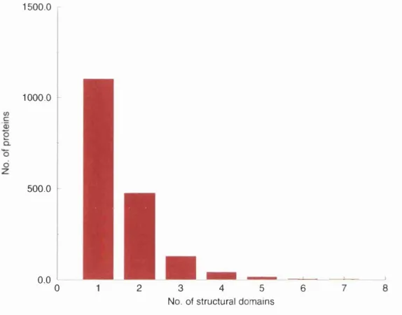
4.1 Domain number distribution... 81
4.2 Percentage of solvent exposed residues assigned by D S S P ... 82
4.3 Residue propensities for core, exposed and domain interface...85
4.4 Random walk m o d e l ... 88
4.5 Frequency of domains as a function of hydrophobicity and length... 90
4.6 Measure of randomness I - Wald-Wolfowitz runs test ... 92
4.7 Measure of randomness II - Random w a l k ... 93
4.8 Hydrophobic run d is tr ib u tio n ... 94
4.9 Further applications of the random walk m o d e l ... 97
5.1 Linker length d istr ib u tio n ... 104
5.2 Amino acid propensities of the linkers; grouped by s i z e ...107
5.3 Amino acid propensities of the linkers; grouped by number of linkers . . . 107
5.4 Residue C a -e x te n t... 112
5.5 Amino acid propensities of the linkers; grouped by structure I ... 114
5.6 Amino acid propensities of the linkers; grouped by structure I I ... 115
5.7 Ward clustering of linker c o m p o sitio n ...123
5.8 Principle component analysis - Eigen v a lu e s ...123
5.9 Xaa-Pro linker propensities versus trans to cis rate c o n s ta n ts ... 127
5.10 Linker database file for linker I b e f A . l ...131
6.1 Flow diagram of DOMAINATION... 136
6.2 Method of domain c u t t in g ...138
6.3 Domain prediction accuracy as a function of E-value cu t-o ff...147
6.4 Significant sequences found by m e t h o d s ... 153
6.5 Histogram of percentage low complexity within local gapped-alignments . 155 7.1 Multi-level smoothing w in d o w ... 161
7.2 Example SCOOBYJDO probability m atrix...162
7.4 Distribution of domain length and average hydrophobicity...166
7.5 Prediction sensitivity... 168
7.6 SCOOBY_DO plot for titin ... 170
8.1 Overview of the SnapDRAGON m eth o d ... 177
8.2 SnapDRAGON domain boundary distribution for lhfhl_refl ...184
8.3 SnapDRAGON domain boundary distribution for 2 p ia _ r e fl... 186
8.4 SnapDRAGON domain boundary distribution for lu k y _ refl... 187
8.5 Error in domain and boundary number prediction... 192
8.6 Domain and boundary number assigned versus real distribution... 194
8.7 The average normalised distance between predicted boundary and closest real linker p e r -p r o te in ...197
8.8 Error in domain and boundary number prediction... 202
8.9 Domain and boundary number: predicted versus r e a l ... 203
8.10 Sensitivity versus reference chain le n g th ...207
9.1 F vl a lig n m e n t... 217
9.2 SCOOBY_DO plots for Fvl, ERV-L, MLV gag and HIV-1 gag ... 218
9.3 Backbone structure of gag CA protein from anemia v i r u s ... 219
List o f Tables
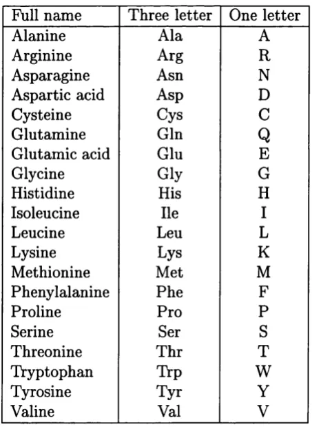
1 Standard one and three letter amino acid c o d e s ... 15
1.1 Domain databcises... 36
5.1 Amino acid pair propensities over all l i n k e r s ...109
5.2 Amino acid pair propensities for medium sized lin k e rs ... 110
5.3 Amino acid pair propensities for large sized linkers... I l l 5.4 Amino acid pair propensities for non-helical l i n k e r s ...117
5.5 Amino acid pair propensities for helical lin k e rs ... 118
5.6 Dyad sequence codes for l i n k e r s ... 119
5.7 tests between linker sets ... 121
6.1 The number of homologous sequences found and missed I ...150
6.2 The number of homologous sequences found and missed I I ... 152
7.1 Average score prediction per protein ... 167
8.1 Domain boundary prediction on a set of 25 3D_ALI alignments . . . . 183
8.2 Accuracy of linker prediction per p r o t e i n ...195
8.3 Accuracy of linker p re d ic tio n ... 206
A bbreviations
3D three dimensional
A adenine
ADM average distance map
AIR aminoimidazole ribonucleotide synthetase
BLAST basic local alignment search tool
bp base pair
C cytosine
Ca alpha-carbon
CA capsid
GASP critical assessment of techniques for protein structure
prediction
CD circular dichroism
COGs cluster of orthologous groups
COOH carboxyl group
DNA deoxyribonucleic acid
DRAGON distance régularisation algorithm for geometry optim isation
DSSP dictionary of secondary structure of proteins
PASTA fast alignment
F v l friend virus susceptibility 1
G guanine
GAR glycinamide ribonucleotide synthetase/ transferase
HIV hum an immunodeficiency virus
HMM hidden markov model
HPLC high performance liquid chromatography
IN integrase
INDIGO integrated domain prediction algorithm
MA m atrix
MHR m ajor homology region
MLV murine leukemia virus
mRNA messenger RNA
NO nucleocapsid
NCBI National Center for Biotechnology Information
NH2 amino group
NMR nuclear magnetic resonance
NRDB non-redundant protein sequence database
PASS prediction of autonomous folding units based on sequence
PDB Brookhaven protein data bank
PH pleckstrin homology domain
PIC pre-integration complex
PSSM position specific scoring m atrix
RDM real distance map
RMSD root mean square deviation
RNA ribonucleic acid
RT reverse transcriptase
SA solvent accessibility
SCOOBY_DO sequence hydrophobicity predicts domains
SCOP structural classification of proteins
SDS-PAGE sodium dodecyl sulphate polyacrylamide gel electrophoresis
SIV simian immunodeficiency virus
SMART simple m odular architecture research tool
SSAP structure and sequence alignment program
SU surface protein
T thymine
TM transm em brane
TU N EID threading using neural network with one-dimensional profile
tRNA transfer RNA
U uracil
Table 1: Standard one and three letter amino acid codes
Full name Three letter One letter
Alanine Ala A
Arginine Arg R
Asparagine Asn N
Aspartic acid Asp D
Cysteine Cys C
Glutamine Gin
Q
Glutamic acid Glu E
Glycine Gly G
Histidine His H
Isoleucine He I
Leucine Leu L
Lysine Lys K
Methionine Met M
Phenylalanine Phe F
Proline Pro P
Serine Ser S
Threonine Thr T
Tryptophan Trp W
Tyrosine Tyr Y
Original publications
1 George RA. and Heringa J. (2000) The REPRO server: finding protein internal sequence repeats through the web. Trends Bioc Sci. 25, 515-517
2 George RA., Kleinjung J. and Heringa J. (2001) Predicting protein structural domains from sequence data. Bioinformatics and Genomes: Current Perspectives. Horizon Scientific Press, (in press)
3 George RA. and Heringa J. (2001) SnapDRAGON - a method to delineate protein structural domains from sequence data. Subm itted to J Mol Biol.
4 George RA. and Heringa J. (2001) Protein domain identification and improved sequence similarity searching using PSI-BLAST. Subm itted to Proteins.
5 George RA, and Heringa J. (2001) An analysis of protein domain linkers: their classification and role in multiple domain folding.
P art I
C hapter 1
D om ains, m odules and th e m eaning o f life
Am ongst the carbon compounds there is an abundance of evidence to prove the existence of internal tendencies or molecular properties which may and do lead to the evolution of more and more complex chemical compounds. A nd it is such synthetic processes, occurring amongst the molecules of colloidal and allied substances, which seem so often to engender or give ‘origin’ to a kind of matter possessing that subtle combination of properties to which we are accustomed to apply the epithet
‘living’. Bastian (1872)
All cellular life requires two types of macromolecule, nucleic acids and proteins.
Nucleic acids contain the ‘blueprint’ to manufacture proteins, the essential active
agents in biochemistry and w ithout which almost none of the m etabolic processes
th a t we associate with life would take place. Many proteins play a structural role
providing the filamentous architecture within cells and the m aterials th a t are used
in, for example, hair, nails, tendons and bones.
Domains, modules and the meaning o f life
The nucleic acids, DNA and RNA, are each assembled from four different nucleotides
and proteins are assembled from twenty different L-amino acids. In each case, the
sequence in which the individual sub-units are assembled is the critical feature th at
determines the final function.
The genetic code is an alphabet of four letters, adenine (A), cytosine (C), guanine
(G) and thymine (T), linked together by 5’-3’ phosphodiester bonds. Each molecule
of DNA can form a double helix from two complementary antiparallel strands of
nucleotides held together by hydrogen bonds between G-C and A-T base pairs (bp)
(Watson and Crick, 1953).
The synthesis of proteins involves copying a specific region of DNA, the gene, into
RNA. Like DNA, RNA is composed of a linear sequence of nucleotides, but the sugar
phosphate backbone of RNA contains ribose instead of a deoxyribose sugar, and the
base thymine is replaced by uracil (U). The synthesis of a single stranded RNA
molecule from DNA is called transcription and results in a direct copy of the gene.
RNA transcripts th a t direct the synthesis of protein molecules are called messenger
RNA (mRNA). W ithin any cell at any particular time the level of transcription of
a particular gene into mRNA can be controlled, the specific binding of regulatory
proteins onto DNA plays a major role in this regulation. The mRNA acts as a
carrier of the genetic information to the ribosome, the translational machinery.
In eukaryotes the mRNA undergoes some modifications before leaving the
nucleus. This includes removal of non-coding regions in the mRNA called introns.
Domains, modules and the meaning o f life
called ‘splicing’.
Each of the four bases are used in combination to form a genetic dictionary of
triplet ‘words’. Each nucleotide triplet, called a codon, specifies one of the twenty
amino acids. Since there are four nucleotides, there are 64 possible nucleotide
triplets. This leads to a degeneracy of the genetic code, where each amino acid
has several possible codons. The translation of mRNA into protein requires small
transfer RNA (tRNA) molecules th a t carry the amino acids to the mRNA. The
tRN A molecule has an anti-codon sequence, which base pairs to its complementary
codon in the mRNA.
The process of translation occurs at the ribosome, a huge complex of more than
50 proteins and RNA molecules. The polymerase activity of the ribosome appears
to be based solely on RNA and for this reason it has been called a ribozyme (Nissen
et a i, 2000). High resolution structures for the ribosome have only recently been
determined (Ban et a i, 2000; Wimberly et a i, 2000; Yusupov et a i, 2001).
The ribosome essentially holds the mRNA in place so th a t the codons may be
matched up with the appropriate anti-codon on the tRNA, ensuring th a t the correct
amino acid is inserted into the growing polypeptide chain. The ribosome travels
along the mRNA molecule in a 5’-3’ direction, translating the nucleotide sequence
into an amino acid sequence, one codon at a time. When the ribosome reaches the
end of the mRNA, both it and the newly synthesised carboxyl (COOH) end are
released into the cytoplasm. The flow of genetic information through the conversion
Domains, modules and the meaning o f life
use RNA to carry their genetic information and are the exception to the rule.
There are about 35,000 protein-coding genes in humans, which comprises less
th an 2% of the entire genome. However, alternative splicing of the mRNA will
generate a much larger set of proteins (International Human Genome Sequencing
Consortium, 2001). It is estim ated th a t more than 30% of human genes will undergo
alternative splicing (Hanke et a i, 1999).
1.1
Protein structure is hierarchical
The proteins we observe in nature have arisen through a selective pressure to
perform specific functions, the functional properties of which are determined by
the overall 3D-protein structure. Unlike the ordered structure of DNA, proteins may
appear highly complex and irregular. Such complexity enables recognition of various
molecules by detailed 3D interactions. The appearance of a protein in solution is
often globular, with the polypeptide chain passing in and out of the central core of
the molecule.
1.1.1 Primary structure
Although the overall structure of a protein molecule may appear irregular there is
an underlying hierarchical order to its complexity. The first level is the sequence
of amino acids, called its ‘prim ary structure’. Amino acids are joined together to
form a polypeptide chain created by the condensation of the COOH group of one
Domains, modules and the meaning o f life
acids lies in the side chain attached to the alpha-carbon (Ca) atom. According
to the chemical nature of the side chains amino acids are usually divided into
three categories (Brândén and Tooze, 1991). The first are those th a t have strictly
hydrophobic side chains, shown using their three letter codes; Ala, Val, Leu, He,
Phe, Pro and Met. The four charged residues. Asp, Glu, Lys and Arg form a second
class. The final class consists of those with polar side chains; Ser, Thr, Cys, Asn,
Gin, His, Tyr and Trp. The amino acid Gly only has a hydrogen atom as a side
chain and can be put in the hydrophobic category or assigned to a fourth category.
The variety of amino acids leads to enormous versatility in the chemical properties
of proteins.
1.1.2 Secondary structure
The prim ary structure of a protein encodes its uniquely folded 3D conformation
(Anfinsen et a l, 1961). The most im portant factor governing the folding of a protein
is the distribution of polar and non-polar side chains (Cordes et al., 1996). Folding
is driven by the burial of hydrophobic side chains into the interior of the molecule
so to avoid contact with the aqueous environment. Generally proteins have a core
of hydrophobic residues surrounded by a shell of hydrophilic residues. Since the
peptide bonds themselves are polar they are neutralised by hydrogen bonding with
each other when in the hydrophobic environment. This gives rise to regions of the
polypeptide th a t form regular 3D structural patterns called ‘secondary structure’.
Domains, modules and the meaning o f life
in the polypeptide backbone: namely the C a, carboxyl carbon (O’) and the amide
nitrogen atoms (N). Their positions, and hence secondary structure, can be defined
by the angles of rotation about the bonds connecting the three atoms: ÿ, 'ip, and lj.
The torsion or dihedral angle w defines rotation around the peptide bond C ’j-Nj+i,
the angle (j) defines the rotation around the N —C a bond and angle 'ip defines the
rotation around the C a-C ’i bond, where i is the zth amino acid. There are two main
types of secondary structure:
• a-helical: Main chain C ’= 0 (residue i) and N-H (residue z+4) atom s are
hydrogen bonded to each other causing the polypeptide backbone to turn
about itself forming a rigid cylinder. There are about 3.6 residues per turn,
which corresponds to a distance of 5.4Â. (p and ^p angles are approximately
-60° and -50° respectively. The rotation of the chain is right handed.
• yd-sheet: This structure is built up from several fully extended, continuous
regions of the polypeptide chain called ^-strands. /5-strands are usually from
four to ten residues in length and aligned adjacent to each other such th a t
hydrogen bonds can form between the C’= 0 atom of one yd-strand and the
N-H of another. /5-sheets can have their strands parallel, antiparallel or mixed.
Some simple combinations of secondary structure elements have been found
to frequently occur in protein structure and are referred to as ‘super-secondary
stru ctu re’ or motifs. For example, the yd-hairpin m otif consists of two adjacent
antiparallel /5-strands joined by a small loop. It is present in most antiparallel /5
Domains, modules and the meaning o f life
common super-secondary structure is the jd-a-jd motif, which is frequently used to
connect two parallel ^-strands. The central a-helix connects the C-termini of the
first strand to the N-termini of the second strand, packing its side chains against
the /9-sheet and therefore shielding the hydrophobic residues of the /9-strands from
the surface.
1.1.3 Tertiary structure
Several motifs pack together to form compact, local, semi-independent units called
domains (Richardson, 1981). The overall 3D structure of the polypeptide chain
is referred to as the protein’s ‘tertiary structure’. Domains are the fundamental
units of tertiary structure, each domain containing an individual hydrophobic core
built from secondary structural units connected by loop regions. The packing of the
polypeptide is usually much tighter in the interior th an the exterior of the domain
producing a solid-like core and a fluid-like surface (Zhou et a i, 1999). In fact, core
residues are often conserved in a protein family, whereas the residues in loops are
less conserved, unless they are involved in the protein’s function. Protein tertiary
structure can be divided into four main classes based on the secondary structural
content of the domain (Levitt and Chothia, 1976).
• All-o; domains have a domain core built exclusively from a-helices. This class
is dom inated by small folds, many of which form a simple bundle with helices
Domains, modules and the meaning o f life
sheets packed against each other. Various patterns can be identified in the
arrangem ent of the strands, often giving rise to the identification of recurring
motifs, for example the greek key motif (Hutchinson and Thornton, 1993).
• a+/3 domains are a mixture of all-a and all-^ motifs. Classification of proteins
into this class is difficult because of overlaps to the other three classes and
therefore is not used in the GATH domain database (Orengo et a i, 1997).
• a//3 domains are made from a combination of P-a-P motifs th a t predominantly
form a parallel ^^-sheet surrounded by am phipathic a-helices. The secondary
structures are arranged in layers or barrels.
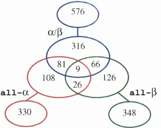
Figure 1.1 shows the distribution of domain class assigned to proteins held
within the OATH structural domain database (Orengo et a i, 1997). Only 1% of
m ultidom ain proteins contain a domain from each of the three classes all-a, all-/9
and ol/P . The m ajority of proteins, 48%, contain an a /P domain. About half of
the m ultidom ain proteins th a t contain an all-o; or all-yd domain also have a domain
from another class, but only a third of proteins with an a jjd domain contain other
classes of domains. Combinations of all-o; and all-/? domains are the least observed
in m ultidom ain proteins, whereas combinations of all-o; and a / p domains are the
most common.
1.1.4 Quarternary structure
Many proteins have a quarternary structure, which consists of several polypeptide
Domains, m odules and the m eaning o f life
576
a/(3
316
126
108
all-(3
330
348
Figure 1.1: Distribution of domain class in multidomain proteins. Domains are organised into their class as described in the CATH domain database (Orengo et ai, 1997). The central Venn diagram contains multidomain proteins, while the individual sets surrounding the Venn diagram contain single domain proteins. No pair of sequences have more than 35% sequence identity.
protein is called a subunit. Hemoglobin, for example, consists of two a and two /? subunits. Each of the four chains has an all-o; globin fold with a heme pocket.
Domain swapping is a mechanism for forming oligomeric assemblies (Bennett
et ai, 1995). In domain swapping, a secondary or tertiary element of a monomeric protein is replaced by the same element of another protein. Domain swapping can range from secondary structure elements to whole structural domains. It also represents a model of evolution for functional adaptation by oligomerisation, e.g. oligomeric enzymes th at have their active site at subunit interfaces (Heringa and Taylor, 1997).
Domains, modules and the meaning o f life
1.2 The significance of domains in proteins
The concept of the domain was first proposed in 1973 by Wetlaufer after X-ray
crystallographic studies of hen lysozyme (Phillips, 1966), papain (Drenth et a l, 1968)
and by limited proteolysis studies of immunoglobulins (Porter, 1973; Edelman, 1973)
(see Section 2.3.1 for more information on limited proteolysis). Wetlaufer defined
domains as stable units of protein structure th a t could fold autonomously. Domains
have been defined as units of:
• compact structure (Richardson, 1981);
• function and evolution (Bork, 1991);
• folding (Wetlaufer, 1973).
Each definition is valid and will often overlap, i.e. a compact structural domain
th a t is found amongst diverse proteins is likely to fold independently within its
structural environment. In a multidomain protein, each domain may fulfil its own
function independently, or in a concerted manner with its neighbours. Domains can
either serve as modules for building up large assemblies such as virus particles or
muscle fibres, or can provide specific catalytic or binding sites as found in enzymes or
regulatory proteins. Therefore, domains can be units of function as well as structure.
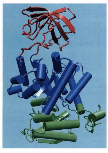
An appropriate example is pyruvate kinase, a glycolytic enzyme th a t plays an
im portant role in regulating the flux from fructose-1,6-biphosphate to pyruvate.
Domains, m odules and the meaning o f life
Oi/jd nucleotide binding domain (Figure 1.2). Each domain recurs in diverse sets of proteins.
i
»
A
Figure 1.2: Pyruvate kinase; PDB code Ipkn (Larsen et al, 1994)
Domains, modules and the meaning o f life
The central a//3-barrel substrate binding domain is one of the most common
enzyme folds. It is seen in many different enzyme families catalysing completely
unrelated reactions (Hegyi and Gerstein, 1999). The a /^ -b a rre l is commonly called
the TIM -barrel named after triose phosphate isomerase, which was the first such
structure to be solved (Banner et a l, 1975). It is currently classified into 26
homologous families in the CATH domain database (Orengo et a l, 1997). The
TIM -barrel is formed from a sequence of p-a-P motifs closed by the first and
last strand hydrogen bonding together, forming an eight stranded barrel. There
is debate about the evolutionary origin of this domain. One study has suggested
th a t a single ancestral enzyme could have diverged into several families (Copley and
Bork, 2000), while another suggests th a t a stable TIM -barrel structure has evolved
through convergent evolution (Lesk et a i, 1989).
The TIM -barrel in pyruvate kinase is ‘discontinuous’, meaning th a t more than
one segment of the polypeptide is required to form the domain. This is likely to be
the result of the insertion of one domain into another during a protein’s evolution.
The inserted /5-barrel regulatory domain is ‘continuous’, made up of a single stretch
of polypeptide. It has been shown from known structures th a t about a quarter of
structural domains are discontinuous (Jones et a l, 1998; Holm and Sander, 1994).
Covalent association of two domains represents a functional and structural
advantage since there is an increase in stability when compared with the same
structures non-covalently associated (Ghelis and Yon, 1979). Other, advantages
Domains, modules and the meaning o f life
otherwise be unstable in aqueous environments, and a fixed stoichiometric ratio of
the enzymatic activity necessary for a sequential set of reactions (Ostermeier and
Benkovic, 2000).
The presence of multiple domains in proteins gives rise to a great deal of fiexibility
and mobility. One of the largest observed domain motions is the ‘swivelling’
mechanism in pyruvate phosphate dikinase. The phosphoinositide domain swivels
between two states in order to bring a phosphate group from the active site of the
nucleotide binding domain to th a t of the phosphoenolpyruvate/pyruvate domain
(Herzberg et a i, 1996). The phosphate group is moved over a distance of 45Â
involving a domain motion of about 100° around a single residue. Domain motions
are im portant for (Gerstein et a i, 1994):
• catalysis;
• regulatory activity;
• transport of metabolites;
• formation of protein assemblies and
• cellular locomotion.
In enzymes, the closure of one domain onto another captures a substrate by an
induced fit, allowing the reaction to take place in a controlled way. Such motions
can be observed when two or more crystallographic 3D structures of a protein are
Domains, modules and the meaning o f life
(1994) led to the classification of two basic types of domain motion; hinge and shear.
Only a relatively small portion of the chain, namely the inter-domain linker and
side chains undergo significant conformational changes upon domain rearrangement
(Janin and Wodak, 1983).
A study by Hayward (Hayward, 1999) found th a t the term ini of a-helices and
/^-sheets form hinges in a large number of cases. Many hinges were found to involve
two secondary structure elements acting like hinges of a door, allowing an opening
and closing motion to occur. This can arise when two neighbouring strands within
a ^-sheet situated in one domain, diverge apart as they join the other domain. The
two resulting term ini then form the bending regions between the two domains,
a-helices th a t preserve their hydrogen bonding network when bent are found to behave
as mechanical hinges, storing ‘elastic energy’ th a t drives the closure of domains for
rapid capture of a substrate (Hayward, 1999).
The interconversion of helical and extended conformations at the site of a domain
boundary is not uncommon. In calmodulin, torsion angles change for five residues
in the middle of a domain linking a-helix. The helix is split into two, almost
perpendicular, smaller helices separated by four residues of an extended strand
(Meador et a l, 1992; Ikura et a l, 1992).
Shear motions involve a small sliding movement of domain interfaces, controlled
by the amino acid side chains within the interface. Proteins displaying shear motions
often have a layered architecture: stacking of secondary structures. The inter
Domains, modules and the meaning o f life
1.3
Domains are units of structure
Evolution gives rise to families of related proteins with similar sequence and
structure. However, sequence similarities can be extremely low between proteins
th a t otherwise share the same structure. Protein structures may be similar because
proteins have diverged from a common ancestor. Alternatively, it is possible th at
some folds may be more favoured than others as they represent stable arrangements
of secondary structures and over the course of evolution some proteins may converge
towards these types of fold. There are currently about 15,000 experimentally
determined protein 3D structures deposited within the Protein D ata Bank (PDB)
(Berman et a i, 2000). However this set contains many near identical structures.
It is im portant to be able to classify proteins by structural family, creating a non
degenerate database in which each family is represented once, to help understand
relationships between protein sequence and structure. Comparison of related
protein structures allows conserved structural motifs to be identified and brings
an understanding of a protein’s evolution.
1.3.1 Domain definition from structural co-ordinates
The im portance of domains as structural building blocks and elements of evolution
has brought about many autom ated methods for their identification and classifi
cation in proteins of known structure. Autom atic procedures for reliable domain
Domains, modules and the meaning o f life
number of protein structures is increasing. Although the boundaries of a domain
can be determined by visual inspection, construction of an autom ated m ethod is not
straightforward. Problems occur when faced w ith domains th a t are discontinuous
or highly associated (Sowdhamini and Blundell, 1995). The fact th a t there is no
standard definition of what a domain really is has m eant th a t domain assignments
have varied enormously, with each researcher using a unique set of criteria (Swindells,
1995).
A structural domain is a compact, globular sub-structure with more inter
actions within it th an with the rest of the protein (Janin and Wodak, 1983).
Therefore, a structural domain can be determined by two visual characteristics;
its compactness and its extent of isolation (Tsai and Nussinov, 1997). Measures
of local compactness in proteins have been used in many of the early m eth
ods of domain assignment (Rossmann et a i, 1974; Crippen, 1978; Rose, 1979;
Go, 1978) and in several of the more recent methods (Holm and Sander, 1994;
Islam et a i, 1995; Siddiqui and Barton, 1995; Zehfus, 1997; Taylor, 1999). One of
the first algorithms (Crippen, 1978) used a C a-C a distance m ap together with a
hierarchical clustering routine th a t considered proteins as several small segments,
10 residues in length. The initial segments were clustered one after another based
on inter-segment distances; segments with the shortest distances were clustered and
considered as single segments thereafter. The stepwise clustering finally included
the full protein. Go (1978) also exploited the fact th a t inter-domain distances are
Domains, modules and the meaning o f life
represented as diagonal plots in which there were distinct patterns for helices,
extended strands and combinations of secondary structures.
The m ethod by Sowdhamini and Blundell (1995) clusters secondary structures in
a protein based on their C a -C a distances and identifies domains from the pattern in
their dendrograms. As the procedure does not consider the protein as a continuous
chain of amino acids there are no problems in treating discontinuous domains.
Specific nodes in these dendrograms are identified as tertiary structural clusters
of the protein, these include both super-secondary structures and domains.
The DOMAK algorithm is used to create the 3Dee domain database (Siddiqui
and Barton, 1995). It calculates a ‘split value’ from the number of each type of
contact when the protein is divided arbitrarily into two parts. This split value is
large when the two parts of the structure are distinct.
The m ethod of Wodak and Janin (1981) was based on the calculated interface
areas between two chain segments repeatedly cleaved a t various residue positions.
Interface areas were calculated by comparing surface areas of the cleaved segments
with th a t of the native structure. Potential domain boundaries can be identified at a
site where the interface area was at a minimum. Other methods have used measures
of solvent accessibility to calculate compactness (Rashin, 1985; Islam et a i, 1995;
Zehfus and Rose, 1986).
The PUU algorithm (Holm and Sander, 1994) incorporates a harmonic model
used to approximate inter-domain dynamics. The underlying physical concept is
Domains, modules and the meaning o f life
will occur between domains. This algorithm is used to define domains in the FSSP
domain database (Holm and Sander, 1997).
Swindells (1995) developed a method, DETECTIVE, for identification of
domains in protein structures based on the idea th a t domains have a hydrophobic
interior. Deficiencies were found to occur when hydrophobic cores from different
domains continue through the interface region.
1.3.2 Domain structure databases
Several m ethods of structural classification have been developed to introduce some
order to the large amount of redundant d a ta present in the PDB. The most widely
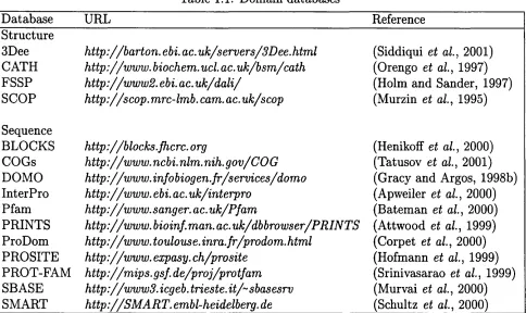
used and comprehensive databases are CATH, 3Dee, FSSP and SCOP (Table 1.1),
which use four unique methods to classify protein structures and consequently have
discrepancies between their assignments (Hadley and Jones, 1999). The domain
databases differ in their detailed organisation. For example, the top level of the
hierarchy in SCOP and CATH is protein class. However, SCOP and CATH differ
in the number of structural classes used.
CATH
A consensus approach using the three domain assignment algorithms, PUU, DE
TECTIV E and DOMAK as well as visual inspection, has been used to create the
CATH structural domain database (Jones et a i, 1998). The CATH database (release
Table 1.1: Domain databases
CO
O i
Database URL Reference
Structure
3Dee http://barton, ebi. ac. uk/servers/3Dee. html (Siddiqui et a l, 2001) CATH http://w w w . biochem.ucl. ac.uk/bsm /cath (Orengo et a l, 1997) FSSP h ttp :// www2. ebi. ac. u k/d a li/ (Holm and Sander, 1997)
SCO ? http://scop.mrc-lmb.cam.ac.uk/8cop (Murzin et a l, 1995)
Sequence
BLOCKS h ttp ://blocks, fhcrc. org (Henikoff et a l, 2000)
COGs http://w w w . ncbi. nlm. nih. gov/COG (Tatusov et a l, 2001) DOMO http://www.infobiogen.fr/services/domo (Gracy and Argos, 1998b)
InterPro http://w w w . ebi. ac.uk/interpro (Apweiler et a l, 2000)
Pfam http://www.sanger. ac.uk/Pfam (Bateman et a l, 2000)
PRINTS http://w w w . bioinf.man. ac.uk/dbbrowser/PRINTS (Attwood et a l, 1999)
ProDom http://w w w . toulouse. inra.fr/prodom. html (Corpet et a l, 2000)
PROSITE http://w w w . expasy. ch/prosite (Hofmann et a l, 1999)
PROT-FAM http : / / mips. gsf. de/proj/p ro tfam (Srinivasarao et a l, 1999)
SBASE http://w w w 3. icgeb. trieste. it/~sbasesrv (Murvai et a l, 2000)
SMART h ttp ://S M A R T , embl-heidelberg. de (Schultz et a l, 2000)
I
.1
s 01
S-<D
S
i
I'
Domains, modules and the meaning o f life
• Class;
• Architecture;
• Topology/fold;
• Homologous superfamily;
• Sequence family.
A rchitecture is the overall shape of a domain as defined by the packing of
secondary structural elements, but ignoring their connectivity. The topology-
level consists of structures with the same number, arrangem ent and connectivity
of secondary structure based on structural superposition using SSAP structure
comparison algorithm (Taylor and Orengo, 1989b). A homologous superfamily
contains proteins having high structural similarity and similar functions, which
suggests th a t they have evolved from a common ancestor. Finally, the sequence
family level consists of proteins with sequence identities greater th an 35%, again
suggesting a common ancestor.
CATH classifies domains into approximately 700 fold families, ten of these folds
are highly populated and are referred to as ‘super-folds’. Super-folds are defined
as folds for which there are a t least three structures w ithout significant sequence
Domains, modules and the meaning o f life
3Dee
3Dee structural domain repository (Siddiqui et a l, 2001) stores alternative domain
definitions for the same protein and organises the domains into sequence and
structural hierarchies. Most of the database creation and update processes are
performed automatically. However, some domains are manually assigned. It contains
non-redundant sets of sequences and structures, multiple structure alignments for all
domain families, secondary structure and fold name definitions. The current 3Dee
release is now two years old and contains 18,896 structural domains.
FSSP
FSSP (Holm and Sander, 1997) is a complete comparison of all pairs of protein
structures in the PDB. It is the basis for the Dali Domain Dictionary (Dietmann
et a i, 2001), a numerical taxonomy of all known structures in the PDB. The taxon omy is derived fully autom atically from measurements of structural, functional and
sequence similarities. The database is split into four hierarchical levels corresponding
to super-secondary structural motifs, the topology of globular domains, remote
homologues (functional families) and sequence families.
The top level of the fold classification corresponds to secondary structure
composition and super-secondary structural motifs. Domains are assigned to one
of five ‘attra cto rs’, which can be characterised as all-o;, all-^d, a / p , a-P meander and
antiparallel /^-barrels. Domains which are not clearly defined to a single attracto r
Domains, modules and the meaning o f life
34,038 domains from 25,808 structures classified into 2,777 sequence families. The
database contains definition of structurally conserved cores and a library of multiple
alignments of distantly related protein families.
SCOP
The SCOP database (Structural Classification of Proteins) is a manual classification
of protein structure (Murzin et a i, 1995). The classification is at the domain level
for many proteins, but in general, a protein is only split into domains when there
is a clear indication th a t the individual domains may have existed as independent
proteins. Therefore, many of the domain definitions in SCOP will be different to
those in the other structural domain databases. The principal levels of hierarchy
are family, superfamily and fold, split into the traditional four domain classes,
all-a, all-/?, a-\~P and a//?. Release 1.55 of the SCOP database contains 13,220
PDB entries, 605 fold types and 31,474 domains.
1.4 Domains are units of evolution
‘Nature is a tinkerer and not an inventor’ (Jacob, 1977), new sequences are adapted from pre-existing sequences rather than invented. Domains are the common m aterial
used by nature to generate new sequences, they can be thought of as genetically
mobile units. Many domain families are found in all three forms of life, Archaea,
Bacteria and Eukarya. Domains th a t are repeatedly found in diverse proteins are
Domains, modules and the meaning o f life
associated with clotting, fibrinolysis, complement, the extracellular m atrix, cell-
surface adhesion molecules and cytokine receptors (Campbell and Downing, 1994).
The m ajority of genomic proteins, two-thirds in unicellular organisms and
more than 80% in metazoa, are multidomain proteins created as a result of gene
duplication events (Apic et a l, 2001). Many domains in multidomain structures
could have once existed as independent proteins. More and more domains in
eukaryotic multidomain proteins can be found as independent proteins in prokaryotes
(Davidson et a l, 1993). For example, vertebrates have a multi-enzyme polypeptide
containing the GAR synthetase, AIR synthetase and GAR transformylase modules
(GARs-AIRs-GARt) ^. In insects, the polypeptide appears as GARs-(AIRs)2-GARt,
in yeast GARs-AIRs is encoded separately from G ARt, and in bacteria each domain
is encoded separately (Henikoff et a i, 1997).
M ultidomain proteins are likely to have emerged from a selective pressure during
evolution to create new functions. Various proteins have diverged from common
ancestors by different combinations and associations of domains. M odular units
frequently move about, within and between biological systems through mechanisms
of genetic shuffling:
• transposition of mobile elements including horizontal transfers (between
species) (Bork and Doolittle, 1992);
• gross rearrangements such as inversions, translocations, deletions and duplica
tions;
Domains, modules and the meaning o f life
• homologous recombination;
• slippage of DNA polymerase during replication.
It is likely th a t all these mechanisms have contributed to the proliferation,
dispersal and loss of protein domains. Some domains are more promiscuous than
others, appearing in a diverse set of protein families and organisms. For example,
the ABC transporter domain constitutes one of the largest domain families th at
appear in all organisms (Henikoff et a i, 1997). Many other families th a t appear in
all organisms show much less proliferation. These include m etabolic enzymes and
components of translational apparatus.
The simplest multidomain organisation seen in proteins is th a t of a single domain
repeated in tandem . The domains may interact with each other or remain isolated,
like beads on string. The giant 30,000 residue muscle protein titin comprises about
120 fibronectin-III-type and Ig-type domains (Politou et a i, 1996). In the serine
proteases, a gene duplication event has led to the formation of a two y0-barrel domain
enzyme (McLachlan, 1979). The repeats have diverged so widely th a t there is no
obvious sequence similarity between them. The active site is located a t a cleft
between the two ^-barrel domains, in which functionally im portant residues are
contributed from each domain. Genetically engineered m utants of chymotrypsin
serine protease were shown to have some proteinase activity even though their active
site residues were abolished and it has therefore been postulated th a t the duplication
event enhanced the enzyme’s activity (McLachlan, 1979).
Domains, modules and the meaning o f life
the kinesins and ABC transporters. The kinesin m otor domain can be at either end
of a polypeptide chain th a t includes a coiled-coil region and a cargo domain (Moore
and Endow, 1996). ABC transporters are built w ith up to four domains consisting
of two unrelated modules, ATP-binding cassette and an integral membrane module,
arranged in various combinations.
Not only do domains recombine, but there are many examples of a domain
having been inserted into another. Sequence or structural similarities to other
domains dem onstrate th a t homologues of inserted and parent domains can exist
independently. An example is th a t of the ‘fingers’ inserted into the ‘palm ’ domain
within the polymerases of the Pol I family (Russell, 1994).
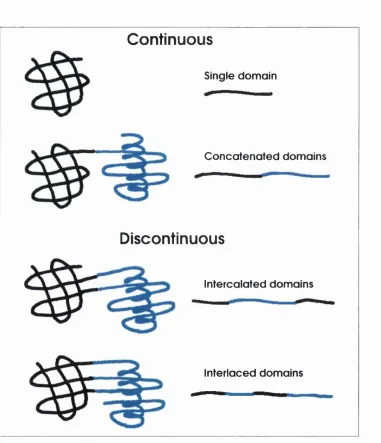
Figure 1.3 displays the connectivity between domains, ordered by their degree of
complexity (Das and Smith, 2000). Although the fourth case represents a two-
domain protein, there is a small chance th at the two domains could naturally
exist as single entities, i.e. the mechanism of its formation would require multiple
simultaneous m utations or an internal translocation event. According to this
rule, there should always be at least one continuous domain in a m ultidomain
protein. This is the main difference between definitions of structural domains and
evolutionary/ functional domains. An evolutionary domain will be limited to one or
two connections between domains, whereas structural domains can have unlimited
connections, within a given criterion of the existence of a common core. Several
Domains, m odules and the meaning o f life
Continuous
Single domain
C oncatenated domains
Discontinuous
intercalated domains
Interlaced domains
Figure 1.3: The connectivity between multidomain proteins (adapted from Das and Smith (2000)). Cartoon representations of protein 3D structure appear on the left and protein sequence on the right, a) This is a single domain with no connections, b) A two domain protein with a single connection, c) A two domain protein with two connections, resulting from an insertion of one domain into another, d) Finally a two domain protein with three inter-domain connections.
Domains, modules and the meaning o f life
1.4.1 Domain sequence databases
At present, there are nearly 100 published genome sequences. This mass of sequence
d ata requires autom ated annotation, with accurate assignment of biological function.
Since many proteins have multiple domains and, therefore, will sometimes have
multiple functions, there is a need to characterise new sequences at the domain level.
Many databases of protein sequence motifs and domains have been developed for
this purpose. Among these are BLOCKS, COGs, DOMO, Pfam, PRINTS, ProDom,
PROSITE, PROT-FAM, SBASE, SMART and InterPro (see Table 1.1).
B LO C K S
Blocks are short, multiply aligned, ungapped segments corresponding to the most
highly conserved regions of proteins. The rationale behind searching a database
of blocks is th a t information from multiply aligned sequences is present in a
concentrated form, reducing noise and increasing sensitivity to distant relationships.
The BLOCKS database (Henikoff et a i, 2000) is autom atically generated by looking
for the most highly conserved regions in groups of proteins documented in various
domain databases. Version 13.0 of the BLOCKS database consists of 8,656 sequence
blocks generated specifically from proteins in the PROSITE database.
COGs
The COGs (Clusters of Orthologous Groups) database is a phylogenetic classification
Domains, modules and the meaning o f life
consists of bacterial and archaeal genomes. Incorporation of the larger genomes of
multicellular eukaryotes into the COG system is achieved by identifying eukaryotic
proteins th a t fit into already existing COGs. Eukaryotic proteins th a t have orthologs
within different COGs are split into their individual domains. The COGs database
currently consists of 3,166 COGs including 75,725 proteins from 44 genomes.
DOMO
The DOMO database (Gracy and Argos, 1998b) contains domain multiple align
ments autom atically generated from successive sequence analysis steps including
similarity search, domain delineation, multiple sequence alignment and m otif con
struction. It has full coverage of the SWISSPROT and FIR sequence databases
(Bairoch and Apweiler, 2000). The database currently holds 99,058 domains
clustered into 8,877 multiple sequence alignments.
Pfam
Pfam is a collection of protein domain family alignments and prohle-Hidden Markov
Models (HMM) (Bateman et a i, 2000). Pfam is composed of two parts, PfamA
and PfamB. PfamA is the curated section of Pfam and contains manually crafted
multiple alignments and profile-HMMs for 3,071 domain families (version 6.6).
PfamB families are basically those th a t are in the ProDom domain database th a t
Domains, modules and the meaning o f life
P R IN T S
PRINTS is a database of protein fingerprints (Attwood et a i, 1999). A fingerprint is
a group of conserved motifs used to characterise a protein family. Fingerprints can
encode protein folds and functionalities more fiexibly and powerfully than a single
motif. Release 31.0 of PRINTS contains 1,550 entries, encoding 9,531 individual
motifs.
ProDom
ProDom (Corpet et al., 2000) is a database of protein domain families autom ati
cally generated from SW ISSPROT and TrEMBL sequence databases (Bairoch and
Apweiler, 2000) using a novel procedure based on recursive PSI-BLAST searches
(Altschul et a i, 1997). Release 2001.2 of ProDom contains 283,772 domain families,
101,957 having at least two sequence members. ProDom-CG (complete genome)
is a version of the ProDom database, which holds only d a ta originating from the
complete genome sequencing projects.
P R O SIT E
PROSITE (Hofmann et a i, 1999) is a good source of high quality annotation for
protein domain families. A PROSITE sequence family is represented as a pattern
or profile. The profiles provide a means of sensitive detection of common protein
domains in new protein sequences. PROSITE, release 16.46, contains signatures
Domains, modules and the meaning o f life
docum entation providing background information on the structure and function of
these proteins.
PR O T-FA M
PROT-FAM (Srinivasarao et a l, 1999) is a curated database of homology clusters
produced and m aintained within the context of the F IR sequence database (George
et a l, 1996). Sequences are clustered into protein superfamilies, if the homology between members covers the entire sequence, and homology domains, regions of
local similarity in proteins. PROT-FAM currently contains 8,538 superfamilies and
374 homology domains.
SB A SE
SBASE domains are protein sequence segments with known structure a n d /o r func
tion (Murvai et a l, 2000). The boundaries of the domains are either previously
defined within publications or determined by homology to domains with known
boundaries such as those given in the PROT-FAM and Pfam databases. The entries
in release 9.0 of SBASE are clustered into 2,425 statistically validated domain groups
(SBASE-A) and 739 non-validated groups (SBASE-B).
S M A R T
SMART (a Simple Modular Architecture Research Tool) contains profile-HMMs
and alignments for each domain family (Schultz et al, 2000). Alignments are based
Domains, modules and the meaning o f life
PSI-BLAST analysis (Altschul et a i, 1997). Alignments are checked manually for
potential false positives or misassembled protein sequences derived from genomic
sources. D atabase release 3.3 has 594 domain families found in signalling, extracel
lular and chromatin-associated proteins. The families are extensively annotated with
respect to phyletic distributions, functional class, tertiary structures and functionally
im portant residues. User interfaces to this database allow searches for proteins
containing specific combinations of domains in defined taxa.
InterPro
Because the underlying construction and analysis methods of the above domain
family databases are different, the databases inevitably have different diagnostic
strengths and weaknesses. The InterPro database (Apweiler et a i, 2000) is a
collaboration between many of the domain database curators. It aims to be a
central resource reducing the am ount of duplication between the databases. Release
3.2 of InterPro contains 3,939 entries, representing 1,009 domains, 2,850 families,
65 repeats and 15 post-translational modification sites. Entries are accompanied
by regular expressions, profiles, fingerprints and HMMs which facilitate sequence
Domains, modules and the meaning o f life
1.5
Domains are autonomous folding units
1.5.1 Protein folding - the unsolved problem
Since the seminal work of Anfinsen over forty years ago (Anfinsen et a l, 1961),
the goal to completely understand the mechanism by which a polypeptide rapidly
folds into its stable native conformation remains elusive. Many experimental folding
studies have contributed much to our understanding, b u t the principles th a t govern
protein folding are still based on those discovered in the very first studies of folding.
Anfinsen showed th a t the native state of a protein is thermodynamically stable, the
conformation being at a global minimum of its free energy.
Folding is a directed search of conformational space allowing the protein to fold
on a biologically feasible tim e scale. ‘Levinthal’s paradox’ states th a t if an averaged
sized protein would sample all possible conformations before finding the one with
the lowest energy, the whole process would take billions of years (Levinthal, 1968).
Proteins typically fold within 0.1 and 1000 seconds, therefore the protein folding
process must be directed some way through a specific folding pathway. The forces
th a t direct this search are likely to be a combination of local and global influences
whose effects are felt at various stages of the reaction (Dill, 1999).
Advances in experimental and theoretical studies have shown th a t folding can be
viewed in term s of energy landscapes (Leopold et a l, 1992; Dill and Chan, 1997),
where folding kinetics is considered as a progressive organisation of an ensemble