Method
Lentiviral vector transduction of spermatozoa as a tool
for the study of early development
Anil Chandrashekran
a,⇑, Ihsan Isa
a, Jayesh Dudhia
e, Adrian J. Thrasher
b,
Nicholas Dibb
a, Colin Casimir
c, Carol Readhead
d, Robert Winston
a aDepartment of Surgery and Cancer, Division of Cancer, Imperial College London, Hammersmith Campus, Institute of Reproductive and Developmental Biology (IRDB), Du Cane Road, London W12 0NN, UK
b
Molecular Immunology Unit, UCL-Institute of Child Health, 30 Guilford Street, London WC1N 1EH, UK c
Department of Natural Sciences, School of Science & Technology, Middlesex University, The Burroughs, London NW4 4BT, UK d
Biological Imaging Center, California Institute of Technology, Beckman Institute, 139-74, Pasadena, CA 91135, USA
eDepartment of Clinical Science Services, The Royal Veterinary College, Hawkshead Lane, North Mymms, Hatfield, Herts AL9 7TA, UK
a r t i c l e
i n f o
Article history: Received 2 October 2013 Revised 13 February 2014 Accepted 19 February 2014
Keywords: Lentiviral vectors Spermatozoa Transduction Development Transgenics In vitro fertilisation
a b s t r a c t
Spermatozoa and lentiviruses are two of nature’s most efficient gene delivery vehicles. Both can be genetically modified and used independently for the generation of transgenic animals or gene fer/therapy of inherited disorders. Here we show that mature spermatozoa can be directly trans-duced with various pseudotyped lentiviral vectors and used in in vitro fertilisation studies. Lentiviral vectors encoding Green Fluorescent Protein (GFP) were shown to be efficiently processed and expressed in sperm. When these transduced sperm were used in in vitro fertilisation studies, GFP expression was observed in arising blastocysts. This simple technique of directly transducing spermatozoa has potential to be a powerful tool for the study of early and pre-implantation devel-opment and could be used as a technique in transgenic develdevel-opment and vertical viral transmission studies.
Ó2014 The Authors. Published by Elsevier B.V. on behalf of the Federation of European Biochemical Societies. This is an open access article under the CC BY-NC-ND license (http://creativecommons.org/licenses/by-nc-nd/3.0/).
1. Introduction
Many genetic processes that occur in the early embryo could be much more easily studied if male germ cells rather than female could be utilised for genetic modification. The accessibility and easy availability of spermatozoa make them attractive candidates for use in such studies. However, although sperm themselves are ideally suited to gene transfer, introducing genetic material into sperm has proven less straightforward[1–3].
Lentiviruses are one of nature’s most efficient gene delivery vehicles. They have been used successfully to treat various human
genetic disorders (acquired and inherited)[4–7], as tools for re-search into gene function and regulation/knock down, and more recently as vehicles for reprogramming of differentiated somatic cells to an embryonic state. As such, the genetic payload engi-neered into these lentiviral vectors has ranged from short interfer-ing RNAs, to transcription factors and microRNAs[8–10].
To generate safe gene transfer vectors the lentiviral genome is modified by removing non-essential genes encoding most of the viral accessory and regulatory genes, and splitting the genome of the virus into separate transcriptional units (trans complementing for expression of viral structural and some regulatory components and cis acting for vector development encoding the transgene/re-porter construct and packaging signal). An additional safety mech-anism ensuring that only the transgene is expressed in transduced cells, is provided by deletion of enhancer elements present in the 30 Long Terminal Repeat (LTR) of the vector[11,12]. Reporter genes or the genetic payload can be engineered into these vectors. Commonly utilised promoters driving the reporter genes or genetic
payload include Phosphoglycerate kinase promoter (PGK),
Elongation factor 1 alpha promoters (EF-1) and Cytomegalovirus
http://dx.doi.org/10.1016/j.fob.2014.02.008
2211-5463/Ó2014 The Authors. Published by Elsevier B.V. on behalf of the Federation of European Biochemical Societies. This is an open access article under the CC BY-NC-ND license (http://creativecommons.org/licenses/by-nc-nd/3.0/).
Abbreviations:GFP, Green Fluorescent Protein; LTR, Long Terminal Repeat; PGK, Phosphoglycerate kinase promoter; EF-1, Elongation factor 1 alpha promoter; CMV, Cytomegalovirus promoter; UCOE, ubiquitous chromatin opening element pro-moter; VSV-g, vesicular stomatitis virus; 293T, Human embryonic kidney cells; 7-AAD, 7-Aminoactinomycin D; IVF, in vitro fertilisation; AZT, azidodeoxythimidine
⇑Corresponding author. Current Address: Department of Clinical Science Ser-vices, The Royal Veterinary College, Hawkshead Lane, North Mymms, Hatfield, Herts AL9 7TA, UK. Tel.: +44 07788743730.
E-mail address:[email protected](A. Chandrashekran).
promoter (CMV). Recently, the use of methylation resistant pro-moters such as ubiquitous chromatin opening element promoter (lacking in enhancer activity) (UCOE) has been successfully utilised in gene transfer protocols, whilst maintaining relative expression levels of reporter and increasing the safety profile of the vectors
[13,14]. The resulting self-inactivating lentiviral vectors can also be readily pseudotyped with diverse variety of viral envelope pro-teins, thus altering the natural tropism of the vectors for various cell types. The envelope most commonly utilised in lentiviral transduction protocols is the G-glycoprotein that originates from the vesicular stomatitis virus (VSV-g). VSV-g-pseudotyped lentivi-ral vectors have been shown to transduce a wide variety of somatic cells.
We thus hypothesised that spermatozoa, though terminally dif-ferentiated haploid cells, might still function as targets for lentivi-ral vector transduction, and therefore enable the exploitation of such genetically modified spermatozoa as tools for the study of early development.
2. Methods
2.1. Pig and mouse spermatozoa
Boar spermatozoa were generously donated by the Pig Improve-ment Company, UK. Swim up population of B6/CBA1 mouse sperm was obtained following cervical dislocation of the animal and exci-sion of the epididymis and vas deferens. All procedures involving animals were carried according to the regulations set out by the Home Office, Animals in Scientific Procedures Act (PPL 70/6320).
2.2. Generation of pseudotyped lentiviral vectors
Human embryonic kidney cells (293T), 293T cells stably expressing murine or porcine membrane-bound Stem Cell Factor (293T-M-mbSCF or 293T-P-mbSCF respectively) was used as pack-aging cells to generate various pseudotyped lentiviral vectors. Murine and porcine membrane bound Stem Cell Factor were iso-lated from the testis of both species by RT-PCR amplification and insertion into a TA Cloning vector (Invitrogen). The isolated SCF cDNA fragments were then sub-cloned into pRep8 (Invitrogen) expression vectors. Establishment of 293T cells over-expressing these membrane-bound Stem Cell Factors was done as we de-scribed previously for human SCF[15], resulting in the murine and porcine constructs 293T-M-mbSCF and 293T-P-mbSCF, respec-tively. Briefly, 15
l
g of vector plasmid (pRRL.ppt.hPGK.eGFP.W-PRE.SIN18)[11]or pHR’SINcPPT-UCOE-E[13], 10l
g of packaging construct plasmid (pD8.74) and 5l
g envelope plasmid-VSV-G or10
l
g envelope plasmid-Ecotropic, GALV-Ó or RD114 [16] wasused in a calcium phosphate transfection mix (Invitrogen) and transfected in a 10 cm tissue culture plate in the presence of
25
l
M chloroquine (Sigma). The transfection mix was removed18 h later and cells washed twice in complete media. 8 ml of fresh medium was added to the plate and viral laden supernatants har-vested 24 h later, centrifuged at low speed (1200 rpm for 5 min), filtered through a 0.45
l
filter and frozen at80°C. A second round of harvest was carried out by replacing the old media with 8 ml of fresh medium and incubation carried out a further 24 h after which the supernatant was again cleared by low speed centrifugation, fil-tered through a 0.45l
filter and frozen at80°C[17].Lentiviral particles were concentrated using polybrene and chondroitin sulphate method [18] and viral pellet resuspended (one hundredth of the original volume) in capacitating medium. Lentiviral particles were titered on 293T cells or NIH 3T3 cells depending on the envelope utilised and analysed by flow cytometry
[17,19]. Concentrated lentiviral vector particles ranged from 108to 109infectious particles per ml.
2.3. Trans methionine-35S radiolabeling and immunoprecipitation of
GFP from lentivirally transduced spermatozoa
Porcine spermatozoa were used to determinede novoprotein synthesis. One ml of sperm (108) cells were washed twice in PBS and sperm pellet resuspended in 780
l
l of capacitating[20]media containing amino acids (lacking methionine) with penicillin and streptomycin. 200l
l of VSV-g pseudotyped viral supernatant wasadded to the mixture. The media was supplemented with 20
l
l(30
l
Ci/mL) Trans methionine-35S label (NEN) and incubation car-ried out for 4 h, at 37°C and 5% CO2in atmosphere. Sperm were then washed twice in cold PBS and lysed using RIPA buffer (con-taining protease inhibitors). Total lysates (normalised to protein concentrations of 2 mg/ml protein) were either run on a 10% dena-turing polyacrylamide gel or immunoprecipitated using a rabbit polyclonal GFP antibody (1:100 dilution) (Abcam, ab290). Gels were dried down and exposed to radioactive film for 2 weeks.2.4. Lentiviral transduction of spermatozoa
Porcine sperm transduction was carried by initially spinning 1 ml of sperm (1108sperm cells). The sperm pellet was
resus-pended in 100
l
l capacitating medium (adapted from bovinesperm capacitating medium)[20]and 200
l
l of viral supernatant (multiplicity of infection, MOI = 1) and incubated for 48 h at 37°C and 5% CO2in atmosphere. GFP analysis was done by flow cytometry and was performed by 7-Aminoactinomycin D (7-AAD) exclusion of dead cells. Transduced cells were compared to mock population.Mouse spermatozoa were obtained following cervical disloca-tion of an adult male mouse. Both epididymis (including the vas deferens) were carefully excised and placed in one well of a 4 well embryo culture plate (Nunc) containing 200
l
l Krebs ringer solu-tion (Sigma) supplemented fresh with 0.2 mM Calcium Chloride, 3 mg/ml BSA and 25 mM Sodium Bicarbonate. A 30 g needle was used to puncture various parts of the epididymis and along the vas deferens to allow sperm to swim out. 200l
l of viral superna-tant was added to the plate as well and incubated for 3 h at 32°C and 5% CO2in atmosphere. Control (mock) experiments were done in the same way except viral supernatant was omitted and replaced with a further 200l
l Krebs ringer solution. Mock and transduced swim up population of sperm was centrifuged, super-natant discarded and sperm pellet resuspended in 100l
l Krebs– Ringer solution. Transduced sperm were then either visualised by confocal microscopy (Zeiss-510 inverted microscope, 40 lens) or used in IVF studies (murine study only).2.5. In vitro fertilisation (IVF) studies
2.6. Genomic DNA extraction, PCR and LAM-PCR analysis from sperm
DNA was extracted from transduced and mock sperm samples (Qiagen). 50 ng DNA was used as template for PCR detection of the Woodchuck Post Regulatory Element Sequence (WPRE) encoded within the lentiviral vector. The primers used were:
WPRE-F 50-A C T G T G T T T G C T G A C G C A A C-30and WPRE-R 50-C A A C A C C A C G G A A T T G T C A G-30
The cycling conditions were: an initial denaturation step at 94°C for 2 min, followed by 34 cycles at 94°C for 1 min, 59°C for 1 min and 72°C for 1 min. A final cycle was carried out with
the extension step of 72°C extended to 10 min. 20% of PCR
products were resolved on a 2% agarose gel and a 174 bp product indicated the amplified WPRE DNA from the lentiviral vectors.
LAM-PCR was performed as previously described[21].
3. Results
3.1. Gene expression in porcine spermatozoa
To establish that mature spermatozoa are capable of de novo
protein synthesis we performed radioisotope labelling of actively synthesised proteins and immunoprecipitation studies. When pig sperm were cultured in methionine-free medium supplemented
with 35S labelled methionine, under conditions favouring
capacitation[20], newly synthesised proteins could be detected by autoradiography following polyacrylamide gel electrophoresis (Fig. 1A). This observation was consistent with previous findings that mature spermatozoa are thought to translate nuclear encoded genes by mitochondrial type ribosomes contained in sperm[20].
IP from sperm -GFP
S35 labelled sperm proteins
1 2 3 5 6 7
Lane:
293T-H2B-GFP: 4 hours post infection 293T-H2B-GFP stable (3 weeks post infection
)
M
MOI 100 10 5 1 1 5 10
48kDa
60kDa
50kDa
40kDa
30kDa
A
B
C
Fig. 1.De novoprotein synthesis and GFP precipitation from vector transduced spermatozoa. (A) Newly synthesised proteins could be detected following culture of porcine sperm with radioactively labelled methionine. Gel blots were dried to completion onto filter paper, exposed to an X-ray film for 2 weeks and X-ray film developed using an automatic developer. (B) 293T cells were incubated with VSV-g pseudotyped lentivectors (with histone 2b-gfp as the reporter) at MOIs ranging from 1–100 in the presence of 35S labelled methionine for 4 h, GFP could be immunoprecipitated quantitatively with an anti GFP antibody. On the other hand, 293T cells that had been transduced with same vector and propagated for 3 weeks in culture and then subjected to radiolabeling (4 h) and immunoprecipitation with antibody to GFP also revealed the 48 kDa histone2b-gfp marker but did not differ quantitatively. (C) In spermatozoa, GFP could also be immunoprecipitated as seen in lane 3. Mock transduced and protein G beads only (no antibody) controls are in lanes 1 and 2 respectively. Total35
Furthermore, when the VSV-g pseudotyped lentiviral vector encoding Histone 2B-fused Green Fluorescent Protein (H2B-GFP) (Fig. 2A top panel) was used to transduce 293T cells and porcine spermatozoa, newly synthesised GFP could be immunoprecipitated from both types of transduced cells (Fig. 1B and C, lane 3).
3.2. Lentiviral transduction efficiencies in porcine spermatozoa
Using this vector (Fig. 2A top panel), the transduction efficiency on porcine spermatozoa was established by flow cytometry. GFP expression was determined in combination with 7-AAD staining
5’ LTR WPRE 3’ LTR (E Δ)
EGFP UCOE
cPPT Psi
SD SA
5’ LTR WPRE 3’ LTR (E Δ)
EGFP hPGK
cPPT Psi
SD SA
H2B
38%
A
B
Fig. 2.Schematic map of the lentiviral vectors utilised in this study. (A) This vector is self-inactivating in transduced cells due to deletions of the enhancer region in the 30LTR.
to exclude dead cells. Up to 38% GFP-positive spermatozoa were detected (Fig. 2B, bottom right panel).
Our laboratory has previously demonstrated that retroviral transduction of somatic cells can be enhanced[22], or specifically targeted[15]by engineering retroviral packaging cells to express (in their plasma membrane) a ligand for a receptor present on the surface of the target cells. Retroviruses produced from such engineered packaging cells incorporate the ligand into their sur-face, resulting in particles with enhanced binding activity on target cells[17]. When utilised in conjunction with a ubiquitously trans-ducing envelope, such as VSV-g, these engineered virus particles demonstrate significantly enhanced transduction efficiencies[17]. Spermatozoa have been reported to express c-kit [23], the receptor for stem cell factor (SCF) ligand. We confirmed this find-ing by immunostainfind-ing porcine spermatozoa and demonstratfind-ing distinct c-kit expression on the acrosome (Fig. 3A). Consequently, VSV-g-pseudotyped lentivirus particles produced in packaging cells expressing porcine membrane-bound SCF (pSCF-VSV-g) showed enhanced levels (about 3.5-fold) of transduction on por-cine spermatozoa compared to lentivirus pseudotyped with VSV-g alone (Fig. 3B). Similar results were also obtained on transduction of murine spermatozoa using lentiviruses displaying murine mbSCF[24].
We then tested the use of the enhancerless UCOE promoter to drive GFP expression from vectors (Fig. 2A, bottom panel) in lentiv-irally-transduced porcine spermatozoa. Transduction efficiencies
comparable to those obtained using vectors employing the PGK promoter were observed. Notably though, the range of expression levels from UCOE was significantly reduced in comparison to PGK, and the peak level of expression was approximately an order of magnitude lower (Fig. 3C).
Lentiviral particles pseudotyped with various types of retroviral envelopes, such as (chimaeric)Gibbon-Ape Leukaemia Virus envelope (cGALV) or Feline Immunodeficiency Virus envelope, (RD114) could also be used to infect porcine spermatozoa with varying levels of efficiency (data not shown).
3.3. Validation of GFP expression in spermatozoa
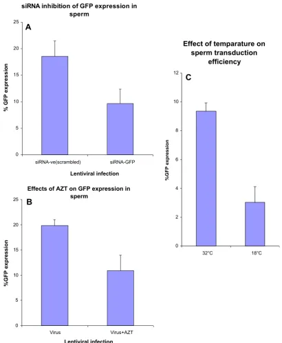
To validate that the fluorescence observed in porcine spermato-zoa was due to lentiviral encoded,de novoGFP synthesis, we per-formed experiments to block the expression of GFP, either with the HIV reverse transcriptase inhibitor, azidodeoxy thimidine (AZT) (Fig. 4B), or by transfection (Escort V, Sigma) with silencing
RNA to GFP (siRNA-GFP) (Ambion) (Fig. 4A). When sperm were
incubated with 1
l
M AZT or 10 nM siRNA-GFP 2 h prior to theaddition of the lentiviral vector, GFP expression, as determined by flow cytometry, was inhibited by about 50% compared to untreated or scrambled siRNA controls (Fig. 4A and B). Further-more, when porcine spermatozoa were incubated with lentiviral vectors at 18°C for 24 h, GFP expression was reduced by at least 3-fold compared to incubations carried out at 32°C (Fig. 4C),
demonstrating that the GFP expression in mature spermatozoa was dependent on lentiviral transduction.
Lentivirally transduced porcine spermatozoa were further ana-lysed for the presence of the lentiviral vector genome in sperm. This was done by PCR amplification of the WPRE sequence encoded within the lentiviral vector genome from lentivirally transduced porcine sperm genomic DNA. The expected 174 bp PCR fragment was successfully amplified (Fig. 5A).
We then investigated whether, following in vitro transduction,
the lentiviral vector genome had integrated into the
spermatozoon DNA. This was determined by performing linear-amplification-mediated polymerase chain reaction (LAM-PCR)
[21]on DNA from transduced pig spermatozoa. From this analysis we found evidence for at least 2 integration sites, one of which appeared to be unique to the pig genome. The first unique
integration sequence was located in the X-chromosome
(Fig. 5B). The other integration site was mapped imperfectly to the porcine homologue of the human (and chimp) Rh D gene.
Two other identifiable LAM–PCR sequences indicated the
presence of episomal (non-integrated) or pre-integration complex forms of the provirus. Despite several attempts, no integration sites could be detected in murine spermatozoa. However, routine detection of pre-integration complexes or episomal forms was observed.
Effects of AZT on GFP expression in sperm
0 5 10 15 20 25
Virus Virus+AZT
Lentiviral infection
%GFP expression
Effect of temparature on
sperm transduction
efficiency
0 2 4 6 8 10 12
32°C 18°C
%GFP expression
siRNA inhibition of GFP expression in
sperm
0 5 10 15 20 25
siRNA-ve(scrambled) siRNA-GFP
Lentiviral infection
% G
F
P
exp
ressi
o
n
A
B
C
3.4. The use of lentivirally transduced spermatozoa in early mouse embryonic development
To further determine whether lentivirally transduced sperma-tozoa could be used for gene transfer to developing embryos; IVF experiments were carried out in mice. ‘‘Swim up’’ populations of mouse spermatozoa from the epididymis and vas deferens were
obtained from 8–12 week old male mice and incubated at 32°C with mSCF-VSV-g pseudotyped lentiviral vectors for 3 h prior to sperm imaging or IVF. Following lentiviral transduction, fluores-cent mouse spermatozoa could also be visualised by confocal imaging (Fig. 6A, bottom two panels). Using standard IVF protocols, transduced sperm gave rise to transgenic blastocysts as shown in
Fig. 6D and E. This figure is from the 3rd experiment carried out. MH1 CO5 seq.
Pig DNA sequence from clone CH242-144C16 on chromosome X, complete sequence
Sperm 73 CAGGAAGTCAAACCACAGAGAGAGGGAGGGAGGGAGGTGTCGGAGAAATT 122 |||||||||||||||||||||||||||||||||||||||||||||||||| Genome 70946 CAGGAAGTCAAACCACAGAGAGAGGGAGGGAGGGAGGTGTCGGAGAAATT 70995
Sperm 123 CCTTTTCGGCAGAGATTTGAAGCGTCTTGTTATATAACAGGATCCGTCTC 172 |||||||||||||||||||||||||||||||||||||||||||||||||| Genome 70996 CCTTTTCGGCAGAGATTTGAAGCGTCTTGTTATATAACAGGATCCGTCTC 71045
Sperm 173 CACTGCAGCGTGATGCAAGAAGCTGCAACAGAACTGAAACACGGGACTTG 222 |||||||||||||||||||||||||||||||||||||||||||||||||| Genome 71056 CACTGCAGCGTGATGCAAGAAGCTGCAACAGAACTGAAACACGGGACTTG 71195
Sperm 223 TGTGTCCCCTGTCCCTGCAGTGGGGAGTCCTCCCAGGCGTTGTTTGCAGG 272 ||||||||||||||||||||||||||||||||||||| |||||||||||| Genome 71116 TGTGTCCCCTGTCCCTGCAGTGGGGAGTCCTCCCAGGTGTTGTTTGCAGG 71145
Sperm 273 CC 274 ||
Genome 71146 CC 71147
MH2 D05 seq.
Pig DNA sequence from clone CH242-144C16 on chromosome X, complete sequence
Sperm 72 CCTGCAAACAACGCCTGGGAGGACTCCCCACTGCAGGGACAGGGGACACA 121 |||||||||||||||||||||||||||||||||||||||||||||||||| Genome 71147 CCTGCAAACAACACCTGGGAGGACTCCCCACTGCAGGGACAGGGGACACA 71098
Sperm 122 CAAGTCCCGTGTTTCAGTTCTGTTGCAGCTTCTTGCATCACGCTGCAGTG 171 |||||||||||||||||||||||||||||||||||||||||||||||||| Genome 71097 CAAGTCCCGTGTTTCAGTTCTGTTGCAGCTTCTTGCATCACGCTGCAGTG 71048
Sperm 170 GAGACGGATCCTGTTATATAACAAGACGCTTCAAATCTCTGCCGAAAAGG 221 |||||||||||||||||||||||||||||||||||||||||||||||||| Genome 71047 GAGACGGATCCTGTTATATAACAAGACGCTTCAAATCTCTGCCGAAAAGG 70998
Sperm 222 AATTTCTCCGACACCTCCCTCCCTCCCTCTCTC 254 |||||||||||||||||||||||||||||||||
Genome 70967 AATTTCTCCGACACCTCCCTCCCTCCCTCTCTC 70935
500bp
100bp
1
2 3 4 5 6
Lanes:
A
B
4. Discussion
In this report, we have shown that mature spermatozoa can be transduced by pseudotyped lentiviral vectors and that these trans-duced spermatozoa can then be utilised efficiently for the study of development. GFP expression in sperm was confirmed and vali-dated by biochemical and molecular analyses.
This study has raised some fascinating biological questions, such as the mode of viral entry, the frequency with which these vectors integrate within the zygote genome and the mechanism by which lentiviral encoded gene products are translated into
protein. Binding of lentiviral vectors to spermatozoa, internalisa-tion and compleinternalisa-tion of reverse transcripinternalisa-tion in sperm appears to be sufficient for transgene expression within sperm and blasto-cysts. The acrosome matrix of sperm may well facilitate the import of the lentiviral vector to the nucleus[25]as it contains proteins that have endosomal like properties [26] and are pH regulated
[26,27]. Mature spermatozoa are thought to translate nuclear encoded genes by mitochondrial type ribosomes contained in sperm[20]. A similar mechanism of translation from exogenously introduced vectors may be operating for transgenes delivered into sperm.
Interestingly, consistent with our previous observations on c-kit+ haemopoietic cells [21], increased transduction efficiencies on sperm resulted from the use of lentiviral particles displaying membrane-bound stem cell factor. The SCF-mediated binding to c-kit on spermatozoa, may also have a significant impact in capac-itating sperm[28,29].
As seen previously in somatic cells, it was also observed that utilising a promoter lacking an enhancer (UCOE) significantly changed the level and pattern of GFP expression, whilst maintain-ing transduction efficiency. Given the random nature of integration of lentiviral vectors, careful consideration must also be given to the choice of promoter utilised[30].
Integration of viral vectors in porcine spermatozoa was con-firmed by LAM-PCR analysis. However, no integration sites in the mouse genome were detected following lentiviral transduction of murine spermatozoa. Internal bands indicating the presence of epi-somes or pre-integration complexes were routinely obtained sug-gesting that GFP expression in mouse spermatozoa probably derived from these. This observation is, perhaps, unsurprising gi-ven the short duration of incubation (3 h) of sperm with the viral vector. The viability of murine sperm was found to decrease signif-icantly after incubation periods longer than 3hrs, rendering them unsuitable for IVF (data not shown). Nevertheless, murine sperma-tozoa incubated with virus for short periods (containing the lentiv-iral vector episome or pre-integration complexes) were still able to transmit the GFP gene to developing embryos via fertilisation. Such a pattern would be consistent with a hypothesis that integration may be occurring soon after the cleavage of the first polar body in the presumptive zygote (and completion of meiosis), following sperm penetration of the oocyte.
This technique of transducing spermatozoa with SCF-displaying lentiviruses should therefore be particularly useful for the estab-lishment of large-animal transgenics, where spermatozoa are readily obtained and are stable for longer periods in culture, there-by facilitating integration of the vector into the genome of the spermatozoon. The relatively simple strategy described in this re-port could be further enhanced and scaled up by flow-sorting of GFP-positive spermatozoa and their use in artificial insemination techniques routinely performed on farm animals.
Our findings imply that transducing spermatozoa directly with pseudotyped lentiviral vectors would be a very powerful tool, not only for the study of early embryonic development, sperm physiol-ogy, transgenesis and fertilisation processes, vertical viral gene transmission [31–33] and epigenetics studies, but also in the development of large-animal transgenics for production of therapeutically important proteins.
Acknowledgements
We thank Ms. Rochelle Diamond (Caltech) for help with FACS analysis. Thanks to Dr. Michael Harkey, Fred Hutchinson Cancer Center, USA for LAM-PCR analysis. Thanks also due to Drs. Mark Fenwick and Jocelyn Mora, Imperial College London for help with confocal microscopy. This work was supported by Atazoa Ltd and, in part the Genesis Research Trust previously known as the Institute of Obstetrics and Gynaecology Trust, UK and Wellcome Trust, UK. Authorship contributions: AC, designed, performed experiments, analysed the data and wrote the paper; II, performed experiments; J.D., A.T., N.D., C.C., R.W., C.R., assisted in the design, performed experiments and contributed to the writing of the paper.
References
[1] Li, C., Mizutani, E., Ono, T. and Wakayama, T. (2009) Production of normal mice from spermatozoa denatured with high alkali treatment before ICSI. Reproduction 137, 779–792.
[2] Li, C., Mizutani, E., Ono, T. and Wakayama, T. (2010) An efficient method for generating transgenic mice using NaOH-treated spermatozoa. Biol. Reprod. 82, 331–340.
[3] Perry, M.M. and Sang, H.M. (1993) Transgenesis in chickens. Transgenic Res. 2, 125–133.
[4] Liechtenstein, T., Perez-Janices, N., Bricogne, C., Lanna, A., Dufait, I., Goyvaerts, C., Laranga, R., Padella, A., Arce, F., Baratchian, M., Ramirez, N., Lopez, N., Kochan, G., Blanco-Luquin, I., Guerrero-Setas, D., Breckpot, K. and Escors, D. (2013) Immune modulation by genetic modification of dendritic cells with lentiviral vectors. Virus Res. 176, 1–15.
[5] Emeagi, P.U., Goyvaerts, C., Maenhout, S., Pen, J., Thielemans, K. and Breckpot, K. (2013) Lentiviral vectors: a versatile tool to fight cancer. Curr. Mol. Med. 13, 602–625.
[6] Zhang, L., Thrasher, A.J. and Gaspar, H.B. (2013) Current progress on gene therapy for primary immunodeficiencies. Gene Ther. 20 (10), 963–969,http:// dx.doi.org/10.1038/gt.2013.21.
[7] Mukherjee, S. and Thrasher, A.J. (2013) Gene therapy for PIDs: progress, pitfalls and prospects. Gene 525, 174–181.
[8] Sakuma, T., Barry, M.A. and Ikeda, Y. (2012) Lentiviral vectors: basic to translational. Biochem. J. 443, 603–618.
[9] Lavial, F., Bessonnard, S., Ohnishi, Y., Tsumura, A., Chandrashekran, A., Fenwick, M.A., Tomaz, R.A., Hosokawa, H., Nakayama, T., Chambers, I., Hiiragi, T., Chazaud, C. and Azuara, V. (2012) Bmi1 facilitates primitive endoderm formation by stabilizing Gata6 during early mouse development. Genes Dev. 26, 1445–1458.
[10] Alder, O., Lavial, F., Helness, A., Brookes, E., Pinho, S., Chandrashekran, A., Arnaud, P., Pombo, A., O’Neill, L. and Azuara, V. (2010) Ring1B and Suv39h1 delineate distinct chromatin states at bivalent genes during early mouse lineage commitment. Development 137, 2483–2492.
[11]Zufferey, R., Dull, T., Mandel, R.J., Bukovsky, A., Quiroz, D., Naldini, L. and Trono, D. (1998) Self-inactivating lentivirus vector for safe and efficient in vivo gene delivery. J. Virol. 72, 9873–9880.
[12]Schwickerath, O., Brouns, G., Thrasher, A., Kinnon, C., Roes, J. and Casimir, C. (2004) Enhancer-deleted retroviral vectors restore high levels of superoxide generation in a mouse model of CGD. J. Gene Med. 6, 603–615.
[13]Zhang, F., Frost, A.R., Blundell, M.P., Bales, O., Antoniou, M.N. and Thrasher, A.J. (2010) A ubiquitous chromatin opening element (UCOE) confers resistance to DNA methylation-mediated silencing of lentiviral vectors. Mol. Ther. 18, 1640–1649.
[14]Zhang, F., Thornhill, S.I., Howe, S.J., Ulaganathan, M., Schambach, A., Sinclair, J., Kinnon, C., Gaspar, H.B., Antoniou, M. and Thrasher, A.J. (2007) Lentiviral vectors containing an enhancer-less ubiquitously acting chromatin opening element (UCOE) provide highly reproducible and stable transgene expression in hematopoietic cells. Blood 110, 1448–1457.
[15]Chandrashekran, A., Gordon, M.Y. and Casimir, C. (2004) Targeted retroviral transduction of c-kit+ hematopoietic cells using novel ligand display technology. Blood 104, 2697–2703.
[16]Takeuchi, Y., Simpson, G., Vile, R.G., Weiss, R.A. and Collins, M.K. (1992) Retroviral pseudotypes produced by rescue of a Moloney murine leukemia virus vector by C-type, but not D-type, retroviruses. Virology 186, 792–794.
[17]Howe, S.J. and Chandrashekran, A. (2012) Vector systems for prenatal gene therapy: principles of retrovirus vector design and production. Methods Mol. Biol. 891, 85–107.
[18]Landazuri, N. and Le Doux, J.M. (2006) Complexation with chondroitin sulfate C and Polybrene rapidly purifies retrovirus from inhibitors of transduction and substantially enhances gene transfer. Biotechnol. Bioeng. 93, 146–158. [19]Sastry, L., Johnson, T., Hobson, M.J., Smucker, B. and Cornetta, K. (2002)
Titering lentiviral vectors: comparison of DNA, RNA and marker expression methods. Gene Ther. 9, 1155–1162.
[20] Gur, Y. and Breitbart, H. (2006) Mammalian sperm translate nuclear-encoded proteins by mitochondrial-type ribosomes. Genes Dev. 20, 411–416. [21]Harkey, M.A., Kaul, R., Jacobs, M.A., Kurre, P., Bovee, D., Levy, R. and Blau, C.A.
(2007) Multiarm high-throughput integration site detection: limitations of LAM-PCR technology and optimization for clonal analysis. Stem Cells Dev. 16, 381–392.
[22]Chandrashekran, A., Gordon, M.Y., Darling, D., Farzaneh, F. and Casimir, C. (2004) Growth factor displayed on the surface of retroviral particles without manipulation of envelope proteins is biologically active and can enhance transduction. J. Gene Med. 6, 1189–1196.
[23]Sandlow, J.I., Feng, H.L. and Sandra, A. (1997) Localization and expression of the c-kit receptor protein in human and rodent testis and sperm. Urology 49, 494–500.
[24]Chandrashekran, A., Sarkar, R., Thrasher, A., Fraser, S.E., Dibb, N., Casimir, C., Winston, R. and Readhead, C. (2014) Efficient generation of transgenic mice by lentivirus-mediated modification of spermatozoa. FASEB J. 28, 569–576. [25]Miyauchi, K., Kim, Y., Latinovic, O., Morozov, V. and Melikyan, G.B. (2009) HIV
enters cells via endocytosis and dynamin-dependent fusion with endosomes. Cell 137, 433–444.
[26]Zhao, L., Shi, X., Li, L. and Miller, D.J. (2007) Dynamin 2 associates with complexins and is found in the acrosomal region of mammalian sperm. Mol. Reprod. Dev. 74, 750–757.
[28]Feng, H., Sandlow, J.I. and Sandra, A. (1998) The c-kit receptor and its possible signaling transduction pathway in mouse spermatozoa. Mol. Reprod. Dev. 49, 317–326.
[29]Feng, H.L., Sandlow, J.I. and Zheng, L.J. (2005) C-kit receptor and its possible function in human spermatozoa. Mol. Reprod. Dev. 70, 103– 110.
[30] Baup, D., Fraga, L., Pernot, E., Van, A.A., Vanherck, A.S., Breckpot, K., Thielemans, K., Schurmans, S., Moser, M. and Leo, O. (2010) Variegation and silencing in a lentiviral-based murine transgenic model. Transgenic Res. 19, 399–414.
[31] Li, F., Li, L., Zhong, Y., Xie, Q., Huang, J., Kang, X., Wang, D., Xu, L. and Huang, T. (2013) Relationship between LTR methylation and gag expression of HIV-1 in human spermatozoa and sperm-derived embryos. PLoS One 8, e54801. [32] Wang, D., Li, L.B., Hou, Z.W., Kang, X.J., Xie, Q.D., Yu, X.J., Ma, M.F., Ma, B.L.,
Wang, Z.S., Lei, Y. and Huang, T.H. (2011) The integrated HIV-1 provirus in patient sperm chromosome and its transfer into the early embryo by fertilization. PLoS One 6, e28586.