Notch signaling regulates murine
atrioventricular conduction and the formation of
accessory pathways
Stacey Rentschler, … , Vickas V. Patel, Jonathan A. Epstein
J Clin Invest. 2011;
121(2)
:525-533.
https://doi.org/10.1172/JCI44470
.
Ventricular preexcitation, which characterizes Wolff-Parkinson-White syndrome, is caused
by the presence of accessory pathways that can rapidly conduct electrical impulses from
atria to ventricles, without the intrinsic delay characteristic of the atrioventricular (AV) node.
Preexcitation is associated with an increased risk of tachyarrhythmia, palpitations, syncope,
and sudden death. Although the pathology and electrophysiology of preexcitation
syndromes are well characterized, the developmental mechanisms are poorly understood,
and few animal models that faithfully recapitulate the human disorder have been described.
Here we show that activation of Notch signaling in the developing myocardium of mice can
produce fully penetrant accessory pathways and ventricular preexcitation. Conversely,
inhibition of Notch signaling in the developing myocardium resulted in a hypoplastic AV
node, with specific loss of slow-conducting cells expressing connexin-30.2 (Cx30.2) and a
resulting loss of physiologic AV conduction delay. Taken together, our results suggest that
Notch regulates the functional maturation of AV canal embryonic myocardium during the
development of the specialized conduction system. Our results also show that ventricular
preexcitation can arise from inappropriate patterning of the AV canal–derived myocardium.
Research Article
Cardiology
Find the latest version:
Notch signaling regulates
murine atrioventricular conduction
and the formation of accessory pathways
Stacey Rentschler,1,2 Brett S. Harris,3 Laura Kuznekoff,4 Rajan Jain,1,2 Lauren Manderfield,1Min Min Lu,2 Gregory E. Morley,4 Vickas V. Patel,2 and Jonathan A. Epstein1,2
1Department of Cell and Developmental Biology and Institute for Regenerative Medicine, and 2Penn Cardiovascular Institute, University of Pennsylvania,
Philadelphia, Pennsylvania, USA. 3Medical University of South Carolina, Charleston, South Carolina, USA. 4New York University, New York, New York, USA.
Ventricular preexcitation, which characterizes Wolff-Parkinson-White syndrome, is caused by the presence of
accessory pathways that can rapidly conduct electrical impulses from atria to ventricles, without the intrinsic
delay characteristic of the atrioventricular (AV) node. Preexcitation is associated with an increased risk of
tachyarrhythmia, palpitations, syncope, and sudden death. Although the pathology and electrophysiology of
preexcitation syndromes are well characterized, the developmental mechanisms are poorly understood, and
few animal models that faithfully recapitulate the human disorder have been described. Here we show that
activation of Notch signaling in the developing myocardium of mice can produce fully penetrant accessory
pathways and ventricular preexcitation. Conversely, inhibition of Notch signaling in the developing
myocar-dium resulted in a hypoplastic AV node, with specific loss of slow-conducting cells expressing connexin-30.2
(Cx30.2) and a resulting loss of physiologic AV conduction delay. Taken together, our results suggest that
Notch regulates the functional maturation of AV canal embryonic myocardium during the development of the
specialized conduction system. Our results also show that ventricular preexcitation can arise from
inappropri-ate patterning of the AV canal–derived myocardium.
Introduction
In the embryonic heart, the atrial myocardium is separated from ven-tricular myocardium by a circumferential band of atrioventricular (AV) canal cardiomyocytes that conduct electrical impulses slowly, resulting in AV delay, which facilitates unidirectional blood flow (1, 2). Embryonic AV canal cells ultimately give rise to myocardium within the lower rim of the atria, myocardium supporting the AV valves, specialized conducting cells of the AV node, and a portion of the left ventricular free wall (3). During early gestation and midgesta-tion in the mouse, AV canal myocytes have an immature contractile network and express low conductance gap junctions, thereby confer-ring slow conduction. They proliferate more slowly than atrial and ventricular myocytes and can be distinguished from adjacent atrial and ventricular myocytes by expression of genes, including Tbx2/3, and bone morphogenetic protein 2 (Bmp2), and by a relative absence of Notch signaling (2, 4–6). During late gestation, a significant num-ber of AV canal myocytes undergo apoptosis (7). At the same time, epicardial cells in the region of the AV junction undergo epithelial-to-mesenchymal transformation and invade the heart to form the annulus fibrosis, an insulating plane of nonconductive tissue sepa-rating the atria and ventricles (8, 9). As a result of these coordinated events, embryonic AV electrical continuity is lost, and conduction from atria to ventricles is funneled through the specialized conduc-tion system of the AV node and His-Purkinje system (10).
The mature AV node is composed of numerous cell types, includ-ing a population of myocytes derived from AV canal myocardium (3). The AV node performs a similar function to that of the embryonic AV canal, in that it delays electrical impulses originating in the atria
before they are allowed to excite the ventricles, thereby allowing coor- dinated contraction of the atria prior to ventricular systole and facili-tating forward blood flow. The developmental processes regulating formation of the AV node from embryonic AV canal cardiomyocytes are just beginning to be elucidated. Bmp and Notch signaling path-ways have been implicated in boundary formation between AV canal and atrium/ventricle and perhaps in maturation of AV canal deriva-tives (2). In the zebrafish heart, for example, knockdown of notch1b results in a failure to develop physiologic AV delay and a prolonged refractory period within the AV canal (11). Expression of activated Notch during early heart formation disrupts AV canal myocardium boundaries and leads to midgestation lethality (12). T-box genes, including Tbx2, Tbx3, and Tbx5, Nkx2.5, and Gata factors have also been implicated (reviewed in ref. 2). For example, Tbx5+/– mice have
reduced expression of Tbx3 and the low conductance gap junction connexin-30.2 (Cx30.2) in the AV canal (13, 14).
Ventricular preexcitation occurs when an electrically active muscular connection, apart from the AV node-His pathway, exists between the atrium and ventricle and can lead to symptomatic tachycardia or sudden death as the first clinical manifestation. Wolff-Parkinson-White syndrome (WPW) is most commonly diag- nosed in children or young adults, with a prevalence of approxi- mately 0.2% (15–17). It is the second most common cause of reen-trant supraventricular tachycardia in the Western world and the most common cause in China (18). The presence of dual AV elec-trical connections (the AV node and the accessory pathway) with different conduction velocities and refractory periods provides the substrate for initiation and perpetuation of reentrant tachycardia. In addition, for reasons that are not well understood, atrial fibril-lation develops in up to one-third of WPW patients at a young age. The occurrence of atrial fibrillation in the presence of an accessory pathway that conducts rapidly can be life threatening, because the rapid ventricular response may lead to ventricular fibrillation.
research article
WPW is most commonly a sporadic and isolated cardiac disorder, although autosomal dominant familial inheritance has been described (19) as has association with congenital heart disease, especially Ebstein’s anomaly (20). There are 2 known genetic associations with WPW in humans. Mutations in the AMP-activated protein kinase γ-2 subunit (PRKAG2) result in a rare form of WPW in the setting of a glycogen storage cardiomyopathy (21–23). Microdeletions of BMP2 have recently been associated with a syndrome of WPW with either cognitive dysfunction or Alagille syndrome (24, 25). In mice, dele-tion of a BMP receptor within the embryonic AV canal leads to a low penetrance of both accessory pathways and AV nodal defects (26, 27). Of interest, the gene encoding a Notch ligand, JAGGED1, known to be associated with Alagille syndrome (28, 29), is located 3.8-MB cen-tromeric to BMP2 and is also deleted in some of these patients. This raises the question of whether altered Notch signaling may contrib-ute to the observed preexcitation in this syndrome.
Here we provide evidence that Notch signaling can modulate late stages of AV canal myocardial maturation. Our data indicate that inhibition of Notch signaling can impede normal development of the AV node and can lead to a selective loss of slow-conducting cells, characteristic of the mature AV node. Conversely, activation of Notch signaling in the mouse induces ventricular preexcitation. Notch-activated mice survive gestation and develop electrophysi-ologic evidence of ventricular preexcitation only after birth. We propose that WPW and other AV conduction disorders may result from aberrations in Notch signaling and abnormal persistence or regression of AV canal myocardial derivatives.
Results
Notch inhibition disrupts AV node development. We sought to determine whether Notch signaling plays a role in the formation and function of the mammalian conduction system. To achieve this, we selectively inhibited Notch signaling within a subset of murine cardiomyocytes through Cre-inducible expression of a dominant-negative truncated form of mastermind-like protein (DNMAML). All 4 mammalian
Notch receptors interact with mastermind-like proteins after ligand-mediated activation and translocation to the nucleus. DNMAML is a well-characterized truncated form of mastermind-like protein that acts as a specific and effective Notch inhibitor by binding to the Notch intracellular domain and preventing recruitment of coactivators (30). We used mice in which DNMAML (knocked into the constitutively active Rosa26 locus) is expressed in a tissue-specific manner after acti- vation by Cre recombinase (31), and we crossed these mice with myo-sin light chain 2vCre/+ animals (Mlc2vCre/+ animals), which express Cre
in embryonic ventricular myocytes and AV nodal cells (32, 33).
Mlc2vCre/+DNMAML
mice are viable, with normal appearing car- diac structures and normal function as assessed by echocardiog-raphy (Supplemental Table 1; supplemental material available online with this article; doi:10.1172/JCI44470DS1). However,
Mlc2vCre/+DNMAML
mice had a significantly smaller AV node vol-ume indexed to heart weight when compared with that of controls (16 ± 5.1 × 10⁶ vs. 30 ± 1.8 × 106 μm3/g; n = three 5-month-old mice
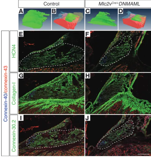
of each genotype; P < 0.01), as demonstrated by 3D reconstruction of the AV node from a representative control and Notch-inacti- vated mutant (Figure 1, A–D, and Supplemental Figure 1). Consis-tent with this observation, Mlc2vCre/+DNMAML mice had fewer cells
expressing the hyperpolarization-activated cyclic nucleotide-gated channel 4 (HCN4), which contributes to the hyperpolarization current of AV nodal tissue and is a marker of cells within the proxi-mal AV node (Figure 1, E and F). Collagen I staining demonstrated a grossly intact annulus fibrosis (Figure 1, G and H).
Cx30.2 is a low conductance gap junction isoform expressed in the murine compact AV node and inferior nodal extension that decelerates electrical impulse propagation, while Cx40 expression within the AV node is confined to the lower AV nodal cells that are contiguous with the His bundle and contribute to faster electrical conduction (3, 34). Myocytes of the compact AV node and inferior nodal extension are derived from AV canal myocardium, while Cx40-positive lower AV nodal cells and the His bundle are derived from ventricular myocardium (3, 34). Mlc2vCre/+DNMAML mice demon-Figure 1
Loss of Notch signaling results in a smaller AV node and alteration of connexin-expressing cells. 3D reconstruction from trichrome-stained images of the AV node of a representative control heart is shown in A and
B (different renderings of the same reconstruction; AV node volume = 8.2 × 106μm3 in this heart) and the AV node of a Mlc2vCre/+DNMAML
mutant is shown in C and D (different renderings of the same recon-struction; AV node volume = 3.5 × 106μm3). (A–D) AV nodal tissue is green, ventricular myocardium is red, tricuspid valve is transparent blue, and (B and D) atrial septum is transparent yellow. (E and F) Immunohistochemistry demonstrates a reduction of HCN4-positive AV nodal cells in Mlc2vCre/+DNMAML mutants compared with that of
con-trol littermates. (G and H) Collagen I staining reveals an intact annulus fibrosis in Mlc2vCre/+DNMAML mutants when compared with control.
(I and J) There is a selective loss of connexin-30.2–positive AV nodal cells
in Mlc2vCre/+DNMAML mutants, with a maintenance of connexin-40–
positive lower nodal cells. (E–J) Connexin-43 is not ectopically upregulated in Mlc2vCre/+DNMAML mutants. Control animals are
litter-mate Mlc2v+/+DNMAML animals. Dashed lines delineate the compact
[image:3.585.41.286.85.340.2]strated a selective loss of Cx30.2-expressing cells within the compact AV node and inferior nodal extension, with a maintenance of Cx40-expressing lower nodal cells (Figure 1, I and J), suggesting a specific loss of AV canal–derived tissue in Notch loss-of-function mutants.
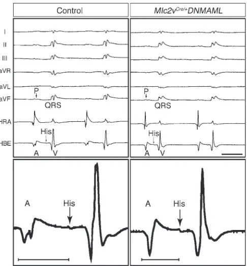
Notch inhibition disrupts AV nodal delay. It has previously been shown that Cx30.2 null mice have accelerated AV conduction, with a concomitantly shortened PR interval compared with that of wild-type mice (35). Global knockout of both Cx30.2 and Cx40 results in restoration of normal AV delay, suggesting that the ratio of connexin isoforms within the AV nodal interface is important for fine-tuning AV delay (36). Given the altered connexin isoform ratio seen in Mlc2vCre/+DNMAML mice, AV conduction was assessed
via surface electrocardiogram (EKG) in anesthetized animals using either pentobarbital (Figure 2 and Table 1) or isoflurane (Supple-mental Results). This analysis revealed a reproducibly shorter PR interval in Notch-inhibited hearts when compared with that of lit-termate control hearts (43.0 ± 4.2 vs. 37.5 ± 2.5 ms; n = 7 control and n = 6 Mlc2vCre/+DNMAML mice at 5 months; P < 0.02), while
the QRS interval and QT interval corrected for murine physiol-ogy (QTm) were unchanged (Table 1). Tissue-specific deletion of
the Notch1 receptor in myocardium (Mlc2vCre/+Notch1flox/flox) also
resulted in shortening of the PR interval (Supplemental Table 2), suggesting that inhibition of Notch1 receptor activity is at least in part responsible for the observed phenotype in DNMAML mice.
Invasive electrophysiology studies demonstrated a shorter atrio-hisian (AH) interval in Notch-inactivated hearts when compared with that of control hearts (35.6 ± 4.5 vs. 28.1 ± 4.5 ms; n = 9 control hearts and n = 8 Mlc2vCre/+DNMAML hearts; P < 0.004), consistent
with accelerated AV conduction, while the remainder of the ven-tricular conduction system parameters appear unaffected (Table 1). During programmed electrical stimulation, the reduction in the AH interval of mutants occurred at every conduction interval tested (Supplemental Figure 2). Another defining characteristic of AV nodal tissue is a relatively long refractory period, or recovery
time after being excited, which has evolved to protect the ventricles from rapidly conducting atrial tachyarrhythmias. Notch-inacti-vated hearts demonstrate a shorter AV effective refractory period (AVERP) when compared with that of control hearts (66.1 ± 4.9 vs. 57.5 ± 7.6 ms, n = 9 control hearts and n = 8 Mlc2vCre/+DNMAML
hearts; P < 0.02), confirming defective AV nodal function.
Notch activation produces accessory pathways. To gain further insight into the role of Notch in the morphogenesis of the AV canal, we used mice in which the Notch1 intracellular domain (NICD, knocked into the constitutively active Rosa26 locus) is expressed in a tissue-specific manner after activation by Cre recombinase (37), and we crossed these mice with Mlc2vCre/+ mice. NICD functions
in a ligand-independent fashion to translocate to the nucleus and recruit coactivators to induce transcription of Notch target genes.
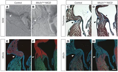
Mlc2vCre/+NICD
[image:4.585.45.288.82.344.2]mice were viable, with no gross structural abnormal-ities evident at birth. However, by the first week of life excess tissue was noted along the right AV junction, which was confirmed by scanning electron microscopy (Figure 3, A and B). Histologic characterization of
Figure 2
Notch inhibition disrupts AV nodal delay. The top panels show repre-sentative 6-lead EKG and intracardiac electrograms from the high right atrium (HRA) and His bundle electrogram (HBE), demonstrating shorter PR and AH intervals in Mlc2vCre/+DNMAML mice when compared with
those in control mice. A, atrial depolarization; P, P wave; V, ventricular depolarization. Scale bar: 50 ms. The bottom panels show 1 represen-tative beat from the His bundle electrogram at higher magnification, with the AH interval demarcated by the bars. The control AH interval was 33 ms, and the Mlc2vCre/+DNMAML AH interval was 22 ms. Control
[image:4.585.308.531.462.671.2]mice are DNMAML littermates of Mlc2vCre/+DNMAML mice.
Table 1
Notch inhibition disrupts AV nodal delay
Electrocardiography
Control (n = 7) Mlc2vCre/+DNMAML (n = 6)
HR (bpm) 406 ± 62 488 ± 47A
PR (ms) 43.0 ± 4.2 37.5 ± 2.5A
QRS (ms) 9.3 ± 2.4 8.0 ± 0.6 QTm (ms) 32.9 ± 3.8 35.0 ± 3.2 Electrophysiology
Control (n = 9) Mlc2vCre/+DNMAML (n = 8)
HR (bpm) 424 ± 63 479 ± 42A
AH (ms) 35.6 ± 4.5 28.1 ± 4.5B
Hd (ms) 4.4 ± 0.8 4.1 ± 0.6 HV (ms) 12.1 ± 1.7 12.0 ± 1.6 SNRT120 (ms) 172 ± 27 166 ± 43
AVERP120 (ms) 66.1 ± 4.9 57.5 ± 7.6A
AERP120 (ms) 39.4 ± 8.5 35.8 ± 7.0
AVWBCL 89.4± 8.5 82.5 ± 6.5 AV 2:1 72.8 ± 9.4 65.0± 7.1 VERP120 (ms) 40.6 ± 9.8 28.8± 7.0
Mean ± SD. AP < 0.05 for control littermate DNMAML versus Mlc2vCre/+
DNMAML mice; BP < 0.01 for control DNMAML versus Mlc2vCre/+
DNMAML mice. HR, heart rate; AH, AH interval; Hd, His duration; HV, His-ventricular interval; SNRT120, sinus node recovery time at drive
train of 120 ms; AVERP120, AVERP at 120 ms; AERP120, atrial effective
refractory period at 120 ms; AVWBCL, AV Wenckebach block cycle length; AV 2:1, AV 2:1 block cycle length; VERP120, ventriculoatrial
research article
the AV junction in Notch-activated hearts revealed muscular connec-tions between the atrial and ventricular myocardium extrinsic to the AV node. Ectopic myocardium spanning the AV junction was most commonly epicardial and occurred on the right side (Figure 3D and Supplemental Figure 3C), but left-sided connections and those cross-ing the annulus fibrosus septally were also often noted (n = 8 postna-tal mice). Muscular connections along the right AV junction expressed the atrial marker myosin light chain 2a (Mlc2a) in the myocardium closest to the atrium and the ventricular marker Mlc2v near the ven-tricle (Figure 3, E–H). Although marker expression does not provide direct evidence for the developmental origin of the tissue, these find- ings are consistent with a hypothesis that functional accessory path-ways derive from AV canal myocardium capable of differentiating into either atrial or ventricular tissue. Furthermore, an expanded region of AV canal–derived AV nodal tissue (Cx30.2 positive, Tbx3 positive, and Cx40 negative) could be found within the base of the interatrial sep-tum (Supplemental Figure 4). We also noted the unusual and striking persistence of myocardium along the atrial surface of the AV valves (Figure 3D and Supplemental Figure 3C). Myocytes derived from AV canal myocardium populate the AV valves during embryogenesis, but this tissue normally regresses by late gestation (38).
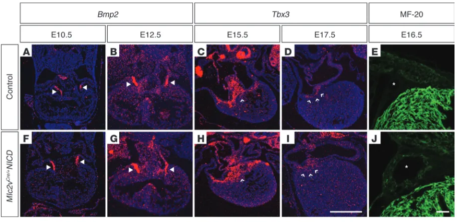
We examined histology and gene expression of staged embryos from E10.5–E18.5 to determine the earliest signs of detectable defects in Mlc2vCre/+NICD specimens. Bmp2
was expressed by AV canal myo-cardium of control embryos at E10.5 and E12.5 (Figure 4, A and B), and expression was unchanged in mutants at these stages (Figure 4,
F and G). Subsequent to E12.5, Bmp2 expression diminished signifi-cantly in this region. Hence, we examined Tbx3 expression at E15.5 and E17.5 to identify AV canal myocardium at these time points. In control embryos (Figure 4, C and D), a sharp boundary of expression was present between the thin rim of remaining AV canal myocardi-um and ventricular tissue. However, in mutant embryos (Figure 4, H and I), Tbx3 expression was slightly expanded and the boundary was irregular, especially at E17.5 in the region of the right posterior AV junction. At E16.5, coronary veins of control embryos could be identified in the AV groove (asterisks, Figure 4E). In mutant embryos, the coronary veins were enlarged and were surrounded by a muscular wall expressing myosin, as detected by MF-20 antibody staining (Fig-ure 4J). Muscularization and structural abnormalities of coronary veins have been identified in humans with WPW, in which this tissue may contribute to accessory pathways (Supplemental Figure 3B and refs. 39–41). Hence, developmental abnormalities were first noted at approximately E15.5 in Mlc2vCre/+NICD embryos and were consistent
with an excess of AV canal–derived tissue.
[image:5.585.87.498.84.337.2]Notch activation produces ventricular preexcitation and atrial arrhythmias in a mouse model of WPW. To determine the functional consequences of the structural abnormalities in Notch-activated mice, serial surface ECGs were performed. At birth, Notch-activated mice had normal electrical activation (n = 7, Supplemental Figure 5A). However, by postnatal day 2, 1 out of 7 mice examined developed electrocardio-graphic changes suggestive of ventricular preexcitation, as evidenced by a short PR interval, wide QRS, and delta wave (data not shown). By
Figure 3
Activation of Notch signaling results in accessory pathway formation. (A and B) Scanning electron microscopy demonstrates excess tissue in the right AV junction of 1-week-old Mlc2vCre/+NICD mice (arrow in B) when compared with that of control mice, which have a distinct AV groove
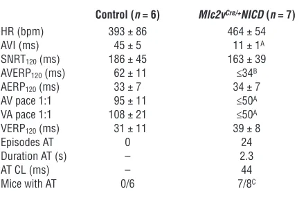
1 week of age, 6 out of 7 mice developed these electrocardiographic changes (Supplemental Figure 5B), and by 8 weeks, 100% of a much larger cohort of Notch-activated mice had electrocardiographic chang-es suggestive of ventricular preexcitation (n > 50). Invasive electro-physiology studies of adult Notch-activated hearts confirmed robust ventricular preexcitation and findings consistent with the presence of rapidly conducting accessory pathways (Figure 5). Abnormally rapid AV conduction was present at baseline and with programmed stimu-lation in all mutant animals examined (n = 7), which was significantly faster than that of littermate controls (Table 2). Even at the shortest cycle length at which our system could pace the atrium of Notch-acti-vated hearts (50 ms or 1,200 bpm), AV conduction remained fixed at a 1:1 ratio, whereas control hearts would exhibit AV conduction block at paced atrial rates greater than approximately 600 bpm (Table 2). Consistent with these findings, the AVERP in Notch-activated hearts was significantly shorter than that in littermate controls (Table 2).
As previously mentioned, WPW patients have a high prevalence of atrial fibrillation, which can lead to rapid activation of the ventricles via the accessory pathway. Notch-activated hearts were also greatly predisposed to the development of atrial tachycardias during programmed electrical stimulation (Table 2), often occur-ring with the introduction of just a single atrial extrastimulus and resulting in a rapid ventricular response.
Further confirmation of ventricular preexcitation is provided by high-resolution optical mapping studies that delineate the spatial and temporal pattern of depolarization throughout the cardiac cycle. In control hearts, optical mapping studies demonstrated that electrical activation began with atrial depolarization, followed
by an AV delay, and then ventricular depolarization. Ventricular depolarization began at the apex of the right and left ventricles and ended near the AV groove (Figure 6, A, B, and E, and Supplemental Video 1). In striking contrast, adult Notch-activated hearts revealed atrial depolarization, followed by immediate ventricular depolariza-tion that traveled from base to apex, without an AV delay (Figure 6, C, D, and F, and Supplemental Video 2). Right-sided posterior breakthrough was noted in all 8 mutant animals that were exam-ined, and 5 of these also showed evidence of additional left- and right-sided breakthroughs, indicating the presence of multiple accessory pathways. The high incidence of right-sided preexcitation was consistent with the preponderance of accessory pathway tissue identified by histology in this location. Echocardiography of adult
Mlc2vCre/+NICD animals revealed normal fractional shortening and
ejection fraction when compared with those of control littermates (Supplemental Table 3), but the contraction pattern of the heart was markedly abnormal, consistent with the activation patterns obtained by optical mapping (Supplemental Videos 3 and 4).
Discussion
In this report, we provide evidence from both gain-of-function and loss-of-function approaches to support a model in which Notch activity is required for proper maturation of AV canal myocardi- um, which normally contributes to the AV node and therefore sig-nificantly impacts cardiac conduction (3, 42). Inhibition of Notch signaling affects the structure and function of the mature AV node by reducing overall size and by producing a specific loss of slowly
conducting Cx30.2-expressing cells derived from AV canal myocar-Figure 4
Developmental analysis reveals late gestation AV canal defects in Mlc2vCre/+NICD embryos. In situ hybridization of staged embryos from E10.5–
E18.5 was performed to determine the earliest signs of detectable AV canal defects in Mlc2vCre/+NICD specimens. (A and B) Bmp2 is expressed in
[image:6.585.66.522.82.304.2]research article
dium, while rapidly conducting Cx40-expressing cells derived from ventricular myocardium (34) are preserved. Tissue-specific dele- tion of the Notch1 receptor in myocardium also resulted in short-ening of the PR interval (Supplemental Table 2), suggesting that inhibition of Notch1 receptor activity is at least in part responsible for the observed phenotype in Mlc2vCre/+DNMAML mice.
The cardiac phenotype produced by our Notch gain-of-function model is strikingly reminiscent of human ventricular preexcitation syndromes, such as WPW. Many of the early reports of this disorder described epicardial bands of ectopic myocardium spanning the AV groove (Supplemental Figure 3A), and early treatments included sur-gical disruption of these epicardial accessory pathways (43). Ectopic tissue spanning the AV boundary in our mouse model was evident by gross inspection and by scanning electron microscopy (Figure 3B). A wide range of additional locations of accessory pathways have been described in humans, including those traversing the annulus fibrosus, epicardial and subendocardial tracts on both right and left sides, and myocardial cuffing of the coronary veins. We identified related exam-ples in our mouse model. To our knowledge, this is the first animal model of preexcitation with a predisposition to atrial arrhythmias, an important cause of morbidity and mortality in patients. Interest-ingly, although subtle histologic and gene expression defects could be identified as early as E15.5, mice did not display electrocardiographic evidence of ventricular preexcitation until several days or more after birth. We attribute this to physiologic mechanisms that contribute to AV delay prior to maturation of the annulus fibrosis as well as the substantial expansion of ectopic myocardial tissue within the region of the AV junction noted to occur after birth (data not shown).
Previously, transgenic mice were generated to model a relatively rare form of WPW, in which the PRKAG2 gene is mutated (44, 45).
These animals also develop ventricular preexcitation in the post-natal period (46), and electrophysiologic defects are associated with abnormally increased glycogen storage in myocardium and cardiac hypertrophy (21). Breeches in the annulus fibrosus allow for electrical communication between atria and ventricles. While these studies allow for further investigation of the underlying mechanisms of ventricular preexcitation in some contexts, trans-genic PRKAG2 mice are probably not optimal models for the more common forms of isolated ventricular preexcitation seen clinically. Further, it is likely that the underlying mechanisms of bypass tract formation are distinct in the Notch and PRKAG2 models, because we observe epicardial accessory pathways and ectopic myocardium, including muscularized coronary veins, which cannot be explained by a breakdown or breech of the annulus fibrosus.
The annulus fibrosus derives from epicardial cells, and recent stud- ies in developing chick embryos demonstrate that mechanical inter-ruption of epicardial cell migration can disrupt annulus fibrosus formation, with resulting ventricular preexcitation (10). Therefore, we propose that ventricular preexcitation can result from at least 2 distinct mechanisms (which are not mutually exclusive). First, defects in formation or maintenance of the annulus fibrosus may allow for electrical continuity between atrial and ventricular tissue. Second, abnormal patterning of AV canal myocardium may result in ectopic myocardial tissue and accessory pathways that can bypass a well-formed annulus. We propose that genetic, epigenetic, or envi- ronmental mechanisms that activate components of the Notch sig-naling cascade may cause patterning defects of AV canal myocardium and WPW. While this possibility, among others, has been previously suggested as a potential cause of WPW, we believe that our data and robust animal model that mimics the human disease provide strong experimental support for this hypothesis. However, as Notch signal-ing is known to be involved in cell-cell signaling, it is possible that activation or inhibition of Notch in AV canal myocardium can lead to indirect effects on neighboring epicardial or endocardial cells, which may contribute to the observed phenotypes. For example, it was recently demonstrated that constitutive Notch1 activation at early developmental stages in murine AV canal myocardium results in abnormal endocardial epithelial-to-mesenchymal transition (47).
Additional support in the future may derive from genetic fate-map-Figure 5
Notch activation leads to ventricular preexcitation. Representative 6-lead EKG tracings and intracardiac electrograms from the high right atrium and His bundle electrogram are shown from a control and Mlc2vCre/+NICD
[image:7.585.43.288.83.281.2]mouse. Note the fused p wave (P) and QRS complexes and widened QRS with delta wave on surface EKG as well as the fused atrial (A) and ventricular (V) electrograms in Notch-activated mice, indicative of robust AV conduction over the accessory pathway. Scale bar: 100 ms. Control mice are NICD littermates of Mlc2vCre/+NICD mice.
Table 2
Notch activation leads to ventricular preexcitation
Control (n = 6) Mlc2vCre/+NICD (n = 7)
HR (bpm) 393 ± 86 464± 54
AVI (ms) 45 ± 5 11± 1A
SNRT120 (ms) 186± 45 163 ± 39
AVERP120 (ms) 62± 11 ≤34B
AERP120 (ms) 33± 7 34± 7
AV pace 1:1 95± 11 ≤50A
VA pace 1:1 108± 21 ≤50A
VERP120 (ms) 31± 11 39 ± 8
Episodes AT 0 24
Duration AT (s) – 2.3
AT CL (ms) – 44
Mice with AT 0/6 7/8C
Mean ± SD. AP < 4 × 10⁻⁵ for control versus Mlc2vCre/+NICD mice; BP < 0.0003 for control versus Mlc2vCre/+NICD mice; CP < 0.01 for control
versus Mlc2vCre/+NICD mice. Controls are littermate NICD mice. AVI, AV
[image:7.585.309.530.553.696.2]ping studies to rigorously determine adult derivatives of AV canal myocardium in Notch-activated hearts. However, technical limita-tions preclude this approach at the present time, since Cre-reporter mice cannot be used to track AV canal myocardial derivatives, because our model uses Cre in alternative tissues to activate Notch.
Notch plays reiterated and complex roles during many stages of cardiovascular development (48), and prior work has suggested that Notch participates in early boundary formation of the AV canal. For example, in chick embryos, activated Notch2 or overexpression of Hesr1 (also known as Hey1) or Hesr2 (also known as Hey2) (down-stream mediators of Notch signaling) can function to repress Bmp2, and this has been proposed as a mechanism by which Notch activity in atria and ventricles can help to restrict Bmp2 expression to the AV canal (6). In the mouse, Hesr1 and Hesr2 are expressed in the forming atria and ventricles, respectively, and misexpression of either gene through- out the heart represses AV myocardium formation and downregu-lates Tbx2 and Bmp2 (5). Overexpression of NICD using the Mesp1Cre/+
driver to activate expression very early during heart formation results in suppression of AV canal myocardium and related marker genes (12). These studies, and others, provide data to support a model in which Notch activity in working atrial and ventricular myocardium suppresses Bmp2 and AV canal myocardial fate, while Bmp2 normally activates Tbx2/3 expression, which instructs AV myocardial matura-tion (42, 49). Our findings are generally consistent with this model, although patterning defects in our animal models did not occur until later in development than seen in the prior studies. It is likely that Notch plays reiterative roles during maturation of the heart and, spe-cifically, of AV canal myocardium, and specific downstream pathways
may vary with developmental stage and context. For example, we did not observe a decrease in Bmp2 or Tbx3 expression in our Notch-acti-vated hearts, as might have been predicted from studies performed at earlier developmental stages (5, 6, 12).
Our analysis of Mlc2vCre/+NICD animals focused on structural
defects likely to be involved in ventricular preexcitation. However, we also uniformly observed the presence of ectopic myocardium along the atrial surface of the AV valve leaflets that we also attrib-uted to persistence of AV canal myocardium. It is interesting to note that a similar pattern of ectopic myocardium has been fre-quently reported in patients with Ebstein’s anomaly (50), which is strongly associated with WPW. Although we did not observe other features of Ebstein’s anomaly in our mouse model, such as atrialization of the right ventricle, it will be of interest to deter-mine whether developmental defects of AV canal myocardium are responsible for some features of this rare disorder.
[image:8.585.44.546.82.330.2]In summary, genetic manipulation of Notch activity provides experimental evidence to support a developmental model to explain common forms of WPW and ventricular preexcitation syndrome. AV canal myocardium, which functions in the embryo to delay AV conduction, undergoes late gestation remodeling, regresses, and gives rise to the AV node. We propose that abnor- mal patterning of AV canal–derived myocardium, under the influ-ence of active Notch signaling, can result in functional accessory pathways and arrhythmias. Further delineation of the molecular cascades responsible for normal remodeling and maturation of AV canal myocardium will inform the genetic and developmental understanding of WPW and related disorders.
Figure 6
Activation of Notch signaling leads to ventricular preexcitation. Electrical activation maps from the posterior surface of the heart, with 1-ms con-tour lines for (A and B) control and (C and D) Mlc2vCre/+NICD hearts. Note the different time scales. Atrial activation in control hearts is followed
research article
Methods
Mice. Mlc2vCre/+, DNMAML, and NICD
mice were maintained on a mixed back-ground and were genotyped using Cre-specific primers (5′ -ATTCTCCCACC-GTCAGTACG-3′ and 5′-CGTTTTCTGAGCATACCTGGA-3′) and primers specific for the ROSA26 locus (5′-AAAGTCGCTCTGAGTTGTTAT-3′, 5′-GCGAAGAGTTTGTCCTCAACC-3′, and 5′ -GGAGCGGGAGAAATG-GATATG-3′). Littermate animals were compared in all experiments unless otherwise noted. All animal protocols were approved by the University of Pennsylvania Institutional Animal Care and Use Committee.
Histology, immunohistochemistry, and in situ hybridization. AV node volume was measured in 3 control Mlc2v+/+DNMAML and 3 Mlc2vCre/+DNMAML
adult hearts using Masson’s trichrome staining of sections through the entire AV node, including the compact AV node, inferior nodal exten-sion, and lower nodal cells (34). The presence of connective tissue located between AV nodal tissue and the surrounding atrial and AV ring myocardi- um as well as the distinct morphology of the AV nodal cells allowed identi-fication of the AV node. Quantification of AV nodal area was performed on every third section using ImageJ software (http://rsbweb.nih.gov/ij/), and AV node volume was subsequently calculated. 3D reconstruction was per-formed by importing the images into Amira 5.3.1 (Visage Imaging GmbH). Sections were aligned, and the labeling function was used to identify and draw discrete cardiac subcompartments, including atrial tissues, tricuspid valve, ventricular myocardium, and AV node, using a drawing tablet. The resulting labeled fields were then rendered into 3D surfaces with minimal smoothing to prevent data loss.
Immunohistochemistry was performed on frozen sections with anti-bodies recognizing HCN4 (H-300, Santa Cruz Biotechnology Inc.), Cx40 (C-20, Santa Cruz Biotechnology Inc.), Cx43 (C8093, Sigma-Aldrich), Cx30.2 (gift from Klaus Willecke, University of Bonn, Bonn, Germany), and Collagen I (MD Bioscience). Secondary antibody fluorescent con-jugates included anti-rabbit Alexa 488 (Invitrogen), anti-goat Alexa 633 (Invitrogen), and anti-mouse TRITCRedx100 (Jackson ImmunoRe- search Laboratories Inc.). Images were captured by sequentially scan-ning 3 channels using a Leica SP5 laser scanning confocal microscope. MF-20 antibody staining was performed on paraffin-embedded sections with antibody from the Hybridoma Bank. Radioactive in situ hybrid-izations were performed on samples that were fixed overnight with 4% paraformaldehyde, dehydrated through an ethanol series, paraffin embedded, and sectioned using previously described probes for Mlc2a and
Mlc2v (51), Bmp2 (52), and Tbx3 (53). Histology, immunohistochemistry, and in situ hybridization images were analyzed using Adobe Photoshop. Control and mutant images were treated identically in all cases in which brightness and contrast were altered.
Echocardiography . Mice were anesthetized using an integrated isoflurane-based system. Two-dimensional images were obtained at 180 frames per second, using a 30-MHz probe (RMV 707B, Visual Sonics) in the paraster- nal long- and short-axis views to guide M-mode analysis at the midven-tricular level. Left ventricular fractional shortening, ejection fraction, and wall dimensions were computed from M-mode measurements.
EKG analysis. EKGs were performed on mice anesthetized with inhaled isoflurane for less than 5 minutes (Supplemental Figure 5 and Supple-mental Table 2), except during invasive electrophysiology experiments (see below). Lead II EKGs were recorded at a sampling rate of 4,000 Hz (ADInstruments). EKG intervals were measured by averaging 100 beats using the LabChart software package (ADInstruments); measurements were taken by an investigator who was blinded to the genotypes of the mice.
Electrophysiology. Protocols for the in vivo mouse electrophysiology have been described in detail (46, 54). Mice were anesthetized with pentobarbi-tal (33 mg/kg i.p.) and laid down on a heating pad to maintain core body
temperature, and limb leads were placed subcutaneously to obtain multi-lead ECGs. An octapolar 1.7-French electrode catheter (CIBer mouse-EP, NuMED) was placed in the right atrium and ventricle under electrogram guidance through a jugular vein cutdown. A programmed digital stimula- tor (DTU-215, Fisher Scientific) delivered electrical impulses at approxi- mately twice diastolic threshold, while surface ECG and intracardiac elec-trograms were displayed on a multichannel oscilloscope recorder (Bard Electrophysiology Inc.) and analyzed offline. Standard pacing protocols were used to assess atrial and ventricular conduction and refractoriness. Measurement of intervals was performed by 2 independent investigators who were blinded to the genotype of the mice.
Scanning electron microscopy. These experiments were carried out at CDB/CVI Microscopy Core (School of Medicine, University of Pennsylvania). Samples were fixed overnight in 4% paraformaldehyde and dehydrated in a graded ethanol series, followed by immersion twice for 10 minutes in 100% HMDS (Sigma-Aldrich), followed by 30 minutes of air drying, as described previously (55). Then samples were mounted on stubs and sputter coated with gold pal-ladium. Specimens were observed and photographed using a Philips XL20 scanning electron microscope (FEI) at 10 kV accelerating voltage.
Optical mapping. Perfused hearts were optically mapped as described previously (56, 57). In brief, mice were heparinized (10,000 IU i.p.) and euthanized with 100% CO2, followed by cervical dislocation. Hearts were
extracted through a median sternotomy, and the aorta was cannulated and Langendorff perfused with a constant pressure of 68–74 mmHg (1.5–2.0 ml/min) with 38°C oxygenated (95% O2 5% CO2) modified Tyrode’s
solution containing 1.8 mM CaCl2, 1.0 mM MgCl2, 1.2 mM KH2PO4,
130 mM NaCl, 4.7 mM KCl, 24 mM NaHCO3, 11.1 mM glucose, 0.052 g/l
albumin, pH 7.4. Blebbistatin (5 mM) was used to limit motion artifacts (58). After 10 minutes of perfusion and equilibrium, hearts were injected with 20 mM voltage-sensitive fluorescent dye di-4-ANNEPS (Invitrogen), and uniform fluorescent saturation was reached in 10 minutes. High- resolution optical mapping studies were performed on an upright micro-scope (BX51WI, Olympus Inc.) outfitted with a high-speed CMOS camera (MiCAM ULTIMA L, SciMedia Ltd.).
Statistics. Student’s 2-tailed t test analysis was used to evaluate differences between groups in all cases, except when comparing the number of mice with atrial tachycardia, for which a χ2 analysis was used. Data are represented as
mean ± SD. P values of less than 0.05 were considered statistically significant.
Acknowledgments
We would like to thank the members of the Epstein laboratory (especially Arun Padmanabhan) for many helpful discussions and technical support. We also thank Klaus Willecke for connex-in-30.2 antibody. This work was supported by the University of Pennsylvania, Division of Cardiology T-32, and the Department of Medicine Measey Foundation Award to S. Rentschler, NIH National Center for Research Resources grant 5P20RR016434- 09 to B.S. Harris, American Heart Association (AHA) Physician-Scientist/Post-Doctoral fellowship AHA 0825548D to R. Jain, NIH grant HL076751 to G.E. Morley, University of Pennsyl-vania Research Fund Award to V.V. Patel, and funds from the WW Smith Endowed Professorship, the NIH (U01HL100405, R01HL095634), and the AHA DeHaan Cardiac Myogenesis Cen-ter to J.A. Epstein.
Received for publication July 22, 2010, and accepted in revised form November 1, 2010.
1. Bakker ML, Christoffels VM, Moorman AF. The cardiac pacemaker and conduction system devel-ops from embryonic myocardium that retains its primitive phenotype. J Cardiovasc Pharmacol. 2010;56(1):6–15.
2. Christoffels VM, Smits GJ, Kispert A, Moorman AF. Development of the pacemaker tissues of the heart. Circ Res. 2010;106(2):240–254.
3. Aanhaanen WT, et al. The Tbx2+ primary myocar- dium of the atrioventricular canal forms the atrio-ventricular node and the base of the left ventricle. Circ Res. 2009;104(11):1267–1274.
4. Horsthuis T, et al. Gene expression profiling of the forming atrioventricular node using a novel tbx3-based node-specific transgenic reporter. Circ Res. 2009;105(1):61–69.
5. Kokubo H, Tomita-Miyagawa S, Hamada Y, Saga Y. Hesr1 and Hesr2 regulate atrioventricular boundary formation in the developing heart through the repres-sion of Tbx2. Development. 2007;134(4):747–755. 6. Rutenberg JB, Fischer A, Jia H, Gessler M, Zhong
TP, Mercola M. Developmental patterning of the cardiac atrioventricular canal by Notch and Hairy-related transcription factors. Development. 2006; 133(21):4381–4390. 7. Hahurij ND, et al. Accessory atrioventricular myo-cardial connections in the developing human heart: relevance for perinatal supraventricular tachycardias. Circulation. 2008;117(22):2850–2858. 8. Gittenberger-de Groot AC, Vrancken Peeters MP, Mentink MM, Gourdie RG, Poelmann RE. Epicar-dium-derived cells contribute a novel population to the myocardial wall and the atrioventricular cushions. Circ Res. 1998;82(10):1043–1052. 9. Zhou B, von Gise A, Ma Q, Hu YW, Pu WT. Genetic
fate mapping demonstrates contribution of epi-cardium-derived cells to the annulus fibrosis of the mammalian heart. Dev Biol. 2010;338(2):251–261.
10. Kolditz DP, et al. Epicardium-derived cells in devel- opment of annulus fibrosis and persistence of acces-sory pathways. Circulation. 2008;117(12):1508–1517. 11. Milan DJ, Giokas AC, Serluca FC, Peterson RT, Mac-Rae CA. Notch1b and neuregulin are required for specification of central cardiac conduction tissue. Development. 2006;133(6):1125–1132. 12. Watanabe Y, et al. Activation of Notch1 signal-ing in cardiogenic mesoderm induces abnormal heart morphogenesis in mouse. Development. 2006; 133(9):1625–1634.
13. Moskowitz IP, et al. The T-Box transcription factor Tbx5 is required for the patterning and maturation of the murine cardiac conduction system. Development. 2004;131(16):4107–4116.
14. Munshi NV, et al. Cx30.2 enhancer analysis identi-fies Gata4 as a novel regulator of atrioventricular delay. Development. 2009;136(15):2665–2674. 15. De Bacquer D, De Backer G, Kornitzer M. Prevalences
of ECG findings in large population based samples of men and women. Heart. 2000;84(6):625–633. 16. Goudevenos JA, Katsouras CS, Graekas G, Argiri
O, Giogiakas V, Sideris DA. Ventricular pre-exci-tation in the general population: a study on the mode of presentation and clinical course. Heart. 2000;83(1):29–34.
17. Munger TM, et al. A population study of the natu-ral history of Wolff-Parkinson-White syndrome in Olmsted County, Minnesota, 1953–1989. Circulation. 1993;87(3):866–873.
18. Wan Q, Wu N, Fan W, Tang YY, Jin L, Fang Q. Clini-cal manifestations and prevalence of different types of supraventricular tachycardia among Chinese. Chin Med J (Engl). 1992;105(4):284–288.
19. Doevendans PA, Wellens HJ. Wolff-Parkinson-white syndrome: a genetic disease? Circulation. 2001;104(25):3014–3016.
20. Follath F, Hallidie-Smith KA. Unusual electrocar-diographic changes in Ebstein’s anomaly. Br Heart J. 1972;34(5):513–519.
21. Arad M, et al. Constitutively active AMP kinase mutations cause glycogen storage disease mim-icking hypertrophic cardiomyopathy. J Clin Invest. 2002;109(3):357–362.
22. Gollob MH, et al. Identification of a gene respon-sible for familial Wolff-Parkinson-White syndrome. N Engl J Med. 2001;344(24):1823–1831.
23. Blair E, et al. Mutations in the gamma(2) subunit of AMP-activated protein kinase cause familial hypertrophic cardiomyopathy: evidence for the central role of energy compromise in disease patho-genesis. Hum Mol Genet. 2001;10(11):1215–1220. 24. Le Gloan L, et al. A 8.26Mb deletion in 6q16 and
a 4.95Mb deletion in 20p12 including JAG1 and BMP2 in a patient with Alagille syndrome and Wolff-Parkinson-White syndrome. Eur J Med Genet. 2008;51(6):651–657.
25. Lalani SR, et al. 20p12.3 microdeletion predis-poses to Wolff-Parkinson-White syndrome with variable neurocognitive deficits. J Med Genet. 2009;46(3):168–175. 26. Stroud DM, et al. Abnormal conduction and morpholo- gy in the atrioventricular node of mice with atrioventric-ular canal targeted deletion of Alk3/Bmpr1a receptor. Circulation. 2007;116(22):2535–2543. 27. Gaussin V, et al. Alk3/Bmpr1a receptor is required for development of the atrioventricular canal into valves and annulus fibrosus. Circ Res. 2005; 97(3):219–226.
28. Li L, et al. Alagille syndrome is caused by muta-tions in human Jagged1, which encodes a ligand for Notch1. Nat Genet. 1997;16(3):243–251. 29. Oda T, et al. Mutations in the human Jagged1 gene
are responsible for Alagille syndrome. Nat Genet. 1997;16(3):235–242.
30. Maillard I, et al. Mastermind critically regulates Notch-mediated lymphoid cell fate decisions. Blood. 2004;104(6):1696–1702.
31. High FA, et al. An essential role for Notch in neu-ral crest during cardiovascular development and smooth muscle differentiation. J Clin Invest. 2007; 117(2):353–363.
32. Chen J, Kubalak SW, Chien KR. Ventricular mus-cle-restricted targeting of the RXRalpha gene reveals a non-cell-autonomous requirement in car-diac chamber morphogenesis. Development. 1998; 125(10):1943–1949.
33. Pashmforoush M, et al. Nkx2-5 pathways and congenital heart disease; loss of ventricular myo-cyte lineage specification leads to progressive cardiomyopathy and complete heart block. Cell. 2004;117(3):373–386.
34. Aanhaanen WT, et al. Developmental origin, growth, and three-dimensional architecture of the atrioventricular conduction axis of the mouse heart. Circ Res. 2010;107(6):728–736.
35. Kreuzberg MM, Willecke K, Bukauskas FF. Connex- in-mediated cardiac impulse propagation: connex-in 30.2 slows atrioventricular conduction in mouse heart. Trends Cardiovasc Med. 2006;16(8):266–272. 36. Schrickel JW, et al. Normal impulse propagation in
the atrioventricular conduction system of Cx30.2/ Cx40 double deficient mice. J Mol Cell Cardiol. 2009; 46(5):644–652.
37. Stanger BZ, Datar R, Murtaugh LC, Melton DA. Direct regulation of intestinal fate by Notch. Proc Natl Acad Sci U S A. 2005;102(35):12443–12448.
38. de Lange FJ, et al. Lineage and morphogenetic analy-sis of the cardiac valves. Circ Res. 2004;95(6):645–654. 39. Ho SY. Accessory atrioventricular pathways: getting to the origins. Circulation. 2008;117(12):1502–1504. 40. Schumacher B, Tebbenjohanns J, Pfeiffer D, Omran
H, Jung W, Luderitz B. Prospective study of retro- grade coronary venography in patients with pos-teroseptal and left-sided accessory atrioventricular pathways. Am Heart J. 1995;130(5):1031–1039. 41. Sun Y, et al. Coronary sinus-ventricular
acces-sory connections producing posteroseptal and left posterior accessory pathways: incidence and elec-trophysiological identification. Circulation. 2002; 106(11):1362–1367.
42. Bakker ML, et al. Transcription factor Tbx3 is required for the specification of the atrioventricular conduc-tion system. Circ Res. 2008;102(11):1340–1349. 43. Hanon S, Shapiro M, Schweitzer P. Early history of the
pre-excitation syndrome. Europace. 2005;7(1):28–33.
44. Sidhu JS, et al. Transgenic mouse model of ventric-ular preexcitation and atrioventricular reentrant tachycardia induced by an AMP-activated protein kinase loss-of-function mutation responsible for Wolff-Parkinson-White syndrome. Circulation. 2005; 111(1):21–29.
45. Arad M, et al. Transgenic mice overexpressing mutant PRKAG2 define the cause of Wolff-Parkin- son-White syndrome in glycogen storage cardiomy-opathy. Circulation. 2003;107(22):2850–2856. 46. Patel VV, et al. Electrophysiologic characterization
and postnatal development of ventricular pre-exci-tation in a mouse model of cardiac hypertrophy and Wolff-Parkinson-White syndrome. J Am Coll Cardiol. 2003;42(5):942–951.
47. Luna-Zurita L, et al. Integration of a Notch-depen-dent mesenchymal gene program and Bmp2-driven cell invasiveness regulates murine cardiac valve for-mation. J Clin Invest. 2010;120(10):3493–3507. 48. High FA, Epstein JA. The multifaceted role of
Notch in cardiac development and disease. Nat Rev Genet. 2008;9(1):49–61.
49. Christoffels VM, Hoogaars WM, Tessari A, Clout DE, Moorman AF, Campione M. T-box tran-scription factor Tbx2 represses differentiation and formation of the cardiac chambers. Dev Dyn. 2004;229(4):763–770.
50. Anderson KR, Zuberbuhler JR, Anderson RH, Beck-er AE, Lie JT. Morphologic spectrum of Ebstein’s anomaly of the heart: a review. Mayo Clin Proc. 1979;54(3):174–180.
51. Kuo CT, et al. GATA4 transcription factor is required for ventral morphogenesis and heart tube formation. Genes Dev. 1997;11(8):1048–1060. 52. Kaartinen V, Dudas M, Nagy A, Sridurongrit S, Lu
MM, Epstein JA. Cardiac outflow tract defects in mice lacking ALK2 in neural crest cells. Development. 2004;131(14):3481–3490.
53. Chapman DL, et al. Expression of the T-box family genes, Tbx1-Tbx5, during early mouse development. Dev Dyn. 1996;206(4):379–390.
54. Ismat FA, et al. Homeobox protein Hop functions in the adult cardiac conduction system. Circ Res. 2005;96(8):898–903.
55. Braet F, De Zanger R, Wisse E. Drying cells for SEM, AFM and TEM by hexamethyldisilazane: a study on hepatic endothelial cells. J Microsc. 1997;186(pt 1):84–87.
56. Rentschler S, et al. Visualization and func-tional characterization of the developing murine cardiac conduction system. Development. 2001;128(10):1785–1792.
57. Morley GE, et al. Reduced intercellular coupling leads to paradoxical propagation across the Pur-kinje-ventricular junction and aberrant myo-cardial activation. Proc Natl Acad Sci U S A. 2005; 102(11):4126–4129.