4201
ACCURATE SEGMENTATION OF PSORIASIS DISEASES
IMAGES USING K-MEANS ALGORITHM BASED ON
CIELAB (L*A*B) COLOR SPACE
1TANYA SHAKIR JARAD, 1ALI J. DAWOOD
1Department of Computer Science, College of Computer Science and Information Technology, University
of Anbar, Anbar, Iraq
E-mail: [email protected], [email protected]
ABSTRACT
Context: Psoriasis turned out to be one of the debilitating and enduring inflammatory skin diseases. Often
misinterpreted as a casual skin disease, it is estimated that approximately 125 million people worldwide suffers due to this infection. The case is made worse when there is no known cure in the status quo. The communal category of psoriasis has been considered as abruptly demarcated scaly and erythematous plaque at patient’s skin. This disease could ensue anywhere on the human body. Problem: Diagnosis of psoriasis requires an experienced specialist in the field of dermatology because of the presence of other skin diseases similar to a large extent which lead to majority cases of an error in diagnostic. As doctors are still mere human and depends on factors such as eye and physical touch that is not error free. In addition, the drugs for psoriasis disease contain quantities of Chemical materials dangerous to other body organs that may put the functionality of critical organs such as the liver and spleen in jeopardy. Meanwhile, over-treatment leads to loss of life of the patient so it must be re-diagnosis multiple times until the confirmation of a high proportion of the dangerous disease. Time is not the greatest threat for this disease rather the accuracy of diagnosis is much crucial and the accuracy of diagnostic plays a pivotal role in combating this atrocious disease. Regular re-diagnosis is considered a must in order to ensure the survivability of patients from the threat it poses. However, re-diagnosis often consumed a great amount of financial expenditure just to ensure that it is indeed a disease of psoriasis and that the appropriate treatment is given may only lead to another issue which is a financial deficiency. Approach: In this paper, the researcher is interested in separating the image and concentrate on the lesion region and extricating disease district. The process itself is an enormous challenge in light of the fact that there is no discovery of this minute segmentation algorithms division executes and all in all dataset. The proposed strategy is based on K-Means clustering as initial segmentation and gets a divided region, including areas of diseased and the proposed K-Means based on CIE Lab L*a*b color spaces instead of using Red, Green and Blue (RGB) color space. Post segmentation based on color feature will be filtered out as non-interesting objects. Finding: The findings from this study have shown that: Firstly the method is depending on the L*a*b color spaces instead of using RGB color spaces, secondly, the method is based on color feature to select disease region of psoriasis or the correct object. The results of this research confirmed that this method works effectively where we have been implementing this method on a database containing 80 medical images of RGB psoriasis diseases image and shows the accuracy of this method was at 95% when we did a comparison between our method and other ways to find that the proposed strategy gives more effective results in the segmentation. The researcher compared accurate segmentation of K-Means cluster formation with color spaces L*a*b on medical imaging and K-Means cluster formation with color spaces RGB on the same images.
Keyword:Color image segmentation, Psoriasis disease diagnosis, K-Means algorithm.
1. INTRODUCTION
Psoriasis disease has been referred as protracted interminable skin ailment and over 125 million people around the world suffering from this lesion, and in status quo, there is no known cure. The most widely recognized kind of psoriasis is
4202 apparent and visible to the naked eyes. It starts with reddish scaling papules that combine to make a shape of round to oval plaque that is simply recognized contiguous at the skin surface. This scale could be observed to be gleaming whitish, also possess bleeding locations during expel. The differences towards psoriasis morphology has been recorded such as Acute eruptive-psoriasis, Chronic plaque-psoriasis, Erythrodermic-psoriasis, Pustular-psoriasis, Reiter's-syndrome, HIV- induced-psoriasis and Light sensitive-induced-psoriasis. In this study, the researcher concentrates on the chronic plaque psoriasis as this type has many attributes such as it’s an all-around characterized plaque that is the most widely recognized as the introduction of psoriasis. Injuries can show up at any place at cutaneous surfaces. These augment towards specific sizes, thereby have a tendency to stay stable for quite a long duration. A transitory brown coloured, whitish or reddish macula relic on the removal of plaque. It is clear that this disease appears to be enduring ailment, thereby critical in observing their state at the patient for choosing the appropriate remedy measures. The rate of the abatement at range of psoriasis in patients would demonstrate change record of these treatments [2]. The technique which parts psoriasis images to typical and unusual skin areas has been implemented. The region of psoriasis vulgaris could be thereby predestined.
2. RELATED WORK
Many researchers have conducted many studies and researches in medical image segmentation to solve psoriasis disease based on many factors such as according to differences intensity and color. According to Taur et al [2], They have utilised an orthogonal subspace method to classify the segment the regions of psoriasis vulgaris. The proposed methods are used for assessing the treatment of psoriasis vulgaris. These fundamental methods constitute feature extraction and psoriasis dissection techniques. They divide psoriasis imagery to the ordinary skin and psoriasis regions utilising orthogonal subspace classifiers and element vectors consolidating fluffy surface range and two-dimensional histograms at shade immersion spaces. Rate in decline at region of psoriasis of patients would demonstrate effectiveness in treatments. Lu et al [3] presented a new system used for assessing psoriasis seriousness. It utilises a detailed visual of simple scales for scoring depth and scrabbling of psoriasis sores and redness, PASI score is slanted and experience the ill effects of poor between and
intra-onlooker concordance. As a fundamental factor of building up a solid assessment strategy for psoriasis, a system is introduced for segmenting scaling in 2-D advanced pictures. The method is accepted to be the first tolimit scaling specifically in 2-D computerized images. The SVM training is gathered specifically from images that are being broken down yielding this technique to be extremely flexible for varieties in lighting and skin type. These techniques appeared towards giving solid segmentations being assessed in imageries that comprise of various lighting condition, skin type and psoriasis type. To develop an automatic system for psoriasis diagnosis Shrivastava et al [4] displayed the main survey of innovation requesting in psoriasis alongside its present practices, difficulties and evaluation methods. The paper likewise directs top to bottom examination of the current writing for every single clinical parameter of Psoriasis Area and Severity Index (PASI) such as region, erythema, layering and depth. They recommend sections in hazard evaluation utilising a choice of emotionally supportive network for stratification of psoriasis in huge populaces. An adjusted knowledge has been introduced in every one of the segments of the plan, in particular: include extraction, highlight choice, infection stratification and general CAD execution assessment. Taur et al [5] presented involuntary strategies for psoriasis image segmentation utilising neuro-fuzzy methods. This could be utilised as parts of a treatment assessment framework. They proposed the psoriasis imageries which are divided to ordinary skins, thereby covers strange areas naturally. These regions of every section in patients with various purposes could be evaluated accurately. This information is utilised for getting quantifiable measures in calculating the progress of treatments. This basic strategy required comprises of component extraction and picture division (arrangement) techniques. The two dimensional histograms of the tone and immersion parts in shading image and fluffy surface range at dim level images has been utilized towards component vector for finding homogeneous locales. For correlation, a shading grouping calculation which was utilised to section digitised dermatoscopic pictures is likewise executed.
3. MATERRIALS AND METHODS
3.1 Materials
4203 amounted in a total of 48 abnormal and 32 normal cases.
3.2 Methods
3.2.1 The proposed framework
The study involves the utilisation of computer-based framework for selecting and perceiving. It can be expected to fill the need of making an apparatus for choosing the appropriate support in recognisable proof of first untimely psoriasis cases. This kind of choice support is a multi-disciplinary development merging therapeutic image handling techniques that require specialists' to figure out how to improve the accuracy of psoriasis by distinguishing pieces of proof. In this way extraordinarily diminishing the false negative and positive affirm ages and upgrading the recognisable proof of genuine positives and true negative cases (affectability and specificity).
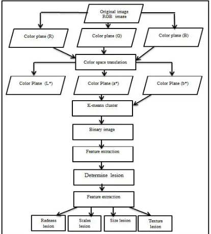
[image:3.612.90.297.462.692.2]The proposed method will include two steps such as the segmentation step and the feature extraction and classification. The accuracy and robustness of the first stage must be carefully measured because of the fact that the feature abstraction or cataloguing stage depend upon segmentation process. We have converted RGB images in L*a*b color spaces with K-Means cluster formation for tracking lesion disease "psoriasis" through color images. The researcher’s proposed method for detector of the psoriasis is in the stages of the proposed framework. The processed can be divided into many major stages as presented in
Figure 1.
Figure 1: Proposed system for the psoriasis diagnosis.
3.2.1.1 Image segmentation
Image segmentation is the most critical approach among practically all mechanised image acknowledgment frameworks. There are several algorithms utilised for image segmentation, and some of them portioned an image contingent upon the object while only a few can portion consequently [6]. Image segmenting turns out as indispensable stage for the medical domain [7]. Fundamentally, image segmentation strategies can be categorised into three classifications such as the edge-based strategies, locale based techniques, and pixel-based strategies [8,9]. On the basis of the following two properties image segmentation algorithms are classified:
1- Discontinuity: As in the discontinuity of an image is partitioned when a sharp change in the intensity is encountered during the procedure. As an example, the algorithms depending on this technique include edge/boundary detection techniques.
2- Similarity / Homogeneity: in Similarity, an image is partitioned on the basis of certain predefined criteria which may be color, size, and many others. For example, algorithms based on this technique include threshold, region growing, splitting, merging and so on.
4204 it can be a contribution from the client. K-Means algorithm is iterative in nature [11].
3.2.1.2 Color spaces
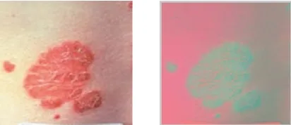
Segmentation is the procedure towards dividing images to combination of object. Color-based segmentation act as significant contrivance in partitioning object by relying upon color categories [12]. Fig. 2 shows an original psoriasis lesion skin example images in RGB formats, which at RGB format could be transformed to another spaces as L*a*b color space, see Figure 2.
Figure 2: RGB image converted to L*a*b color space.
Additional color spaces is L*a*b space that comes out as color-contra space. Lightness could be characterized at L-dimension, but further two dimensions are color-contra proportions. This is on the basis of non-linearly trampled CIE XYZ
color space matches [13]. The more
differentiation there is between the normal and the abnormal skin in the picked color space, the more sensitive the segmentation will be, hence will be translated into the color dimensions for better identification. So as to acquire a color portrayal that improves the difference between the normal and the abnormal skin, a few works have been dedicated to change the first RGB picture into a more evident color space much of the time standard and free of the image. The most well-known and broadly utilised color spaces are the CIEL*a*b* and CIE L*u*v*, which is portrayed in. These changes have been appeared to create great segmentation comes about for some dermatological determinations [14][15][16].
3.2.1.3 K-Means clustering
Cluster formation turns out as a method for dividing sets of information to precise quantity of collections or categories. Additionally, one of the most sought after method where k-means clustering. Here, the divided parts are treated as collections of information to k-quantity collection of information [17,18]. This classification is given a group of information to k quantity of separate
group. K-means procedure comprises of two distinct stages. First, it computes the k centroid and at second stage, it grosses every point to clusters that possesses adjacent centroid from their corresponding information points. Dissimilar approaches prevail for defining these distances of adjacent centroid and the possible approaches are Euclidean distances. This approach comprises of two distinct phases, during initial phase this method computes the k-centroid, thereby at succeeding phases it proceeds with every points of the cluster which possesses neighbouring centroid from conforming information point [19]. When grouping is completed it re-calculates the fresh centroid for every cluster and on the basis of the centroid, fresh Euclidean distance shall be considered among every center and every data points, thereby allocates the points in the clusters that possess minimum Euclidean distance. Every cluster at the partition shall be well-defined by their member objects and centroid. The centroid of every group shall be considered as point to which the totality of distance from every objects in that cluster is diminished. Therefore, K-means shall be considered as iterative procedure that diminishes the totality of distance from every object to its cluster centroid. We shall deliberate imagery with a tenacity of x×y and the imagery is clustered to k-number of clusters. p(x, y) is the input pixel to be clustered and ck is the cluster center. The procedure for k-means 2 clustering as follows,
1. Quantity of cluster k and center is initialized. 2. Euclidean distances d amongst midpoint and every pixel of the image is evaluated by means of relation as follows,
(1) 3. Every pixel is assigned to the adjoining center on the basis of the distance d.
4. Ultimately, pixel is assigned and fresh location of the center is re-calculated by means of relation as follows.
(2)
5. The procedure is repeated unless tolerance or error value is satisfied.
6. Re-shape cluster pixels to image.
[image:4.612.92.298.251.340.2]4205 if the first centroid has been arbitrarily selected, it would acquire dissimilar outcome for dissimilar first center. Therefore, initial centers has been prudently selected thereby the anticipated segmentation is attained. Also, the computational difficulty turns out to be additional term that needs to be considered in planning K-means cluster formation. This trusts upon quantity of information essentials, the quantity of groups and quantity of iterations. Skin colouring has been disintegrated to melanin constituent and hemoglobin constituent [1]. These two constituents associate openly with skin colour and is employed for distinguishing among erythema and further non-inflamed skin like standard skin and moles. Here, we have employed k-means cluster formation procedure with k=2 for dividing psoriasis picture to divisions of one class of psoriasis lesion and additional standard skin.
3.2.1.4 Clustering pixels into image
After the segmenting process produced a binary image for each cluster, the pixel location of the clusters are then fetched from the original image and put zeros except for those pixels locations, see Figure 3.
(a) (b) (c) Figure 3: (a) Binary image after apply K-Means clustering algorithm. (b). (c) Clustering pixels into
the image from the original image.
3.2.1.5 Extraction psoriasis lesion
Subsequently, after the image segmentation process on the two clusters are done, here comes another challenge: how extraction the lesion object and filtering out from the non-interesting object of the images psoriasis. To distinguish between lesion pixels and non-lesion, the researcher used a color histogram algorithm for red channel of the two clusters of RGB color space
as Figure 4. On a dataset of 40 images indicated
that the difference between the red band exhibits a good contrast to discriminate between the lesion and the normal healthy skin. The Calculation of standard deviation is extremely important to be correctly interpreted for the data. On top of when the mean needs to be interpreted for accuracy. The model mean is normal and calculated as quantity of every detected consequences in sample
divided by overall quantity of proceedings. The parameter is employed as representation for sample mean. In scientific terms, it could be expressed mathematically as,
(3)
(4) Where represents color value of i-th colour constituent of j-th picture pixel and N corresponds to overall quantity of pixels at the picture. , (i=1,2,3) indicate mean and standard deviation of each channel of images correspondingly.
[image:5.612.316.527.269.429.2]
(a) (b)
Figure 4: Column (a) shows a histogram of red channel of RGB colors for lesion cluster. Column
(b) shows a histogram of red channel of RGB colors for the non-lesion cluster.
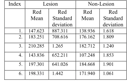
The highest mean and standard deviation indicated the psoriasis lesion. The following Table 1 shows the differences in red color densities between two clusters lesion and the non-lesion by using the mean and standard deviation functions.
Table 1: shows the differences of color densities between two clusters.
Index Lesion Non-Lesion Red
Mean Standard Red deviation
Red
Mean Standard Red deviation
1. 147.623 887.311 138.936 1.618
2. 183.251 708.616 176.162 1.809
3. 210.285 1.265 182.712 1.240
4. 143.836 652.211 107.248 1.853
5. 197.301 641.026 184.668 1.901
6. 198.331 1.442 171.940 1.061
[image:5.612.90.296.393.481.2] [image:5.612.320.528.580.714.2]4206 The extraction feature turns out to be most energetic constituents in computer aided analysis (pCAD) scheme. Considering optimum cataloguing, most operational structures in terms of degree of discernment amongst classes are prudently acquired. These features have been mined by relating scientific processes on pixel of images and thereby lessening the unusual dataset. Plentiful feature extraction methods are existing in literatures that could be suitable for dermatology image. Now, four foremost classes of features, textures, colors, redness and chaotic have been deliberated. Nonetheless, previously to feature extraction the phase of hair elimination procedure like conventional Dull Razor algorithm [20] is useful in the pre-filtering stage. The anticipated technique employed quantity of features, as psoriasis is lingering ailment. It is significant to trail the disorder of the patient for selecting correct treatments. The anticipated technique of feature extraction is employed for evaluating the treatment of psoriasis and discovers any change of the lesion. Features are approximate to scores that depending on the doctor such as color, area, scales and texture features for discovering changing on psoriasis before and after treatment.
3.2.3 Color features
As there is acceptable colour unevenness of low to high harshness of psoriatic lesions, colour features might deliver respected evidence regarding stratification of psoriasis. Figures like mean, standard deviations, variances, entropies, skew, colour irregularity, chromatic variance in color plane and kurtosis of every colour constituent at numerous colour space has been anticipated in [21– 24]. The greatest widespread and protuberant four colour space i.e., RGB, HSV, YCbCr and CIEL*a*b has been deliberated. RGB is representation of red–green–blue colour spaces, HSV that signifies hues, saturations and value colour spaces, YCbCr characterises luminance and chrominance colour spaces and CIEL*a*b signifies lightness and colour constituent spaces. As every colour spaces encompasses three colour constituents, the aggregate of 24 colour features (4 colour space*3 colour component*2 statistic) has been mined from four colour space for every sample images [25]. The proposed had used CIEL*a*b colour space for segmentation process and RGB for feature extraction.
3.2.3.1 Area assessment lesion feature
This valuation towards psoriasis lesions chiefly involves three object at images
(background, vigorous skin and psoriatic lesion). This contains two stages: (i) subdivision of regions of Interest (ROI) that is complete skin regions comprising of psoriatic lesion, and (ii) subdivision of psoriatic lesion. Formerly, percentages of psoriatic lesion areas are deliberated by captivating the ratio of areas of the psoriatic lesions to the areas of ROI [26]. The area psoriasis lesion is more an interactive factor affecting in the treatment any change on the lesion can calculate from number pixels of the segment. To calculated percentage of the area lesion of the total area of psoriasis we are applying the following function
Where cable from monitor changes in the area lesion after the treatment and getting improvement.
3.2.3.2 Redness lesion feature
Feature distinguishing and categorizing redness of skin is deliberated and structures in psoriasis lesions, that contributes for complete structure of psoriasis picture cataloguing is considered. Trusting these at minds, three features are mined that indicates redness in skin (fierceness of red-green, fierceness of red-blue and redness to be ratios of mean values of R, G and B, where μR, μG and μB characterize mean values of R, G and B color constituent of RGB color spaces.
The proposed had red channel from color histogram algorithm gave very high contrast from other channels green and blue of RGB color space, so calculate the mean red μR of red channel applying the following function
3.2.3.3 Scales lesion feature
Apparent removal in whiter scale that makes psoriasis lesions denser could be referred to scaliness. Very little mechanisms are existing at literature towards scaliness valuation of psoriasis lesions. A research effort existing in the literature put forward by Lue et al. [3] deliberates the procedure in segmenting the scaling of 2D digital psoriasis image. This procedure comprises of dual phases: (i) feature extraction phase and (ii) subdivision of scale. At feature extraction phase, ascending difference chart has been established for (5)
4207 enhancing contrast of scaling at erythema by means of mult scale cente surround filters. Moreover, banks of Gabor filter has been employed for texture examination for segmenting scaling images from standard skin images when color variance amongst these two is smaller. During the second phase, erythema and shady pixel is detached by the use of threshold procedure in yielding of the scaling dissimilarity map
.
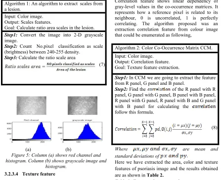
This scale feature in lesions represents white (brightness) colors and notices the variations intimate to lesions, thereby the anticipated procedure appropriates the grouping of image bands to be principal constituent towards Independent Histogram and convert to grayscale images by means of the threshold employed with uppermost threshold values of histograms, every pixel with values among (240-255) has been estimated, see Figure 5. Subsequently, by calculating the number of pixels that have inedsity among rang (240-255), then divided on area of extracted lesion. The procedure is explained subsequently as, to extracted area the scales.Algorithm 1: An algorithm to extract scales from a lesion.
Input: Color image. Output: Scales features.
Goal: Calculate ratio area scales in the lesion. Step1: Convert the image into 2-D grayscale image.
Step2: Count No.pixel classification as scale (brightness) between 240-255 density.
Step3: Calculate the ratio scale area
[image:7.612.81.528.362.727.2]
(a) (b)
Figure 5: Column (a) shows red channel and histogram. Column (b) shows grayscale image and
histogram.
3.2.3.4 Texture feature
Texture examination is a vigorous part in investigation of pattern appreciation. Diversity of methods have been employed in calculating textural resemblance. This anticipated color co-occurrence matrix (CCM) depiction in texture structures for exactly representing the color, spatial requirement of texture at images. Here, color co-occurrence matrix (CCM) has been created on the basis of location and space among the image pixel. Expressive figures have been removed in these co-occurrence matrix, categorizing the depiction of texture as the rudimentary texture pattern is administrated by intervallic incidence in definite color, these co-occurrence in colors during pre-defined comparative locations is sensible degree in attendance of textures and periodicities of pattern. We have employed CCM strategy towards texture explanation in experimentations using 14 numerical features mined from them and correlation feature is employed. Numerous texture features like entropy, energy, contrasts, inverse differences, homogeneities and correlations are mined in co-occurrence matrix in the colour of images.
Correlation feature shows linear dependency of gray-level values in the co-occurrence matrices. It represents how a reference pixel is related to its neighbour, 0 is uncorrelated, 1 is perfectly correlating. The algorithm proposed was an extraction correlation feature from colour image that could be enumerated as following,
Algorithm 2: Color Co-Occurrence Matrix CCM. Input: Color image.
Output: Correlation feature. Goal: Texture feature extraction.
Step1: In CCM we are going to extract the feature from R panel, G panel and B panel.
Step2: Find the of the R panel with R panel, G panel with G panel, B panel with B panel, R panel with G panel, R panel with B and G panel with B panel for calculating the
follow this formula.
Where are mean and
standard deviations of
Here we have extracted the area, color and texture features of psoriasis image and the results obtained are as shown in Table 2.
Table 2: Features the psoriasis lesion. (7)
4208
Feature Psoriasis Area Lesion 0.2-0.9
Mean Red 143.836 - 210.285 Area Scales Ratio 0.0033-0.7750
Correlation -0.0260 - 0.0673
4. SEGMENTATION RESULT
Here, these employed images are gotten
from psoriasis skin disease Image Database towards Image Processing database. They are in
JPG image formats apprehended from RGB design in resolution of 200 x 200 pixel for the color image. The proposed system reads the
color image and then converts them towards
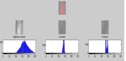
L*a*b color spaces; the L*a*b seems decent assortment of segmentation in psoriasis image. The two constituents (a and b) at L*a*b color spaces are designated for experiments. Then we process segmentation the image by means of K-Means cluster formation procedure for determining diseased parts of healthy part in images towards psoriasis which could be
enumerated Figure 6.
We initially consider K for K-Means as 2 for the segmented images with two indicates offering a good separation of psoriasis from the image, we compare the algorithm k-means with RGB color space directly without conversion to another color space it gives inaccurate and inefficient results as shown in Figure7. Where the segmentation is not clear, this leads to difficult and overlapping features extracted from the segment and lack of knowledge of the lesion region of healthy skin, retail be inaccurate.
Why does the researcher use the K-Means cluster formation procedure in L*a*b color space instead of K-Means algorithm with RGB color space in image segmentation subsequent to the conversion of images at L*a*b color spaces? The researcher can easily distinguish visually the main colours terms of an image that contains only the first two colours which are the colour of the lesion and the second healthy skin colour. We can distinguish the colors from each other. L*a*b* color spaces (otherwise called CIELAB or CIE L*a*b*) empowers us in determining the pictorial contrasts. The L*a*b* shading spaces are gotten in CIE XYZ tristimulus value. L*a*b* spaces contains the brilliance layer 'L*', chromaticity layer 'a*' indicating the color drop alongside the red green
axis, and chromaticity layer 'b*' representing the color drop in blue yellow axis. Every color information has been observed at 'a*' and 'b*' layers. We could module these differences among dual color employing Euclidean distance metric. This feature is not available with RGB color space. We can successfully achieve segmentation for psoriasis disease images with 95% success rate as this percentage is much higher in comparison to when we utilised the method using an RGB color space as shown in
Table 3.
[image:8.612.314.534.279.387.2]Table 3: Performance of algorithm.
Figure 6: The histogram for three channels.
Comparison of accuracy segmentation
results are
amongst RGB, L*a*b color
space
using
the K-Means cluster formation
procedure
and
segmenting manually.
(a) (b) (c)
Figure 7: (a) Segmentation K-Means with L*a*b colour space. (b) Segmentation K-Means with RGB
color space. (c) Segmentation manually.
5. CONCLUSION
We conclude that the proposed method as a means of assembling segmentation process using K-Means algorithm on images Psoriasis is a new style with the L * a * b color spaces, this method is successful in detecting and lesion segmentation psoriasis on all the images in almost the database. We strongly recommend our method for future research conducts, where we utilise another segmentation algorithms which is more sophisticated, for example, genetic algorithm or
C-Accurate
[image:8.612.313.522.440.539.2]4209 means algorithm or perhaps utilise a different colou space, such as HSV or YCbCr to give the most accurate results and better segmentation process on images of diseases skin. In conclusion, the methods introduced by the researcher may not only move our efforts in combating diseases forward but more importantly, could save many precious lives in the process.
REFRENCES:
[1] Lu, Juan. Objective assessment of psoriasis treatment through skin images. Diss. University of Melbourne, Department of Computing and Information Systems, 2014.
[2] Taur, Jinshiuh S., et al. "Segmentation of
psoriasis vulgaris images using
multiresolution-based orthogonal subspace techniques." IEEE Transactions on Systems,
Man, and Cybernetics, Part B
(Cybernetics) 36.2 (2006): 390-402.
[3] Lu, Juan, et al. "Automatic segmentation of scaling in 2-d psoriasis skin images." IEEE transactions on medical imaging 32.4 (2013): 719-730.
[4] Shrivastava, Vimal K., et al. "First review on psoriasis severity risk stratification: An
engineering perspective." Computers in
biology and medicine63 (2015): 52-63.
[5] Taur, Jin-Shiuh. "Neuro-fuzzy approach to the segmentation of psoriasis images." The Journal of VLSI Signal Processing 35.1 (2003): 19-27.
[6] Vij, Sugandhi, Sandeep Sharma, and Chetan
Marwaha. "Performance evaluation of color image segmentation using K means clustering
and watershed technique." Computing,
Communications and Networking Technologies (ICCCNT), 2013 Fourth International Conference on. IEEE, 2013.
[7] Mohammed, Mazin Abed, et al. "Automatic segmentation and automatic seed point selection of nasopharyngeal carcinoma from microscopy images using region growing based
approach." Journal of Computational
Science 20 (2017): 61-69.
[8] Chen, Chang Wen, Jiebo Luo, and Kevin J. Parker. "Image segmentation via adaptive K-mean clustering and knowledge-based morphological operations with biomedical applications." IEEE transactions on image processing 7.12 (1998): 1673-1683.
[9] Tsai, Chwei-Shyong, and Chin-Chen Chang.
"An improvement to image segment based on human visual system for object-based
coding." Fundamenta Informaticae 58.2
(2003): 167-178.
[10]Akhtar, Nadeem, Nishi Agarwal, and Armita Burjwal. "K-mean algorithm for Image Segmentation using Neutrosophy." Advances
in Computing, Communications and
Informatics (ICACCI, 2014 International Conference on. IEEE, 2014.
[11]Samundeeswari, E. S., P. K. Saranya, and R.
Manavalan. "Segmentation of Breast
Ultrasound image using Regularized K-Means (ReKM) clustering."Wireless Communications,
Signal Processing and Networking
(WiSPNET), International Conference on. IEEE, 2016.
[12]Ivanovici, Mihai, Noël Richard, and Dietrich Paulus. "Color image segmentation." advanced color image processing and analysis. Springer New York, 2013. 219-277.
[13]Baldevbhai, Patel Janakkumar, and R. S.
Anand. "Color image segmentation for medical images using L* a* b* color space." IOSR Journal of Electronics and Communication Engineering 1.2 (2012): 24-45.
[14]Baldevbhai, Patel Janakkumar, and R. S.
Anand. "Color image segmentation for medical images using L* a* b* color space." IOSR Journal of Electronics and Communication Engineering 1.2 (2012): 24-45.
[15]Xu, Lang, et al. "Segmentation of skin cancer images." Image and Vision Computing 17.1 (1999): 65-74.
[16]Ercal, Fikret, et al. "Neural network diagnosis of malignant melanoma from color images." IEEE Transactions on biomedical engineering 41.9 (1994): 837-845.
[17] Khan, Shehroz S., and Amir Ahmad. "Cluster center initialization algorithm for K-means clustering." Pattern recognition letters 25.11 (2004): 1293-1302.
[18]Mohammed, Mazin Abed, et al. "Artificial neural networks for automatic segmentation
and identification of nasopharyngeal
carcinoma." Journal of Computational
Science (2017).
[19] Omran, Mahamed GH, Andries P.
Engelbrecht, and Ayed Salman. "An overview of clustering methods." Intelligent Data Analysis 11.6 (2007): 583-605.
[20] Lee, Tim, et al.
"Dullrazor®: A software approach to hair removal from images."Computers in biology and medicine 27.6 (1997): 533-543.
[21] Celebi, M. Emre, et al. "A methodological
4210
images." Computerized Medical Imaging and Graphics 31.6 (2007): 362-373.
[22]Pereira, Silvio M., et al. "Classification of color images of dermatological ulcers." IEEE
journal of biomedical and health
informatics 17.1 (2013): 136-142.
[23] Chang, Wen-Yu, et al. "Computer-aided
diagnosis of skin lesions using conventional digital photography: a reliability and feasibility study." PloS one8.11 (2013): e76212.
[24]Maglogiannis, Ilias G., and Elias P.
Zafiropoulos. "Characterization of digital medical images utilizing support vector machines." BMC Medical Informatics and Decision Making 4.1 (2004): 4.
[25]Shrivastava, Vimal K., et al. "First review on psoriasis severity risk stratification: An
engineering perspective." Computers in
biology and medicine63 (2015): 52-63.