Psychology Honors Theses Department of Psychology
5-7-2016
Gender Influence on Cognitive and Structural
Differences in People with Schizophrenia
Nadia A. Quyyum
Georgia State UniversityFollow this and additional works at:https://scholarworks.gsu.edu/psych_hontheses
This Thesis is brought to you for free and open access by the Department of Psychology at ScholarWorks @ Georgia State University. It has been accepted for inclusion in Psychology Honors Theses by an authorized administrator of ScholarWorks @ Georgia State University. For more information, please [email protected].
Recommended Citation
Quyyum, Nadia A., "Gender Influence on Cognitive and Structural Differences in People with Schizophrenia." Thesis, Georgia State University, 2016.
WITH SCHIZOPHRENIA
A Thesis
Submitted in Partial Fulfillment of the Requirements for Graduation with Undergraduate Research Honors
In the Honors College Georgia State University
2016
by
Nadia Quyyum
Committee:
Dr. Jessica Ann Turner, Thesis Advisor
Sarah Cook, Honors College Associate Dean
WITH SCHIZOPHRENIA by
NADIA QUYYUM
Under the Direction of Jessica Ann Turner, PhD
ABSTRACT
Previous literature has explored sex differences to explain differences in cognition and behavior. If there are sex differences in the brain, how would these differences alter our perception of treatment and diagnosis in a clinical population, such as people with
schizophrenia? In our study, we wanted to find out whether there is an effect of sex on cortical thickness (CT) in the parietal lobule and cognitive measures of verbal memory, verbal learning, attention, spatial reasoning, and working memory in healthy controls (HC) and people with schizophrenia (SZ). We additionally explored relationships between cognition and parietal lobule CT. RESULTS: There is no effect of sex on cognition and CT in the parietal lobule, but we found differences in correlations between CT and cognition in each sex and diagnosis group. DISCUSSION: Further research is necessary to discover whether clinicians need to consider gender in the treatment of people with schizophrenia.
WITH SCHIZOPHRENIA
by
NADIA QUYYUM
A Thesis Submitted in Partial Fulfillment of the Requirements for Graduation with Undergraduate Research Honors
In the Honors College Georgia State University
Copyright by Nadia Quyyum
WITH SCHIZOPHRENIA
by
NADIA QUYYUM
Thesis Advisor: Jessica Ann Turner
Electronic Version Approved: Date:
Honors College
DEDICATION
ACKNOWLEDGEMENTS
TABLES OF CONTENTS
DEDICATION ... iv
ACKNOWLEDGEMENTS ... v
LIST OF TABLES ... vii
LIST OF FIGURES ... viii
INTRODUCTION ... 1
General Sex Differences in Cognition and Brain Structure ... 1
Sex Differences in the Parietal Lobule ... 2
Sex Differences in Cognition and Brain Structure in Schizophrenia ... 2
Purpose of Our Study ... 3
METHOD ... 4
RESULTS ... 5
The Effect of Gender & Diagnosis on Cognition ... 5
The Effect of Gender & Diagnosis on Cortical Thickness ... 12
Partial Correlation between Cognition and Cortical Thickness ... 17
DISCUSSION ... 20
LIST OF TABLES
Table 1. Demographics ... 4 Table 2. MANOVA Results on Effect of Diagnosis on Cognitive Measures ... 6 Table 3. MANCOVA Results on the Effect of Diagnosis on Parietal Lobule Cortical Thickness ... 12 Table 4. Partial correlations of cognitive measures with parietal lobule cortical thickness
measures across gender when controlling for age ... 18 Table 5. Partial correlations of cognitive measures with parietal lobule cortical thickness
LIST OF FIGURES
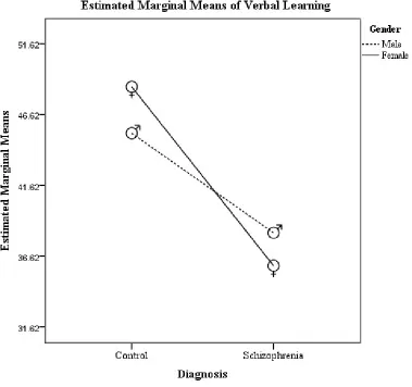
Figure 1. MANOVA result of Verbal Learning. ... 7
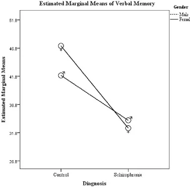
Figure 2. MANOVA result of Verbal Memory. ... 8
Figure 3. MANOVA result of Attention. ... 9
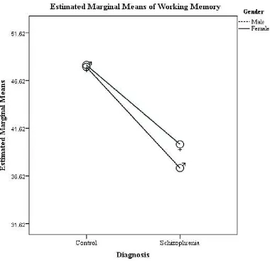
Figure 4. MANOVA result of Working Memory. ... 10
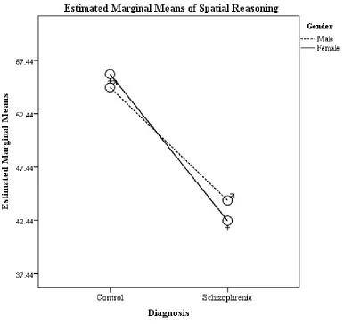
Figure 5. MANOVA result of Spatial Reasoning ... 11
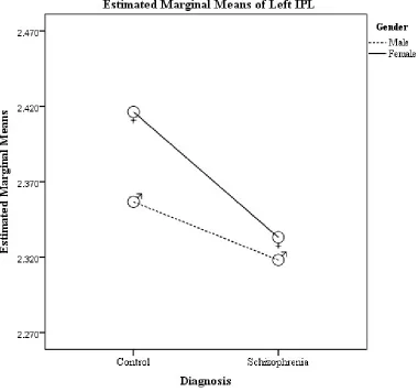
Figure 6. MANCOVA result of Left IPL. ... 13
Figure 7. MANCOVA result of Right IPL. ... 14
Figure 8. MANCOVA result of Left SPL. ... 15
Figure 9. MANCOVA result of Left SMG. ... 16
Figure 10. MANCOVA result of Right SMG. ... 17
INTRODUCTION
Ever since John Gray (1992) proposed that men are from Mars and women are from Venus, researchers have conducted studies to find out if this notion was certainly the case. Over the last 50 years, researchers wanted to explore sex differences in the brain to explain our differences in cognition and behavior. There have been numerous studies conducted on
differences in brain size and tissue, as well as studies that looked into differences in cognition. It is important to investigate these differences in clinical populations, such as people with
schizophrenia, to see whether each sex must be provided with different types of treatment due to the possible differences in the expression of the illness and the severity of the illness between male and female patients.
General Sex Differences in Cognition and Brain Structure
Studies on sex differences as early as the 19th century discovered that male brains were
bigger than female brains. Findings on brain size have remained consistent, further supported by magnetic resonance imaging (MRI) studies on brain tissue. MRI studies have revealed not only sex differences in brain size, but also in cortical thickness, which refers to the combined
thickness of the outer layers composed of gray matter in the cerebral cortex. Compared to men, women have greater cortical thickness (Im et al, 2006; Mutlu et al., 2013; Plessen, Hugdahl, Bansal, Hao, & Peterson, 2014; Sowell et al., 2007). Recent studies have found that cortical thickness is an important measurement of brain structure because the thickness of the cortex might indicate a level of intelligence, due to its relationship with gray matter volume (Choi et al., 2008; Karama et al, 2009; Narr et al, 2007). If cortical thickness positively correlates with
Many studies have explored sex differences in cognitive abilities. Researchers have utilized a wide range of cognitive measures, such as those that assess memory, attention, and verbal learning. Women generally score higher on cognitive measures than men, and they often excel at measures of verbal reasoning. On the other hand, men almost always have the upper hand on spatial reasoning tasks. Researchers have asked and wondered what was the explanation for these cognitive differences. One common theory is that sex differences in brain structure, particularly the parietal lobule, may explain the differences found in cognition.
Sex Differences in the Parietal Lobule
The parietal lobe is a key area for research on sex differences in the brain. As mentioned previously, many studies have found that women’s cortical thickness is greater than men’s in this region. Functional MRI studies have indicated that the parietal lobule activates when performing spatial reasoning tasks (Culham & Kanwisher, 2000; Save & Poucet, 2000; Seurinck,
Vingerhoets, de Lange, 2004). For example, Koscik et al. (2009) studied whether there was a relationship between the scores on the Mental Rotations Test (MRT) and the parietal lobe, and whether there were sex differences in that region. Their results revealed that not only did men score better on MRT than women, but men also had greater surface area in the parietal lobe than women; they concluded that this finding may account for the disadvantage that women have in spatial reasoning tasks. This sexual dimorphism confirms the need for further research on sex differences in that region.
Sex Differences in Cognition and Brain Structure in Schizophrenia
Do these sex differences apply to a clinical population, such as people with
grandeur while additionally displaying negative symptoms of blank affect and social withdrawal. Regarding its effect on the brain, due to the severity of the illness, people with schizophrenia have less gray matter, thinner cortices, and less brain volume than the healthy population (Hulshoff et al., 2002; Kuperberg et al., 2003; Schultz et al., 2010; van Haren et al., 2011). Regarding cognition, people with schizophrenia tend to perform poorly compared with their healthy counterparts, especially in areas of attention, working memory, and executive functioning (Bilders et al, 2002; Caspi et al, 2003; Cornblatt et al, 1985; Gold et al, 1997; McGurk et al, 2004; Pantelis et al, 1999).
Sex differences in people with schizophrenia tend to be similar to that of the healthy population. Women with schizophrenia tend to perform better on cognitive tests than men with schizophrenia (Han et al., 2012; Ittig et al., 2015; Longenecker, Dickinson, Weinberger, & Elvevag, 2010; Roesch-Ely et al., 2009; Torniainen et al., 2011; Vaskinn et al., 2011). This could be due to sexual dimorphisms found in schizophrenia literature regarding the age of onset, which occurs earlier in men, and the effects of estrogen as a protective factor for women (Aloysi, Van Dyk, & Sano, 2006; Heringa, Begemann, Goverde, & Sommer, 2015; Kulkarni et al., 2008; Salokangas, 1993; van der Leeuw et al., 2013). In terms of structure, Frederikse et al. (2000) found that when investigating sex differences between a healthy population and people with schizophrenia, there is a clear distinction found in gray matter volume in the parietal lobe between healthy men and men with schizophrenia; as for the women, there were surprisingly no significant differences between healthy women and women with schizophrenia.
Purpose of Our Study
than generalizing the treatment plan for all people with schizophrenia. In our study, we examined cortical thickness in relation to cognition in people with schizophrenia to see whether our results remain consistent with the results of previous literature, as well as build off of Frederikse et al.’s (2000) findings by focusing on differences found particularly in the parietal lobe. We
additionally wanted to see whether there was an effect of sex on cortical thickness and cognition, or if the interaction between sex and diagnosis would have an effect on both measures.
METHOD
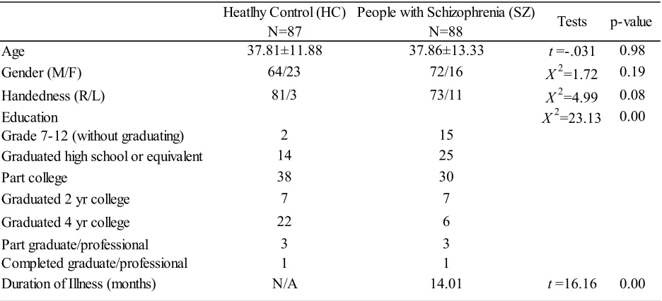
[image:15.612.73.541.416.629.2]Participants. The sample consisted of 175 participants, with 87 identifying as healthy controls (HC) and 88 people with schizophrenia or schizoaffective disorder (SZ). There were a total of 136 men and 39 women in the dataset. Table 1 summarizes the age, gender, handedness, education status, and duration of illness of our participants.
Table 1. Demographics
MRI Acquisition. We obtained the Centers of Biomedical Research Excellence (COBRE) dataset from the Mind Research Network (MRN) in Albuquerque, New Mexico. At MRN, a Siemens TIM Trio 3T scanner was used to collect MRI data using a 12-channel head coil.
Age
Gender (M/F) Handedness (R/L) Education
Grade 7-12 (without graduating) Graduated high school or equivalent Part college
Graduated 2 yr college Graduated 4 yr college Part graduate/professional Completed graduate/professional Duration of Illness (months)
14 73/11 15 37.81±11.88 64/23 81/3 2 25 72/16 3 1 38 7 22 3 1 30 7 6 Heatlhy Control (HC)
N=87 People with Schizophrenia (SZ) N=88 Tests p-value 0.98 0.19 0.08 0.00
t=-.031
X2=1.72
X2=4.99
X2=23.13 37.86±13.33
Cognitive Battery. The COBRE dataset included an extensive cognitive battery. For our study, we obtained the scores of our participants from the Hopkins Verbal Learning Test
Immediate (HVLT-I) & Delay (HVLT-D) to assess both verbal learning and verbal memory; the Continuous Performance Test—Identical Pairs Version (CPT-IP) to assess attention; the
Neuropsychological Assessment Battery (NAB) Mazes Test to assess spatial reasoning; and the Wechsler Memory Scale—Third Edition (WMS-III) to assess working memory.
Cortical Thickness Measures. We utilized Freesurfer, a brain imaging software, to extract cortical thickness measures of the various regions of the parietal lobule: inferior parietal lobule (IPL), superior parietal lobule (SPL), supramarginal gyrus (SMG), precuneus (PCUN), and postcentral gyrus (PoCG).
Statistical Analyses. We utilized SPSS 21 to conduct analyses among cortical thickness measures and cognitive battery scores. We analyzed the program’s marked outliers of cortical thickness by looking at the MRI scans of the participants to see whether the cortical thickness was true to size. A multivariate analysis of covariance (MANCOVA) was conducted to test whether there was an effect of gender or a three-way interaction between gender and diagnosis on cortical thickness in the parietal lobule region while using age as a covariate. A multivariate analysis of variance (MANOVA) to see if there is an effect of gender on cognition. We also conducted partial correlations between each cognitive measure and cortical thickness in all regions of the parietal lobule while co-varying for age across diagnosis and gender.
RESULTS
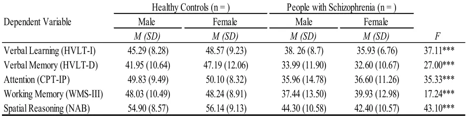
The Effect of Gender & Diagnosis on Cognition
diagnosis on cognition. However, diagnosis type had a statistically significant effect on all cognitive measures. Figures 1-5 provide a graph of each result.
Table 2. MANOVA Results on Effect of Diagnosis on Cognitive Measures
Dependent Variable Male Female Male Female
M (SD) M (SD) M (SD) M (SD) F
Verbal Learning (HVLT-I) 45.29 (8.28) 48.57 (9.23) 38. 26 (8.7) 35.93 (6.76) 37.11*** Verbal Memory (HVLT-D) 41.95 (10.64) 47.19 (12.06) 33.99 (11.90) 32.60 (10.67) 27.00*** Attention (CPT-IP) 49.83 (9.49) 50.10 (8.32) 35.96 (14.78) 36.60 (11.26) 35.33*** Working Memory (WMS-III) 48.03 (10.49) 48.24 (8.91) 37.44 (13.50) 39.93 (12.98) 17.24*** Spatial Reasoning (NAB) 54.90 (8.57) 56.14 (9.13) 44.30 (10.58) 42.40 (10.57) 43.10*** Note: M: mean; SD: standard deviation; F: MANCOVA result; *p < .05; **p < .01; ***p < .001
The Effect of Gender & Diagnosis on Cortical Thickness
[image:23.612.73.540.294.546.2]Results of the MANCOVA are summarized in Table 3. The MANCOVA revealed that there was no significant effect of gender, neither an effect of gender x diagnosis on cognition. Like the results in cognition, diagnosis type had a statistically significant effect on the following regions’ cortical thickness: left IPL (F (1, 170) = 7.47, p = .007; right IPL, (F (1, 170) = 9.79, p = .002; left SPL, (F (1, 170) = 4.07, p = .045; left SMG, (F (1, 170) = 14.35, p = .000; and right SMG, (F (1, 170) = 9.94; p = .002. Figures 6-10 provided a graph for each significant result.
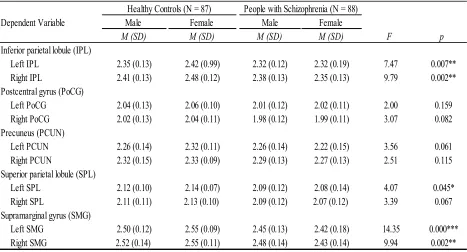
Table 3. MANCOVA Results on the Effect of Diagnosis on Parietal Lobule Cortical Thickness
Dependent Variable Male Female Male Female
M (SD) M (SD) M (SD) M (SD) F p
Inferior parietal lobule (IPL)
Left IPL 2.35 (0.13) 2.42 (0.99) 2.32 (0.12) 2.32 (0.19) 7.47 0.007**
Right IPL 2.41 (0.13) 2.48 (0.12) 2.38 (0.13) 2.35 (0.13) 9.79 0.002**
Postcentral gyrus (PoCG)
Left PoCG 2.04 (0.13) 2.06 (0.10) 2.01 (0.12) 2.02 (0.11) 2.00 0.159
Right PoCG 2.02 (0.13) 2.04 (0.11) 1.98 (0.12) 1.99 (0.11) 3.07 0.082
Precuneus (PCUN)
Left PCUN 2.26 (0.14) 2.32 (0.11) 2.26 (0.14) 2.22 (0.15) 3.56 0.061
Right PCUN 2.32 (0.15) 2.33 (0.09) 2.29 (0.13) 2.27 (0.13) 2.51 0.115
Superior parietal lobule (SPL)
Left SPL 2.12 (0.10) 2.14 (0.07) 2.09 (0.12) 2.08 (0.14) 4.07 0.045*
Right SPL 2.11 (0.11) 2.13 (0.10) 2.09 (0.12) 2.07 (0.12) 3.39 0.067
Supramarginal gyrus (SMG)
Left SMG 2.50 (0.12) 2.55 (0.09) 2.45 (0.13) 2.42 (0.18) 14.35 0.000***
Right SMG 2.52 (0.14) 2.55 (0.11) 2.48 (0.14) 2.43 (0.14) 9.94 0.002**
Healthy Controls (N = 87) People with Schizophrenia (N = 88)
Figure 10. MANCOVA result of Right SMG. There was a significant effect of diagnosis on the
right supramarginal gyrus, (F (1, 170) = 9.94; p = .002.
Partial Correlation between Cognition and Cortical Thickness
A Pearson product-moment correlation coefficient was computed to assess the
relationship between parietal lobule CT and cognitive measures. The results are summarized in Table 4. Across gender, a scatterplot depicts two positive correlations shared by men and women: the right PCUN and CPT-IP, and the left SMG and CPT-IP. In women, various
correlations were found between cortical thickness and verbal memory, and only one correlation found between the right IPL and verbal learning. However, after testing for a Bonferroni
close to significant, positive correlation between the right IPL and HVLT-D, r = 0.41, n = 39, p = .002. Greater cortical thickness in the right IPL was associated with a higher HVLT-D score in women. Figure 11 depicts correlations between female controls and female patients in the relationship between the right IPL and HVLT-D score.
[image:29.612.74.540.378.611.2]Across diagnosis, no significant correlations were found in controls; however, two significant, positive correlations were found in patients between the right precuneus and CPT-IP, r = .26, n = 85, p = .019 and between the left supramarginal gyrus and CPT-IP, r = .22, n = 85, p = .046. Neither correlation passed the Bonferroni correction. Summary of the correlations can be referred to in Table 5.
Table 4. Partial correlations of cognitive measures with parietal lobule cortical thickness measures across gender when controlling for age
Parietal Lobule Regions HVLT-I HVLT-D CPT-IP WMS-III NAB HVLT-I HVLT-D CPT-IP WMS-III NAB Inferior Parietal Lobule (IPL)
Left IPL 0.04 0.02 0.16 0.07 0.06 0.30 0.41* 0.33 -0.01 0.30
Right IPL 0.13 0.09 0.14 0.05 0.15 0.36* 0.51** 0.41* 0.11 0.38*
Postcentral Gyrus (PoCG)
Left PoCG 0.01 -0.01 0.14 0.09 0.14 -0.05 0.18 0.14 -0.11 0.15
Right PoCG 0.08 0.03 0.12 0.06 0.06 -0.02 0.34* 0.20 -0.12 0.18
Precuneus (PCUN)
Left PCUN 0.09 0.10 0.19* 0.06 0.05 0.14 0.28 0.32 0.02 0.29
Right PCUN 0.15 0.13 0.19* 0.06 0.07 0.08 0.18 0.36* 0.05 0.20
Superior Parietal Lobule (SPL)
Left SPL 0.04 0.01 0.19* 0.00 0.08 0.27 0.43* 0.30 0.04 0.48**
Right SPL 0.04 0.02 0.14 0.01 0.10 0.26 0.37* 0.31 0.10 0.28
Supramarginal Gyrus (SMG)
Left SMG 0.09 0.08 0.19* 0.10 0.12 0.19 0.40* 0.43** 0.07 0.25
Right SMG 0.15 0.09 0.15 0.10 0.18* 0.12 0.31 0.34* 0.03 0.31
Male Female
Table 5. Partial correlations of cognitive measures with parietal lobule cortical thickness measures across diagnosis type when controlling for age
Parietal Lobule Regions HVLT-I HVLT-D CPT-IP WMS-III NAB HVLT-I HVLT-D CPT-IP WMS-III NAB
Inferior Parietal Lobule (IPL)
Left IPL 0.14 0.15 0.08 0.00 -0.01 -0.05 0.01 0.14 -0.02 0.06
Right IPL 0.09 0.10 0.06 -0.01 0.07 0.14 0.19 0.17 -0.01 0.18
Postcentral Gyrus (PoCG)
Left PoCG 0.01 -0.01 0.01 -0.06 -0.02 -0.14 -0.02 0.15 0.06 0.17
Right PoCG 0.01 -0.03 0.00 -0.10 0.06 -0.01 0.11 0.13 0.03 -0.01
Precuneus (PCUN)
Left PCUN 0.02 -0.01 0.17 -0.01 -0.06 0.11 0.21 0.22 0.03 0.15
Right PCUN -0.02 -0.03 0.03 -0.07 -0.18 0.16 0.20 .26* 0.06 0.20
Superior Parietal Lobule (SPL)
Left SPL 0.05 0.04 0.08 -0.09 -0.09 0.03 0.08 0.19 -0.04 0.15
Right SPL -0.06 -0.10 0.01 -0.12 -0.12 0.13 0.17 0.19 0.03 0.15
Supramarginal Gyrus (SMG)
Left SMG -0.02 0.04 -0.08 -0.08 -0.08 0.01 0.08 0.22* 0.02 0.12
Right SMG 0.08 0.03 -0.06 -0.07 -0.07 0.05 0.09 0.19 0.07 0.15
Controls Patients
Figure 11. Partial Correlation between the Right IPL and Verbal Memory in Female Participants. After applying the Bonferroni correction, there was a close to significant, positive correlation between the right inferior parietal lobule and verbal memory, r = 0.41, n = 39, p = .002. Across diagnosis type, there were no significant correlations in female controls, r = 0.39, n = 23, p = .070, or female patients, r = .04, n = 16, p = 0.873.
DISCUSSION
al., 2010; Gogos et al., 2010). The effect of diagnosis on cognition and structure is consistent with previous literature regarding the differences in cognition and structure between healthy population and schizophrenic population, with patients showing worse cognitive performance and thinner cortices than healthy controls. In terms of correlations, there was a close to significant correlation between the right inferior parietal lobule and verbal memory in female participants, which makes us question what is significant about the inferior parietal lobule and how it could be involved in verbal memory.
The Inferior Parietal Lobule
The inferior parietal lobule, composed of the angular gyrus and supramarginal gyrus, was originally thought to be involved in motor planning (Cohen et al., 1994; Snyder et al., 1998). Recently, because Freesurfer software separately distringuishes between the inferior parietal lobule and the supramarginal gyrus, we were mostly working with the angular gyrus within the inferior parietal lobule (Desikan et al., 2005). The angular gyrus is part of the Default Mode Network, which is activated when an individual’s mind is simply wandering and not focused on a task (Buckner, Andrews-Hanna, & Schacter, 2008). The angular gyrus has additionally been noted in studies of verbal working memroy, episodic and semantic memory, creativity, and self-awareness (Bellana, Liu, Anderson, Moscovitch, & Grady, 2015; Ciaramelli et al, 2008; Jung et al, 2010; Park, Kirk, & Waldie, 2015; Sui, Chechlacz, & Humphreys, 2012). The inferior parietal lobule is additionally one of the many prominent regions in verbal memory circuitry in women (Abbs, Liang, Makri, Tsuang, & Seidman, 2011).
Limitations
extra female participants in our study could have the potential to alter some of their results we have received in this study. Additionally, the majority of the patients in our sample were, if not all, medicated before completing the cognitive tests, which may have allowed them to perform accordingly than if they were not medicated. Unlike Frederikse et al., we solely focused on cortical thickness rather than measuring differences in gray matter volume, surface area, and asymmetry. Although cortical thickness and gray matter volume are related, they are certainly not the same thing, which may account for why our findings were not similar to that of
Frederikse et al. Perhaps we could include these measures in the future to see whether there are sex differences across those particular domains. We focused specially on the parietal lobule region, which may contribute to only some cognitive processes in the measures that we chose for our study. Considering other regions, such as the frontal or temporal regions, may contribute to differences in correlations between structure and cognition. Additional cognitive measures are also worth questioning to see whether there are other forms of cognition in which we can mark gender differences.
Conclusions
REFERENCES
Abbs, B., Liang, L., Makris, N., Tsuang, M., Seidman, L. J., & Goldstein, J. M. (2011).
Covariance modeling of MRI brain volumes in memory circuitry in schizophrenia: Sex differences are critical. NeuroImage, 56(4), 1865-74.
Albus, M., Hubmann, W., Mohr, F., Scherer, J., Sobizack, N., Franz, U.…Wahlheim, C. (1997). Are there gender differences in neuropsychology performance in patients with first-episode schizophrenia? Schizophrenia Research, 28(1), 39-50.
Aloysi, A., Van Dyk, K., & Sano, M. (2006). Women’s cognitive and affective health and neuropsychiatry. Mt Sinai J Med, 73(7), 967-75.
Baldo, J.V., & Dronkers, N.F. (2006). The role of inferior parietal and inferior frontal cortex in working memory. Neuropsychology, 20(5), 529-538.
Bellana, B., Zhongxiu, L., Anderson, J.A.E., Moscovitch, M., & Gray, C.L. Laterality effects in functional connectivity of the angular gyrus during rest and episodic retrieval.
Neuropsychologia, 80, 24-34.
Bilder, R.M., Goldman, R.S., Volavka, J., Czobor, P., Hoptman, M., Shietman, B.…Lieberman, J.A. (2002). Neurocognitive effects of clozapine, olanzapine, risperidone, and haloperidol in patients with chronic schizophrenia or schizoaffective disorder. Am J Psychiatry, 159(6), 1018-1028.
Buckner, R.L., Andrews-Hanna, J.R., & Schacter, D.L. (2008). The brain’s default network: Anatomy, function, and relevance to disease. Annals of the New Yok Academy of Sciences, 1124(1), 1-38.
Caspi, A., Reichenberg, A., Weiser, M., Rabinowitz, J., Kaplan, Z., Knobler, H….Davidson, M. (2003). Cognitive performance ins schizophrenia patients assessed before and following the first psychotic episode. Schizophrenia Research, 6(2-3), 87-94.
Choi, Y.Y., Shamosh, N.A., Cho, S.H., DeYoung, C.G., Lee, M.J., Lee, J.M….Lee, K.H. (2008). Multiple bases of human intelligence revealed by cortical thickness and neural activation. J Neurosci, 28(41), 10323-10329.
Ciaramelli, C.L., Grady, M., & Moscovitch, M. Top-down and bottom-up attention to memory: a hypothesis (AtoM) on the role of the posterior parietal cortex in memory retrieval.
Neuropsychologia, 46(7), 1828-1851.
Cohen, L., Dehaene, S., Chochon, F., Lehericy, S., & Naccache, L. (1994). Language and calculation within the parietal lobe: A combined cognitive, anatomical and fMRI study. Neuropsychologia, 38(10), 1426-1440.
Cornblatt, B.A., Lenzenweger, M.F., Dworkin, R.H., & Erlenmeyer-Kimling, L. (1985). Positive and negative schizophrenic symptoms, attention, and information processing.
Schizophrenia Bulletin, 11(3), 397-408.
Culham, J.C., & Kanwisher, N.G. (2001). Neuroimaging of cognitive functions in human parietal cortex. Curr Opin Nerobiol, 11(2), 157-163.
Frederikse, M., Lu, A., Aylward, E., Barta, P., Sharma, T., & Pearlson, G. (2000). Sex
Gogos, A., Joshua, N., & Rossell, S.L. (2010). Use of the Repeatable Battery for the Assessment of Neuropsychology Status (RBANS) to investigate group and gender differences in schizophrenia and bipolar disorder. Aust N Z Psychiatry, 44(3), 220-229.
Gold, J.M., Carpenter, C., Randolph, C., Goldberg, T.E., & Weinberger, D.R. (1997). Auditory working memory and Wisconsin Card Sorting Test performance in schizophrenia. Arch Gen Psychiatry, 54(2), 159-165.
Gray, J. (1992). Men are from Mars, women are from Venus: A practical guide for improving communication and getting what you want in your relationships. New York, NY: HarperCollins.
Han, M., Huang, X.F., Chen da, C., Xiu, M.H., Hui, L., Liu, H….Zhang, X.Y. (2012). Gender differences in cognitive function of patients with chronic schizophrenia. Prog
Neuropsychopharmacol Biol Psychiatry, 39(2), 358-363.
Heringa, S.M., Begemann, M.J., Goverde, A.J., & Sommer, I.E. (2015). Sex hormones and oxytocin augmentation strategies in schizophrenia: A quantitative review. Schizophrenia Research, 168(3), 603-613.
Hulshoff Pol, H.E., Schnack, H.G., Bertens, M.G., van Haren, N.E., van der Tweel, I., Staal, W.F….Kahn, R.S. (2002). Volume changes in gray matter in patients with schizophrenia. Am J Psychiatry, 159(2), 244-250.
Ittig, S., Studerus, E., Papmeyer, M., Uttinger, M., Koranyi, S., Ramyead, A., & Riecher-Rossler, A. (2015). Sex differences in cognitive functioning in at-risk mental state for psychosis, first episode psychosis and healthy control subjects. Eur Psychiatry, 30(2), 242-250. Jung, R.E., Segall, J.M., Jeremy Bockholt, H., Flores, R.A., Smith, S.M., Chavez, R.S., & Haier,
R.J. (2010). Neuroanatomy of creativity. Hum Brain Mapp, 31(3), 398-409.
Karama, S., Colom, R., Johnson, W., Deary, I.J., Haier, R., Waber, D.P….Evans, A.C. (2009). Cortical thickness correlates of specific cognitive performance accounted for by the general factor of intelligence in healthy children aged 6 to 10. Neuroimage, 55(4), 1443-1453.
Koscik, T., O’Leary, D., Moser, D.J., Andreasen, N.C., & Nopoulos, P. (2009). Sex differences in partial lobe morphology: Relationship to mental rotation performance. Brain Cogn, 69(3), 451-459.
Kubota, M., van Haren, N.E., Haijma, S.V., Schnack, H.G., Cahn, W., Hulshoff Pol, H.E., & Kahn, R.S. (2015). Association of IQ change and progressive brain changes in patients with schizophrenia. JAMA Psychiatry, 72(8), 803-812.
Kulkarni, J., de Castella, A., Fitzgerald, P.B., Gurvich, C.T., Bailey, M., Bartholomeusz, C., & Burger, H. (2008). Estrogen in severe mental illness: a potential new treatment approach. Arch Gen Psychiatry, 65(8), 955-960.
Longenecker, J., Dickinson, D., Weinberger, D.R., & Elvevag, B. (2010). Cognitive differences between men and women: A comparison of patients with schizophrenia and healthy volunteers. Schizophrenia Research, 120(1-3), 234-235.
McGurk, S.R., Coleman, T., Harvey, P.D., Reichenberg, A., White, L., Friedman, J….Davis, K.L. (2004). Working memory performance in poor outcome schizophrenia: Relationship to age and executive functioning. J Clin Exp Neuropsychol, 26(2), 153-160.
Mutlu, A.K., Schneider, M., Debbane, M., Badoud, D., Eliez, S., & Schaer, M. (2013). Sex differences in thickness, and folding developments throughout the cortex. Neuroimage, 82, 200-207.
Narr, K.L., Woods, R.P., Thompson, P., Szeszko, P., Robinson, D., Dimtcheva, T….Bilder, R.M. (2007). Relationships between IQ and regional cortical gray matter thickness in healthy adults. Cereb Cortex, 17(9), 2163-2171.
Pantelis, C., Barber, F.Z., Barnes, T.R., Nelson, H.E., Owen, A.M., & Robbins, T.W. (1999). Comparison of set-shifting ability in patients with chronic schizophrenia and frontal lobe damage. Schizophrenia Research, 37(3), 251-270.
Park, H.R., Kirk, I.J., & Waldie, K.E. (2015). Neural correlates of creative thinking and schizotypy. Neuropsychologia, 73, 94-107.
Plessen, Hugdahl, Bansal, Hao, & Peterson (2014). Sex, age, and cognitive correlates of asymmetries in thickness of the cortical mantle across the life span. J Neurosci, 34(18), 6294-6302.
Roesch-Ely, D., Hornberger, E., Weiland, S., Hornstein, C., Parzer, P., Thomas, C., & Weisbrod, M. (2009). Do sex differences affect prefrontal cortex associated cognition in
Salokangas, R.K. (1993). First-contact rate for schizophrenia in community psychiatric care. Consideration of the oestrogen hypothesis. Eur Arch Psychiatry Clin Neurosci, 242(6), 337-346.
Save, E. & Poucet, B. (2000). Hippocampal-parietal cortical interactions in spatial cognition. Hippocampus, 10(4), 491-499.
Schultz, C.C., Koch, K., Wagner, G., Roebel, M., Nenadic, I., Schachtzabel, C….Schlosser, R.G. (2010) Complex pattern of cortical thinning in schizophrenia: Results from an automated surface based analysis of cortical thickness. Psychiatry Research: Neuroimaging, 182, 134-140.
Seurinck, R., Vingerhoets, G., de Lange, F.P., & Achten, E. (2004). Does egocentric mental rotation elicit sex differences? Neuroimage, 23(4), 1440-1449.
Sowell, E.R., Peterson, B.S., Kan, E., Woods, R.P., Yoshii, J., Bansal, R….Toga, A.W. (2007). Sex differences in cortical thickness mapped in 176 healthy individuals between 7 and 87 years of age. Cereb Cortex, 17(7), 1550-1560.
Snyder, L.H., Batista, A.P., & Andersen, R.A. (1998). Change in motor plan, without a change in the spatial locus of attention, modulates activity in posterior parietal cortex. J
Neurophysiol, 79(5), 2814-2819.
Sui, J., Chechlacz, M., & Humphreys, G.W. Dividing the self: Distinct neural substrates of task-based and automatic self-prioritization after brain damage. Cognition, 122(2), 150-162. Torniainen, M., Suvisaari, J., Partonen, A.E., Castaneda, A.E., Kuha, A., Perala,
Vaskinn, A., Sundet, K., Simonsen, C., Hellvin, T., Melle, I., & Andreassen, O.A. (2011). Sex differences in neuropsychological performance and social functioning in schizophrenia and bipolar disorder. Neuropsychology, 25(4), 499-510.
van der Leeuw, C., Habets, P., Gronenschild, E., Domen, P., Michielse, S., van Kroonenburgh, M….Marcelis, M. (2013). Testing the estrogen hypothesis of schizophrenia: Associations between cumulative estrogen exposure and cerebral structural measures. Schizophrenia Research, 150(1), 114-120.