orthotic therapy and physical rehabilitation
for chronic plantar fasciitis: a case study
Ivano A Costa,
BSc (Hons), BEd, DC, FCCRS(c)*Anita Dyson,
BSc (Hons), DC, FCCRS(c)**A 15-year-old female soccer player presented with chronic plantar fasciitis. She was treated with acetic acid iontophoresis and a combination of rehabilitation protocols, ultrasound, athletic taping, custom orthotics and soft tissue therapies with symptom resolution and return to full activities within a period of 6 weeks. She reported no significant return of symptoms post follow-up at 2 months. Acetic acid iontophoresis has shown promising results and further studies should be considered to determine clinical effectiveness. The combination of acetic acid iontophoresis with
conservative treatments may promote recovery within a shorter duration compared to the use of one-method treatment approaches.
(JCCA 2007; 51(3):166–174)
k e y wo r d s: acetic acid iontophoresis, plantar fasciitis, heel pain, custom orthotics and foot physical
rehabilitation.
Cas d’une joueuse de soccer de 15 ans présentant une fasciite plantaire chronique. Elle a été traitée avec une iontophorèse d’acide acétique et une combinaison de protocoles rééducatifs, d’ultrasons, de bandages, d’orthèse adaptée et de traitements des tissus mous avec résolution des symptômes et retour aux activités dans une période de six semaines. Elle n’a signalé aucun retour significatif des symptômes après un suivi de deux mois. L’iontophorèse d’acide acétique a présenté des résultats prometteurs et des études supplémentaires doivent être envisagées en vue de déterminer l’efficacité clinique. La combinaison d’iontophorèse d’acide acétique avec des traitements conservateurs est susceptible d’entraîner un rétablissement plus rapide comparé aux approches thérapeutiques à méthode unique.
(JACC 2007; 51(3):166–174)
m o t s c l é s : iontophorèse d’acide acétique, fasciite plantaire, talalgie, orthèse adaptée et rééducation podologique
Introduction
Plantar fasciitis or plantar heel pain is a commonly seen condition and can occur among all age groups, sex, eth-nicity, or activity levels.1 It is most frequently seen in
overweight male runners (BMI > 25 kg/m2) older than 30
years of age.3 Plantar fasciitis is considered to be an
over-use syndrome as it develops over time and is a result of repeated stress that exceeds the body’s inherent capacity to repair and adapt which eventually leads to the failure
of the ligaments, bones and muscles.3 The plantar fascia
acts as a stabilizer of the longitudinal arch, which is very important in the propulsive phase of gait as it serves to make the foot a rigid lever via the “windlass effect” mechanism.4,5 Plantar fasciitis is an inflammation of the
plantar fascia during the acute stages and most common-ly results in heel pain at the medial tubercle of the cal-caneus.6 Tendonitis has 3 stages to its inflammation:
acute – onset to 2 weeks, subacute 2–6 weeks and
chron-* Advanced Health Recovery, 117 Limestone Crescent, North York, Ontario, M3J 2R1. Tel: 416-661-0363. ** Private practice, 7 Jack Court, Markham, Ontario, L3P 3R4. Tel: 416-388-4608.
In partial fulfillment of the Fellowship in Canadian Chiropractic Rehabilitation Sciences. The patient has provided consent to use case information for the report.
ic greater than 6 weeks.7 Given that it can also be
associ-ated with an overuse syndrome, the term – osis would be more applicable than – it is since the former refers to an insidious chronic tendon degeneration due to a partial rupture of the fibres and a mechanical overload while the latter refers to an acute traumatic inflammatory re-sponse.8,9,10,11 Tendonitis is generally the old term that is
now differentiated into tendonitis – inflammatory and considered a relatively more acute condition and tendon-osis – a non-inflammatory and chronic condition.12
Lat-eral radiographs are usually unremarkable; however a small percentage of patients will have a bony spur located at the medial calcaneal tubercle.13 Hiss sites that the
physiological reaction to constant excessive stress loads will cause the formation of new connective tissues pro-gressing from fibrocartilagenous fibers to cartilaginous to bone.14 This adaptive process would normally not
pro-duce pain, however if during any stage of the process the stress levels were to overcome the connective tissue for-mation then micro tearing and inflamfor-mation would de-velop causing the characteristic heel pain syndrome.
Typically, the pain with plantar fasciitis is worse in the morning and improves with walking and stretching or it remains as a constant ache that worsens after a period of rest. The development of plantar fasciitis has been sus-pected to be caused by overuse and an accumulation of mi-crotrauma and can be related to repetition of athletic activity, improper biomechanics, improper training pro-grams, abnormal anatomy, inappropriate footwear, nerve entrapment, tight triceps surae, fat-pad atrophy, repetitive micro trauma, muscle strength imbalances and range of motion deficits.15,16 Hypomobile joints do not absorb
weight-bearing stress and hence manipulation and mobi-lization of both the bony and soft tissues are often recom-mended for the treatment of plantar fasciitis.17 Risk factors
for plantar fasciitis include obesity or sudden weight gain, reduced ankle dorsiflexion, pes planus and occupations that require prolonged periods of weight bearing.1 Usually
the patient profile will exhibit the following characteris-tics: excessive pronation, pes planus, tight gastrocnemius/ soleus complex, participation in running activities with a recent history of maladaptive excessive training demands or plantar foot injury.2 Plantar fasciitis is usually unilateral
and self-limiting however the typical resolution time is an-ywhere between 6–18 months. Early recognition and treatment of plantar fasciitis with conservative therapies is
usually successful in approximately 80% of cases.2,18,19
Unsuccessful cases that have undergone conservative treatments for periods no less than 6 months are typically referred for cortisone injection or surgery with a limited degree of success and the potential for adverse negative re-sults over the long term.19,20,21
Treatments prescribed for acute cases of plantar fascii-tis usually include general stretch/strength exercises, or-thotics, NSAIDs, ice, physical therapy modalities, night splints, activity modification, athletic taping and heel pads.1,19 In a narrative review of randomized controlled
trials, Stuber concluded that the use of joint mobilizations and manipulation, stretching of the plantar fascia and Achilles tendon, orthotics and night splints were recom-mended over other forms of conservative treatment.19 In
cases of chronic heel pain these treatment regimes are typically less successful and require longer periods of re-covery. The use of acetic acid iontophoresis prescribed treatments for chronic heel pain has shown promising results within 3–4 weeks and over an extended follow up period of greater than 2 years.22 Iontophoresis is a
non-invasive drug delivery system that uses a low electrical current to deliver aqueous ionic solutions transdermally to superficial areas. The aqueous acetic acid is ionized to form the negatively charged acetate ion which is then transmitted through the skin. It has been speculated that the physiological responses to chronically inflamed tissue results in higher concentrations of insoluble calcium car-bonate to an injured area which contributes to the ongoing pain cycle and abnormal restructuring of myofascial tis-sue.22 The acetate ion in acetic acid combines with the
cal-cium ion in calcal-cium carbonate to form more soluble calcium acetate which is then able to dissolve within local blood circulation and be removed from the site of injury.23
CaCO3 + 2H(C2H3O2) = Ca(C2H3O2)2 + H
2O + CO2
The following case study illustrates the use of various prescribed treatments and acetic acid iontophoresis in an adolescent female competitive soccer player with a histo-ry of chronic heel pain.
History and presenting complaints
Her symptoms initially began insidiously one year ago and were gradually worsening. She reported that she had previous episodes of heel pain in the past but it would spontaneously resolve after 1–2 days of rest. Her symp-toms were aggravated by activities involving running and prolonged standing. The intensity of pain was 7/10 in the morning and 4/10 throughout the day. Relieving factors included rest and ice. At that point, she was evaluated by her family doctor and referred for x-rays. X-rays were unremarkable for heel spurs or fractures. She was diag-nosed with plantar fasciitis and referred to a podiatrist. The podiatrist prescribed a 3 mm rigid, full length orthot-ic with a neutral heel post and given stretches for the gas-trocnemius/soleus muscles. She was instructed to wear the orthotics for all activities and that her symptoms would resolve within 2–6 weeks.
The patient’s heel pain mildly improved. After a period of 1 year, she reported that her pain was characterized as a constant, dull ache localized over the medial calcaneal tubercle with an intensity of 5/10. In addition, she devel-oped increasing stiffness and a constant ache in her ante-rior thighs bilaterally. She reported that she still had severe pain with her first few steps in the morning and es-pecially after intense running the previous day. Her ante-rior thigh pain was aggravated while playing soccer and limited her playing time to less than 20 minutes. The se-vere heel pain would last 2–4 hours after onset. The ap-plication of ice would reduce the pain intensity but she reported that a constant dull ache was localized to the heel. She was not taking any medications or natural sup-plements. She had a history of exercised-induced asthma and allergies to pollen. Systems review, previous acci-dents / hospitalizations and past illnesses were unremark-able and other than the heel pain she was in good health.
Exam Findings
Vitals
This 5’5” soccer player weighed 145 lbs. She is a non-smoker and reported being in good health.
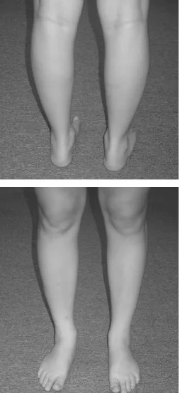
Posture
Standing posture evaluation revealed generally good alignment in the lumbopelvic region. She had bilateral pes planus with mild calcaneal eversion and bilateral functional genu valgus (Figure 1).
Physical Evaluation and Gait
Ankle range of motion was limited in passive dorsiflexion bilaterally. Extension of the 1st metatarsal phalangeal joint was within a normal range of movement but pro-duced pain along the plantar aponeurosis. Severe tender-ness was provoked on palpation over the medial calcaneal tubercles bilaterally (more severe on the right side). Mus-cle hypertonicity was noted bilaterally in the gastrocnemi-us/soleus complex. Taut bands were found in the plantar aponeurosis and fascia. Calluses were apparent in the me-dial aspect of the 1st rays. Joint play revealed restrictions in the subtalar and ankle mortise joints bilaterally.
Active knee range of movement was within normal limits. Resisted knee movements were unremarkable. Mild atrophy was noted in the right vastus medialis ob-lique (VMO) muscle. Contraction of the quadriceps in extension produced a mild lateral deviation of the patella. Thomas and Ely’s tests for hypertonicity of the hip flex-ors and quadriceps muscles were positive bilaterally.
Active hip range of movement was within normal lim-its. Resisted right hip abduction was mildly weak. A trig-ger point was found in the right tensor fascia lata (TFL) muscle. Ober’s test was positive for TFL / iliotibial band hypertonicity.
Gait assessment revealed prominent forefoot overpro-nation with mild foot flaring laterally. One-leg standing functional genu valgus testing was positive bilaterally. Heel and toe walking was normal.
Leg length comparisons were within normal limits. The following orthopedic tests were negative: anterior and posterior drawer, talar tilt, Kleiger’s, Thompson’s, Homan’s, Noble’s compression and Hibb’s.
Clinical impression
Chronic bilateral plantar fasciitis secondary to longstand-ing poor biomechanical foot function with associated muscle hypertonicity of the gastrocnemius/soleus com-plex, tensor fascia lata and mild atrophy of the right vas-tus medialis oblique.
Treatment Plan
Phase 1: Reduce pain intensity/inflammation and patient education
Due to the chronic nature of her heel pain, acetic acid iontophoresis was used at a frequency of 3 times per
week for 2 weeks and then 2 times per week for 2 weeks. The treatments were delivered using the Empi Dupel Ion-tophoresis System with an 80–90 mA·min dosage with 4 ml of 5% acetic acid solution. Acetic acid has a negative ionic polarity so it was added to the delivering pad which was connected with the negative (cathode-black) elec-trode to repel the acetic acid ions through the skin into the underlying tissue. The buffering pad was placed just above the treatment area over the Achilles tendon and was connected to the positive (anode-red) electrode.
The acetic acid iontophoresis was then immediately followed by a 50% pulsed ultrasound at 1.5 W/cm2
inten-sity for 8 minutes (Figure 2).
The patient was instructed to limit playing soccer and running activities for the following 2 weeks. She was un-able to comply with restricted activity due to a number of injured teammates and she continued to play to her own ability during the course of treatment with restricted playing times and limited practices to facilitate recovery. Bilateral plantar fascial taping using the arch taping technique24 was utilized before games and practices to
re-duce tension on the plantar fascia (Figure 3). Ice massage or the use of cold packs for 15–20 minutes was recom-mended daily and after activity to reduce pain and recur-rent inflammation.
Phase 2: Reduce fascial tension,
muscle hypertonicity, joint hypomobility and biomechanical dysfunctions
Once the pain intensity reduced, manual manipulation of soft tissues was used in an attempt to restore normal mus-cle lengths and joint movements. Treatments began uti-lizing light myofascial therapy on the plantar fascia and gastrocnemius/soleus complex. Mobilizations/manipula-tions to hypomobile articulaMobilizations/manipula-tions were performed on the ankle mortise and subtalar joints. As the patient im-proved, deeper myofascial treatments were applied to the plantar fascia, tibialis anterior, fibularis longus/brevis, tensor fascia lata (TFL) and iliotibial (IT) band. Trans-verse frictions to the plantar fascia insertion were applied followed by ice massage. At that time, the patient began using a new pair of full-length custom orthotics with a 2 mm thickness, 2-degree medial extrinsic heel post and full heel cushion.
Phase 3: Muscle stretch/strengthening, ankle proprioception and return to full activity
The patient was given daily stretches utilizing 2 sets of 20–30 seconds hold for the following: plantar fascia, gastrocnemius, soleus, tibialis anterior, quadriceps, ham-strings, iliopsoas, TFL and IT band. Foot towel scrunches and scoops were performed to facilitate the small mus-cles of the foot and stabilize the plantar fascia. Theraband
exercises for ankle inversion/eversion and dorsiflexion/ plantarflexion were performed and progressed from red to green to blue bands at a frequency of 3 times per week with 2–3 sets of 15–20 repetitions. Open chain exercises beginning with leg extensions emphasizing terminal knee extension to enhance VMO muscle activity and ham-string curls were prescribed using the Zinovieff weight-lifting protocol to enhance leg strength. This protocol recommends incorporating 3 sets of 10 repetitions with the first set using the 10 repetitions maximum (RM) weight, ¾ RM weight for the second set and ½ RM weight for the third set. Proprioceptive exercises pro-gressed from 1-leg standing with eyes open to eyes closed, standing wobble board exercises to 1 leg stance with leg bends. Functional lower limb strengthening ex-ercises utilizing half lunges and wall squats were used to facilitate closed chain movement patterns. Progressive jogging from 5 km to 10 km with increasing 1 km in-tervals were prescribed and monitored for symptom ag-gravation. Once the patient was able to jog 10 km and participate in full practices without aggravating any symptoms, she then returned to regular soccer activity without any limitations.
Discussion
Acute plantar fasciitis is an overuse syndrome that us-ually responds quite well with conservative treat-ment.2,4,13,25,26,27,28,29,30 It is an overuse syndrome
whereby the repair process cannot keep up with the stress that the body endures. The three major sources of stress are poor training technique, repetitive overuse and inher-ent biomechanical imbalances.3 Large increments in
itation and orthotics helped this patient return to her regu-lar activities within a period of six weeks.
Treatment using acetic acid iontophoresis had been previously indicated in treating conditions such as myosi-tis ossificans, calcific bursimyosi-tis and calcific tendonimyosi-tis. The rationale for treatment would primarily aim at increasing the solubility of calcium deposits in tendons and soft tissues22,31 to encourage the removal of excess calcium
ions from the injury site into the blood stream. With re-spect to pain generation, current literature focuses be-yond radiographic evidence of pathological calcified or ossified structures and places a greater emphasis on the physiological events that precedes this process. His-topathologic changes of patients who have chronic heel pain include an initial low-grade periosteal inflammation, edema, fibroblastic and inflammatory cell proliferation.22
Calcium deposits infiltrate inflamed, dead, or dying tis-sue despite normal blood calcium levels and normal cal-cium metabolism.32 One theory proposes that denatured
proteins from damaged cells unmask reactive groups that bind with phosphate radicals that attract and bond with calcium ions, which in turn, open collagen bundles caus-ing tissue swellcaus-ing, fat saponification and further tissue disruption.22 Consequently, these calcium ions break
pro-tein cross-linkages with polyaminoglycans like chondroi-tin sulfate disrupchondroi-ting other protein linkages.33 The
continual progression of chronic tissue inflammation due to abnormal stress progresses from a physiological reac-tion to fibrocartilagenous tissue formareac-tion leading to car-tilage deposition and eventual bone spur development.22
Shama and Kominsky noted that of 1,000 patients who had been radiographed, only 132 had evident heel spurs of which, only 39% complained of a history of heel pain.22 Thus, it could be reasoned that bone spurs are the
long-term pathological response to maladaptive tissue dysfunction and that the deposition of dystrophic calcium that occurs prior to osseous formation is the primary fo-cus of chronic pain generation. Japour et al. describes in detail the theoretical biochemical process where the use of acetic acid iontophoresis converts insoluble calcium carbonate in chronically inflamed tissue to calcium ace-tate, which is blood-soluble.22 Pulsed ultrasound was
used to reduce inflammation, perfuse local blood flow and facilitate the removal of the newly formed calcium acetate into the blood and thereby remove it from the lo-calized area of heel pain.
The use of athletic taping provided temporary mechan-ical stability and support for the strained plantar fas-cia.31,34 The amount of actual mechanical support has
been questionable and current literature places a greater emphasis on proprioceptive mechanisms via sensory af-ferent cues through traction of the tape on the skin to re-duce pain intensity and increase muscular and joint support.35 Nonetheless, athletic taping and or bracing are
effective means to limit range of motion, increase propri-oception and reduce pain intensity to injured struc-tures.36,37 Studies have shown that athletic taping was
superior to anti-inflammatories and heel cup treatments1
or NSAIDs in combination with injections.15 Athletic
taping using non-elastic zinc oxide tape to the plantar fas-cial arch as described in Arhheim was initially used to re-duce strain and pain intensity on the plantar fascia.24 In
addition, prophylactic bilateral plantar arch fascial taping was utilized before practices and games to encourage a progressive return to soccer activity and limit the aggra-vation of symptoms. Once the patient had adapted to the new orthotics, the athletic taping was omitted from treat-ment to evaluate her progressive response.
Ample evidence exists, based on subjective pain relief, symptom resolution and patient satisfaction, to support the continued use of orthotics in treating biomechanical injures in the lower limb, particularly in runners.25,27,28
Orthotic intervention is appropriate for those injuries re-sulting from identifiable abnormal biomechanics such as hyperpronation, excessive rearfoot eversion, high ever-sion velocity, increase internal rotation, increased impact and loading rate of vertical ground reaction force, exces-sive supination with increased ankle inversion move-ments and external rotation momove-ments.28,38 In addition,
orthotics may also derive their benefit by altering muscle activation and proprioceptive mechanisms involved in regulating muscle function and dampening soft-tissue vi-brations.28
For plantar fasciitis with associated pes planus, Na-woczenski et al. prescribes a firmer, more rigid orthotic with a medial heel post to help minimize excessive pro-nation.28 Gross et al. suggests that the custom semi-rigid
foot orthotics may maintain medial longitudinal arch height sufficiently to reduce tensile stress within the plantar fascia and provide clinically significant reduc-tions in pain and disability.27 The custom orthotics may
the medial calcaneal tubercle for additional pressure re-lief.28 In addition, straight-last footwear with motion
con-trol features such as reinforced heel counter and medial midsole reinforcement should be recommended for this foot type.28
In prescribing orthotics, the age, weight, foot type, bio-mechanical characteristics and activity level should be considered to determine the degree of rigidity and acces-sory modifications necessary to limit excessive move-ments of the lower limb complex. This 145 pound patient with forefoot overpronation was initially given a very rig-id 3mm thick orthotic with a neutral heel post. Over the course of one year, this orthotic was not successful in sig-nificantly reducing her symptoms and led to the develop-ment of additional problems in the knee, quadriceps and lateral pelvic musculature. Through clinical experience, it was determined that a 2 mm thick orthotic with a 2-degree extrinsic medial heel post and additional heel cushion was appropriate for her weight level and biome-chanical foot dysfunction. This orthotic would be able to provide enough support for the longitudinal arch and lim-it overpronation and rearfoot eversion. Accommodation to the new orthotic took two weeks with a progressive re-duction in lower limb pain and an increased ability to sustain longer running and playing times.
Several manual therapeutic techniques were used to manipulate soft tissue and joint structures to restore nor-mal muscle lengths and joint movements. Initial treat-ments focused on the plantar fascia and gastrocnemius/ soleus complex to reduce tension and muscle hypertonic-ity. Manipulation and mobilization of the ankle mortise, subtalar and tarsal-metatarsal joints as well as axial trac-tion were performed in the presence of restricted motrac-tion. Deeper myofascial treatments focused on the anterior and lateral leg compartment and the TFL/IT band complex. Brantingham et al. describes the benefits of soft tissue therapy on the plantar muscles and fascia in addition to joint manipulation to restore normal myofascial move-ment.17 Cross-frictional massage was used to soften and
reduce fibrotic scar tissue17 in the plantar muscles and
fascia followed by ice massage during the late phases of recovery.
The prescribed stretches were advised to be done twice per day for 20 seconds and included stretches for the gas-trocnemius, soleus, fibularis, TFL and IT band, quadri-ceps, hamstring and iliopsoas muscles. Once the pain
intensity reduced, non-weight-bearing stretching of the plantar fascia as described in DiGiovanni et al. was included in the daily routine.39 Towel scrunches and
foot scoop exercises were used to facilitate the intrinsic muscles of the foot and reduce plantar fascial tension. Foot inversion/eversion and ankle dorsi/plantar flexion strengthening exercises targeting the extrinsic muscles of the lower leg progressed using increasingly resistive Theraband tubing to reduce muscular imbalance and in-crease mechanical stability. These exercises were per-formed every other day and progressed from 2 sets of 15– 20 repetitions to 3 sets over a 4 week period. During the 3rd–6th week, a wall pulley machine was used to perform the same exercises within the clinic. VMO facilitation, weighted knee extension and flexion exercises using a weight ratio of 1.3 to 1 ratio was used to target the quad-riceps and hamstring muscle groups. Strength progres-sion was monitored utilizing the Zinovieff weightlifting protocol. Closed chain modified lunges and wall squats were used to facilitate compound muscle groups and functional movement patterns. Modified jogging was ini-tiated during this phase of recovery and progressed from running 5km to 10 km with 1 km intervals over a period of 2 weeks.
Proprioceptive exercises were used to facilitate intrin-sic and extrinintrin-sic muscles to enhance motor coordination, strength and stability thereby reducing tensile stress on the plantar fascia. Balance exercise improves propriocep-tion during both the rehabilitapropriocep-tion phase and the competi-tion phase of recovery.40,41 The proprioceptive exercises
progressed from 1-leg standing with eyes open to eyes closed; standing wobble board exercises to 1 leg stance wobble board exercises with leg bends.
Conclusion
to resolve the effects of tissue adaptation due to chronic inflammation while improving musculoskeletal abnor-malities and biomechanical imbalances. This integrative approach in the conservative management of chronic plantar fascitis resulted in the resolution of symptoms and a high degree of patient satisfaction within 6 weeks of treatment.
References
1 Kahn J. Principles and Practice of Electrotherapy. New York, Churchill Livingstone; 1987.
2 Brantingham JW. Plantar fasciitis. J Chiropr Tech 1992 Aug; 4(3):75–83.
3 Lynch DM, Goforth WP, Martin JE, Odom RD, Preece CK, Kotter MW. Conservative treatment of plantar fasciitis: a prospective study. J Am Podiatr Med Assoc 1998; 88(8):375–380.
4 Arnheim DD, Prentice WE. Principles in Athletic Training, Eighth Edition. St. Louis: Mosby Year Book; 1993. 5 Dimou ES, Brantingham JW, Wood T. A randomized
controlled trial (with blinded observer) of chiropractic manipulation and achilles stretching vs. orthotics for the treatment of plantar fasciitis. J Am Chiropr Assoc 2004 Sept; 41(9):32–42.
6 Roxas M. Plantar fasciitis: diagnosis and therapeutic considerations. Alternative Med Rev 2005; 10(2):83–93. 7 Hiss J. Functional Foot Disorders. Los Angeles: University
Publishing Company, 1949.
8 Maffulli N, Khan KM, Puddu G. Overuse tendon conditions: time to change a confusing terminology. Arthroscopy: The Journal of Arthroscopic and Related Surgery. 1998 Nov/Dec; 14(8):840–843.
9 Wieder DL. Treatment of traumatic myositis ossificans with acetic acid iontophoresis. Physical Therapy 1992 Feb; 72(2):133–137.
10 Sharma P, Maffulli N. Current concepts review: tendon injury and tendinopathy: healing and tepair. J Bone Jt Surg 2005 Jan; 87-A(1):187–202.
11 Black AS, Kanat IO. A review of soft tissue calcifications. J Foot Surg 1985; 24:243.
12 Verhagen EALM, van der beek AJ, van Mechelen W. The effect of tape, braces and shoes on ankle range of motion. Sports Med 2001; 31(9):667–677.
13 Malliou P, Gioftsidou A, Pafis G, Beneka A, Godolias G. Proprioceptive training (balance exercises) reduces lower extremity injuries in young soccer players. J Back and Musculoskeletal Rehab 2004:101–104.
14 Callahan MP, Denegar CR, Segree CA. The effect of vacuum-molded orthotics on lower extremity overuse injuries. J Sport Rehab 1993; 2:251–260.
15 Nawoczenski DA, Janisse DJ. Foot orthoses in
rehabilitation-what’s new. Clinics in Sports Medicine 2004; 23:157–167.
16 DiGiovanni BF, Nawoczenski DA, Lintal ME, Moore EA, Murray JC, Wilding GE, Baumhauer JF. Tissue-specific plantar fascia stretching exercise enhances outcomes in patients with chronic heel pain. J Bone Jt Surg 2003 Jul; 85-A(7):1270–1277.
17 Fredberg U. Tendinopathy – tendinitis or tendinosis? The question is still open. (Letter). Scandinavian J Med Science in Sports 2004; 14:270–271.
18 Cimbiz A, Bayazit V. Evaluation of balance and muscle strength in physical education students with recovered lower limb injuries. J Back Musculoskeletal Rehab 2004; 111–116.
19 La Porta GA, La Fata PC. Pathologic conditions of the plantar fascia. Clin Podiatr Med Surg 2005; 22:1–9. 20 Leach RE, Seavey MS, Salter DK. Results of surgery in
athletes with plantar fasciitis. Foot and Ankle 1986 Dec; 7(3):156–161.
21 Austin WM. Shin splints with underlying posterior tibial tendonitis: a case report. J Sports Chiropractic & Rehab 1996 Dec; 10(4):163–169.
22 Bergmann JN. History and mechanical control of heel spur pain. Clinics in Podiatric Medicine and Surgery 1990 Apr; 7(2):243–259.
23 Kaya BK. Plantar fasciitis in athletes. J Sport Rehab 1996; 5:305–320.
24 Gross MT, Byers JM, Kraft JL, Lackey EJ, Melton KM. The impact of custom semi-rigid foot orthotics on pain and disability for individuals with plantar fasciitis. J Ortho Sports Phys Ther 2002 Apr; 32(4):149–157.
25 Cordova ML, Ingersoll CD, LeBlanc MJ. Influence of ankle support on joint range of motion before and after exercise: a meta-analysis. J Ortho Sports Phys Ther 2000; 30(4):170–182.
26 You SH, Granata KP, Bunker LK. Effects of
circumferential ankle pressure on ankle proprioception, stiffness, and postural stability: a preliminary investigation. J Ortho Sports Phys Ther 2004 Aug; 34(8):449–459. 27 Perle SM. Rearfoot-forefoot orientation and traumatic
risk for runners. J Sports Chiropr Rehab 1998 Jun; 12(2):99–101.
28 Tillu A. Effect of acupuncture treatment on heel pain due to plantar fasciitis. Acupuncture in Medicine 1998 Nov; 16(2):66–69.
29 Hammer WI. Functional Soft Tissue Examination and Treatment by Manual Methods – New Perspective Second Edition. Maryland: Aspen Publications; 1999.
30 Hyde TE, Gengenbach MS. Conservative Management of Sports Injury. Williams and Wilkins. Maryland: Jones and Bartlett Publishers; 1997.
32 Acevedo JI, Beskin JL. Complications of plantar fascia rupture associated with corticosteroid injection. Foot and Ankle 1998 Feb; 19(2):91–97.
33 Pollard H. Management of plantar fasciitis: a case report. J Sports Chiropr Rehab 1999 Sep; 13(3):93–98.
34 Cunnane G, Brophy DP, Gibney RG, FitzGerald O. Diagnosis and treatment of heel pain in chronic inflammatory arthritis using ultrasound. J Manip Phys Thera 1997 Jan; 20(1):67.
35 Seligman DA, Dawson DR. Customized heel pads and soft orthotics to treat heel pain and plantar fasciitis. Arch Phys Med Rehab 2003 Oct; 84:1564–1567.
36 Japour CJ., Vohra R, Vohra PK, Garfunkel L, Chin N. Management of heel pain syndrome with acetic acid iontophoresis. J Am Podiatr Med Assoc 1999 May; 89(9):251- 257.
37 Almedkinders LC, Temple JD. Etiology, diagnosis, and treatment of tendonitis: an analysis of the literature. Med Science in Sports and Exercise 1998 Aug; 30(8):1183–1190.
38 Powell M, Post W, Keener J, Wearden S. Effective treatment of chronic plantar fasciitis with dorsiflexion night splints: a crossover prospective randomized outcome study. Foot and Ankle 1998; 19(1):10–17.
39 Stuber K, Kristmanson K. Conservative therapy for plantar fasciitis: a narrative review of randomized controlled trials. J Can Chiropr Assoc 2006 Jun; 50(2):118–133.
40 Robbins S, Waked E, Rappel R. Ankle taping improves proprioception before and after exercise in young men. J Sports Med 1995; 29(4):242–247.
41 Taunton JE, Ryan MB, Clement DB, McKenzie DC, Lloyd-Smith, DR. Plantar fasciitis: a retrospective analysis of 267 cases. Physical Therapy in Sport 2002; 3:57–65.