699
© 2018 by the Serbian Biological Society How to cite this article: Ahmed N, Aljuhani N, Salamah S, Surrati H, El-Agamy DS, Elkablawy MA, Ibrahim SRM, Mohamed GA. Pulicaria petiolaris effectively attenuates lipopolysaccharide (LPS)-induced acute lung injury in mice. Arch Biol Sci. 2018;70(4):699-706.
Pulicaria petiolaris
effectively attenuates lipopolysaccharide (LPS)-induced acute lung
injury in mice
Nishat Ahmed1, Naif Aljuhani1, Sarah Salamah2, Heba Surrati2, Dina S. El-Agamy1,3, Mohamed A.
Elkablawy4,5, Sabrin R. M. Ibrahim6,7,* and Gamal A. Mohamed8,9
1Department of Pharmacology and Toxicology, College of Pharmacy, Taibah University, Al-Madinah Al-Munawwarah,
30001, Saudi Arabia
2Pulmonary Research Team, College of Pharmacy, Taibah University, Al-Madinah Al-Munawwarah, 30001, Saudi Arabia 3Pharmacology and Toxicology Department, Faculty of Pharmacy, Mansoura University, 35516, Egypt
4Department of Pathology, College of Medicine, Taibah University, Al-Madinah Al-Munawwarah, 30001, Saudi Arabia 5Department of Pathology, Faculty of Medicine, Menoufia University, Menoufia, 32511, Egypt
6Department of Pharmacognosy and Pharmaceutical Chemistry, College of Pharmacy, Taibah University, Al Madinah Al
Munawwarah 30001, Saudi Arabia
7Department of Pharmacognosy, Faculty of Pharmacy, Assiut University, Assiut 71526, Egypt
8Department of Natural Products and Alternative Medicine, Faculty of Pharmacy, King Abdulaziz University, Jeddah
21589, Saudi Arabia
9Department of Pharmacognosy, Faculty of Pharmacy, Al-Azhar University, Assiut Branch, Assiut 71524, Egypt
*Corresponding author: [email protected]; [email protected]
Received: May 10, 2018; Revised: June 3, 2018; Accepted: June 26, 2018; Published online: July 17, 2018
Abstract: Members of the genus Pulicaria have been used in traditional medicine for alleviating several complaints as they have a rich pool of biometabolites. Acute lung injury (ALI) is a serious disease with an elevated mortality rate. The present investigation aimed to evaluate the total phenolic and flavonoid contents and antioxidant capacity of the metha-nolic extract of P. petiolaris Jaub. and Spach. (PP) (Asteraceae). Moreover, the potential protective potential of PP against lipopolysaccharide-(LPS)-induced ALI was assessed. PP is a rich source of phenolics and flavonoids. The total phenolic content (TPC) was 68.05 mg gallic acid equivalent (GAE)/g dried extract, and the total flavonoid content (TFC) was 45.86 mg quercetin equivalent (QE)/g dried extract. Additionally, PP possessed a promising DPPH-scavenging activity, with an IC50=27 μg/mL. Our results showed that PP lessened LPS-induced lung injury. PP effectively reduced pulmonary edema as it lowered total protein and the lung wet/dry weight (W/D) ratio in the bronchoalveolar lavage fluid (BALF). It also significantly ameliorated the level of lactate dehydrogenase (LDH) in the BALF and improved the histopathological lesions in the lung tissue. LPS-induced inflammatory cell infiltration was greatly depressed in PP-treated animals. PP showed anti-oxidant capacity as it reduced the LPS-induced increase in the lipid peroxidation marker, malondialdehyde (MDA). It also increased the activity of superoxide dismutase (SOD) and the content of reduced glutathione (GSH). This study indicates that PP significantly decreased LPS-induced inflammation in the LPS-mediated ALI murine model, suggesting that it may become a significant preventive strategy for treating nonspecific inflammation of the lungs.
Key words: Pulicaria petiolaris;lung injury; lipopolysaccharide; antioxidant; inflammation
INTRODUCTION
Acute lung injury (ALI) has a high mortality and no effective treatment for it is available. ALI is character-ized by acute respiratory distress syndrome (ARDS), which is accompanied by an acute inflammatory
to produce an experimental model of ALI in mice [1-3]. Administration of LPS leads to the release of many cytokines, such as tumor necrosis factor-α (TNF-α) and interleukins (ILs), which trigger inflammatory responses and alter immune system functioning. LPS induces epithelial cell apoptosis, causes damage to the epithelial cell layers and stimulates the release of reac-tive oxygen species (ROS) that leads to neutrophilic leukocyte aggregation and eventually to lung tissue injury [4,5]. Mechanical ventilation is the only effec-tive therapy for ARDS [6]. Hence, there is a need to search for new therapeutics that could be used for its treatment.
Natural products display remarkable roles, not only in the synthesis, design, and discovery of new drugs, but also as prominent sources of innovative drugs and bioactive substances. The genus Pulicar-ia (tribe Inuleae and family Asteraceae) comprises around 100 species that are widely distributed in Eu-rope, Asia and Africa [7]. The plants of this genus have been used in traditional medicines for treating various ailments, such as back-pain, inflammation, menstrual cramps, intestinal disorders, dysentery and diarrhea [8,9]. They possess different bioactivities: cytotoxic, antipyretic, antioxidant, antispasmodic, antimicrobial, antihistaminic, analgesic, hepatoprotective, antiin-flammatory, cardioprotective and nephroprotective [9-11]. This genus is known to be rich in terpenoids, phenolics, caryophyllene sterols, caryophyllenes and xanthines [9,12]. Moreover, many sesquiterpenes isolated from this genus have been shown to exhibit a wide range of biological activities [9,13,14]. In the course of our biological evaluation of Saudi plants, the protective potential of P. petiolaris against LPS-induced ALI was assessed. Furthermore, its flavonoid and phenolic contents as well as antioxidant activity were evaluated.
MATERIALS AND METHODS
Plant material and extraction
The aerial parts of P. petiolaris were collected from the Jeddah-Taif road, Saudi Arabia in April 2017. The plant was kindly specified by the staff of the Department of Natural Products and Alternative Medicine, King
Ab-dulaziz University, Saudi Arabia, based on the library database and morphological features [15]. This was confirmed by Dr. Emad Al-Sharif, Biology Department, Faculty of Sciences and Arts, Saudi Arabia. A voucher specimen (PP 2017-1) was stored at the Department of Natural Products and Alternative Medicine herbarium, King Abdulaziz University. The air-dried powdered aerial parts of PP (500 g) were extracted with methanol (2 x 1000 mL) using an IKA Ultra-Turrax T 25 digital instrument (IKA Labortechnik, Staufen, Germany). The solvent was removed under reduced pressure and the dried total methanolic extract (TME) (12.6 g) was kept at 4°C until use in biological tests.
Chemicals and reagents
All chemicals were obtained from commercial sources and were of analytical grade. Gallic acid, quercetin, ascorbic acid, sodium carbonate, sodium hydroxide, 2,2-dyphenyl-1-picrylhydrazyl (DPPH) and sodium nitrate were purchased from Sigma Chemical Co. (Germany). The Folin-Ciocalteu’s phenol reagent and aluminum chloride were from Fluka Chemie AG (Buchs, Switzerland). Escherichia coli serotype O111:B4 LPS was obtained from Sigma-Aldrich (St. Louis, MO, USA) and freshly dissolved in normal sa-line.
Preparation of standard solutions
Gallic acid and quercetin (1 g each) were dissolved separately in methanol (100 mL) to obtain standard solutions A and B, respectively.
Determination of the TPC
The Folin-Ciocalteu method was used to assess the TPC of PP [16]. A standard curve for gallic acid was prepared by dilution (0.1, 0.5, 1.0, 2.5 and 5 mg/mL) in methanol from solution A. One hundred μL of each
dilution was mixed with 500 μL distilled H2O and the
Folin-Ciocalteu reagent (100 μL) and left for 6 min.
Then, 500 μL of distilled H2O and 1 mL 7% Na2CO3
Determination of the TFC
The TFC of the TME was estimated by the AlCl3
complex assay [17]. A calibration curve for quercetin (standard) was drawn. Dilutions of 0.1, 0.5, 1.0, 2.5 and 5 mg/mL were prepared in methanol from solu-tion B. From each dilusolu-tion, 100 μL was mixed with
500 μL of distilled H2O and 5% NaNO3 (100 μL) and
left for 6 min. Then, a 10%-AlCl3 solution (150 μL)
was added and kept for 5 min and 200 μL 1M NaOH was added. The absorbance was measured at 430 nm. The same procedure was carried out with PP TME, and the TFC was expressed as the quercetin equivalent (mg QE/g dried extract). All procedures were done in triplicate and the mean values were estimated.
Antioxidant activity
The antioxidant potential was assessed using the DPPH assay as outlined previously [18,19]. One mL of the TME (10, 20, 40, 60, 80, and 100 µg/mL) was mixed with 1 mL of DPPH and kept for 0.5 h. UV absorbance was estimated at 517 nm. Each experi-ment was carried out in triplicate using ascorbic acid (standard). The percentage of free radical scavenging activity was estimated using the following formula:
Antioxidant activity = 100 × (1 - ) absorbance with the sample absorbance of the blank
The IC50 was calculated from the inhibition
per-centages graph against the concentration of extract plot using a nonlinear regression algorithm.
Animals
BALB/c mice (25-30 g) were supplied by the Animal Facility, College of Pharmacy, Taibah University. The animals were maintained in standard cages and al-lowed free access to food and water under standard condition of temperature (25°C) and a dark/light cy-cle. The experimental protocol adhered to the Guide-lines of the Ethical Committee of Taibah University, Saudi Arabia which closely adheres to NIH guidelines.
Experimental design
Mice were divided into 4 experimental groups (n=6). The animals were treated according to the following
regimen: control group: the mice were given the vehi-cle once daily for 5 days; LPS group: the mice received a single LPS intraperitoneal (i.p.) injection (10 mg/kg); PP-treated groups: the animals were given PP (50 and 100 mg/kg) by oral gavage (p.o.) for 5 days before LPS injection; 24 h after LPS injection, the blood was ob-tained from the retro-orbital sinus and the serum was separated and stored at -20°C until further analysis.
The mice were humanely killed with an over-dose of anesthesia using diethyl ether. The chest was opened, and the left lung was clamped. The right lung was lavaged using 0.9% saline. BALF was collected and centrifuged at 2000 x g and 4°C for 15 min. The cells were collected and counted. The BALF supernatants were stored at -80°C. A small piece of the left lung was weighed and homogenized in phosphate buffer (pH 7.4, 0.1 mol/L) in an ice bath. The homogenate was centrifuged at 2000 x g and 4°C for 20 min. The supernatants were kept at -80°C for analysis of oxida-tive stress parameters. The remaining left lung was dissected and washed with ice-cold saline and fixed for 24 h in neutral buffered 10% formalin and submit-ted for histopathological assessment.
Determination of lung W/D ratio
The lung W/D ratio was estimated to assess the de-gree of pulmonary edema as described previously [20]. A part of the left upper lung was blotted dry and weighed to determine “wet” weight. Then, it was placed in an oven (80°C) for 24 h to measure the “dry” weight and the lung W/D ratio was determined.
Determination of total and differential cell counts in BALF
Total cell counts were determined using a hemocy-tometer. Cell pellets were resuspended in 100 μL saline (0.9%), centrifuged onto slides and stained for 8 min with Wright-Giemsa. The differential cell counts were quantified using a light microscope at 40× magnifica-tion by counting a total of 200 cells/slide.
Determination of protein
(Cat. NO. 23225, Thermofisher Scientific, MA, USA), according to the manufacturer’s protocol.
Determination of LDH activity
LDH activity was measured in BALF using a kit (Hu-man, Wiesbaden, Germany), following the manufac-turer’s guidelines. Briefly, the samples were mixed with nicotinamide adenine dinucleotide (NADH), sodium pyruvate and TRIS buffer. The change in absorbance was estimated at 340 nm.
Histopathological analysis of lung
Fixed lung samples were embedded in paraffin wax and sectioned (5 μm). Lung specimens were stained with hematoxylin-eosin (H&E) and examined ran-domly with no knowledge of the group. Histopatho-logical lesions were evaluated on the basis of the de-gree of inflammation, thickening of the alveolar wall and cellular proliferation. Lesions were semiquantita-tively graded as described previously [20].
Determination of oxidative stress
The malondialdehyde (MDA) content is used as an index for lipid peroxidation. SOD activity and the GSH content were assessed and served as indices of the antioxidant capacity of the lung. These param-eters were estimated in the supernatants of the lung homogenates. The MDA content was estimated as de-scribed previously [21]. In brief, MDA was quantified by reaction with thiobarbituric acid (TBA) and the absorption was measured spectrophotometrically at 532 nm.
SOD activity was assessed by observing the SOD-inhabitable autooxidation of pyrogallol [22]. The change in absorbance was recorded at 420 nm.
The GSH assay relies on the reaction of 5,5-dithio-bis-2-nitrobenzoic acid with GSH and the product was measured at 412 nm spectrophotometrically [23].
Statistical analysis
Statistical analysis was performed using one-way anal-ysis of variance (ANOVA) followed by Tukey’s Kramer
multiple comparisons test. The data were expressed as means±SE for 6 mice. P<0.05 was considered as significant.
RESULTS
Flavonoid and phenolic compound contents and antioxidant activity
P. petiolaris is a rich source of phenolics and flavo-noids (TPC: 68.05 mg gallic acid equivalent (GAE)/g dried extract; TFC: 45.86 mg quercetin equivalent (QE)/g dried extract) with a promising DPPH
scav-enging activity (IC50=27 μg/mL), when compared to
ascorbic acid (IC50=32.9 μg/mL). The high phenolic
and flavonoid contents of the PP extract, in addition to antioxidant activity, might provide antiinflamma-tory activity. A positive relation between the flavonoid and phenolic contents and free radical scavenging ac-tivity has been reported [24]. It is noteworthy that the plant extracts with high phenolic contents also possess high flavonoid contents, as reported for other Pulicaria species [12,25].
Effect on LPS-induced pulmonary edema
The LPS injection caused remarkable increases in the total protein content and the lung W/D ratio as compared to control mice (Fig. 1). Pretreatment with PP resulted in a significant amelioration of these two parameters of pulmonary edema when compared to the LPS group.
Fig. 1. PP decreased LPS-induced pulmonary edema. A – Lung
W/D ratio. B – Protein content. Mice were treated with two
dif-ferent doses of the methanolic extract of PP (50 and 100 mg/kg, p.o.) once daily for 5 days prior to LPS injection (10 mg/kg, i.p.).
Data are the mean±SE n=6. *P<0.05vs control group; #P <0.05
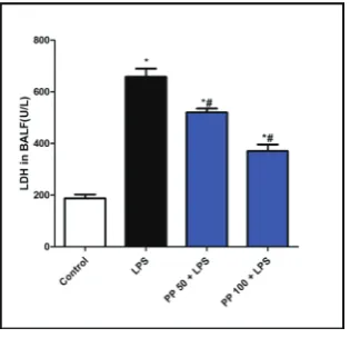
Effect on LDH activity in BALF
As demonstrated in Fig. 2, LPS raised the level of LDH in BALF compared to the control group. The PP treat-ment significantly attenuated the high level of LDH compared to the LPS group.
Effect on lung histopathological examination
Lung sections of normal mice showed normal lung histology, while those of the LPS group showed marked inflammatory cell infiltration alveolar wall thickening. PP-treated animals showed a marked im-provement in lung lesions (Fig. 3).
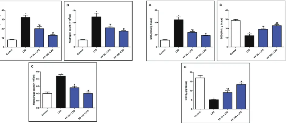
Effect on inflammatory cell counts in BALF
As shown in Fig. 4, LPS administration induced an elevation of inflammatory cell infiltration into lung tissue. Also, there was elevation of the differential and total cell counts, mainly neutrophils, in the BALF in comparison to the control group. The PP pretreat-ment significantly reduced the differential and total cell counts compared to the LPS group.
Effect on oxidative stress and antioxidant markers
LPS injection increased the oxidative stress param-eter and MDA in lung tissue (Fig. 5). LPS attenuated
the antioxidant activity of the lung, as presented by decreased SOD and GSH levels. The PP pretreatment significantly increased SOD activity and GSH levels, which were accompanied by decreased levels of MDA when compared to the LPS group.
DISCUSSION
ALI is characterized by an extensive inflammatory reaction in the lung tissue along with disruption of the alveolar-capillary barrier, which leads to severe impairment of gas exchange [26]. The current study showed the ability of the PP extract to attenuate LPS-induced ALI, which may be attributed to the antioxi-dant ability of PP. PP showed a remarkable ability to
Fig. 3. Lung specimen stained with H&E (×400) showing
attenu-ation of LPS-induced pulmonary damage by PP. Control –
Sam-ple showing normal alveolar capillaries (arrows), thin alveolar
walls (chevrons) and alveolar space (stars). LPS – Sample with
congested alveolar capillaries (arrows), thickened alveolar walls (chevrons), acute inflammatory cellular infiltrates (lightning bolt),
and pink fluid exudates in alveolar spaces (stars). PP 50 + LPS –
sample displaying moderate alveolar capillary congestion (arrows), moderate thickening of alveolar walls (chevrons) with moderate inflammatory cellular infiltrates (lightning bolt), and less pink
exudates in alveolar spaces (stars). PP 100 + LPS – Sample
ex-hibiting greater improvement of all LPS-induced inflammatory responses in the lung tissue. Scale bar 25 μm (×400).
Fig. 2. PP suppressed LDH activity in BALF. Mice were treated with two different doses of the methanolic extract of PP (50 and 100 mg/kg, p.o.) once daily for 5 days prior to LPS injection (10 mg/kg, i.p.). Data are the
mean±SE n=6. *P<0.05vs control group.
attenuate LPS-induced pulmonary edema and decrease LDH activity, as well as inflammatory cell infiltration. The histopathological lesions in the lungs were greatly improved in PP-treated animals. The results further showed that the protective effect of PP could be linked to its ability to inhibit LPS-induced oxidative stress.
Activation of neutrophil and macrophage infiltra-tion into lung tissue participates in the elevainfiltra-tion of permeability of the alveolar/capillary barrier, which results in the development of pulmonary edema. Es-timation of the protein content and lung W/D ratio has been used to evaluate the integrity of the alveolar-capillary barrier and the extent of pulmonary edema [27,28]. The results of the present study revealed that LPS injection resulted in the development of pul-monary edema, as there was significant elevation in the total protein content and lung W/D ratio in the BALF. Additionally, LDH activity was significantly elevated in the BALF in the LPS group of experimen-tal animals, which was a reflection of increased cell death. Interestingly, pretreatment with the PP extract reduced these parameters of lung damage.
Inflammatory cell infiltration and accumula-tion in lung tissue is a pathological hallmark of ALI
[29,30]. Normally, the polymorphonuclear leukocytes (PMNs) play essential roles in the clearance of de-bris and pathogens from the alveolar space. However, excessive and persistent sequestration of PMNs may result in additional damage to the lung tissue through the release of multiple toxic mediators, including proteases, ROS and proinflammatory cytokines, all of which exacerbate ALI [31,32]. During ALI, neu-trophils are the first immune cells recruited into the inflammation site. This is usually accompanied by the increase of lung edema and alveolar-capillary barrier permeability. Activated neutrophils extravasate and migrate into the alveolar space where they secrete chemoattractants, such as leukotriene B4 (LTB4) and recruit more leukocytes to expand the inflammation response. Neutrophil influx into the lung is believed to have a key role in the progression of ALI [33]. Clini-cally, the number of inflammatory cells in the BALF of ARDS patients closely correlates with disease sever-ity [34]. Experimentally, attenuation of inflammatory cell infiltration into lung tissue in LPS-induced ALI results in a reduction of lung damage and improve-ment of capillary-alveolar function [35]. In line with previous investigations, the present study revealed that LPS administration significantly induced
inflamma-Fig. 4.PP ameliorated inflammatory cell infiltration in lung tissue.
A – Total cell count. B – Neutrophil count. C – Macrophage count.
Mice were treated with two different doses of the methanolic ex-tract of PP (50 and 100 mg/kg, p.o.) once daily for 5 days prior to
LPS injection (10 mg/kg, i.p.). Data are the mean±SE n=6. *P <
0.05vs control group; # P<0.05 vs LPS group.
Fig. 5: PP decreased LPS-induced oxidative stress. Mice were treated with two different doses of the methanolic extract of PP (50 and 100 mg/kg, p.o.) once daily for 5 days prior to LPS
in-jection (10 mg/kg, i.p.). Data are the mean±SE n=6. *P<0.05 vs
tory cell infiltration, mainly neutrophils, in the lungs. On the other hand, PP pretreatment markedly de-creased the LPS-induced influx of inflammatory cells into the lungs, leading to attenuation of lung injury. These findings were supported by the histopathologi-cal examination of the lung tissue, which revealed at-tenuation of LPS-induced inflammatory cell infiltra-tion and improvement of pathological lesions after PP pretreatment. Together, these data suggest that the protective activity of PP against LPS-induced ALI may be mediated through inhibition of inflammatory cell influx, mainly of neutrophils, into the lung.
Recent evidence has suggested that the main pathogenic mechanisms of ALI include oxidative stress, cytokine release, inflammation and apoptosis [36]. During ALI, large amounts of ROS are gener-ated, which cause cell membrane lipid peroxidation and the destruction of lung parenchymal cells. ROS overproduction also damages the capillary basement membrane and other stromal components. LPS-in-duced oxidative stress is associated with depressed antioxidant activity of the lung, which aggravates lung damage [27,28,37,38]. Several studies have reported on the antiinflammatory capacities of flavonoids and sesquiterpene lactones of the genus Pulicaria [9,39,40]. It is noteworthy that Mothana et al. [41] previously demonstrated the selective cytotoxicity of the PP methanol extract against human the urinary bladder carcinoma cell line. However, more studies are needed to elucidate the molecular mechanisms that underlie the biological activity of the methanolic extract of PP.
CONCLUSION
The results of the present study confirmed the eleva-tion of MDA, the parameter of lipid peroxidaeleva-tion, and depression of GSH levels and SOD activity in the lungs of LPS-intoxicated animals. PP pretreatment restored the levels of GSH and SOD to near normal values, which eventually resulted in the suppression of oxidative stress and lipid peroxidation. This study pro-vides evidence of the protective activity of PP against LPS-induced ALI in mice, which may be linked to its antioxidant activity due to the presences of phenolic constituents. Thus, this study encourages further
re-search into the molecular mechanisms of PP activity and isolation of bioactive metabolites.
Funding: This research did not receive any grant from funding agencies in the public, commercial, or not-for-profit sectors.
Author contributions: Nishat Ahmed, Naif Aljohani, and Dina S. El-Agamy were responsible for conceiving, designing and car-rying out the laboratory work, and contributed to the writing of
the manuscript. Sarah Salamahand Heba Surrati collected the
literature. Sabrin R. M. Ibrahim and Gamal A. Mohamed collected the plant material, prepared the extract, assessed TPC, TFC, and antioxidant activity and contributed to the writing of the manu-script. Mohamed A. Elkablawy contributed to the practical work and analyzed the data. All authors read and approved the final manuscript. Sabrin R. M. Ibrahim finalized and submitted the manuscript.
Conflict of interest disclosure: None to declare
REFERENCES
1. Matute-Bello G, Frevert CW, Martin TR. Animal models of acute lung injury. Am J Physiol Lung Cell Mol Physiol. 2008;295:L379-99.
2. Matute-Bello G, Downey G, Moore BB, Groshong SD, Mat-thay MA, Slutsky AS, Kuebler WM. An official American Thoracic Society workshop report: features and measure-ments of experimental acute lung injury in animals. Am J Respir Cell Mol Biol. 2011;44:725-38.
3. Yunhe F, Bo L, Xiaosheng F, Fengyang L, Dejie L, Zhicheng L, Depeng L, Yongguo C, Xichen Z, Naisheng Z, Zhengtao Y. The effect of magnolol on the Toll-like receptor 4/nuclear factor κB signaling pathway in lipopolysaccharide-induced acute lung injury in mice. Eur J Pharmacol. 2012;689:255-61. 4. Takashima K, Matsushima M, Hashimoto K, Nose H, Sato M, Hashimoto N, Hasegawa Y, Kawabe T. Protective effects of intratracheally administered quercetin on lipopolysaccharide-induced acute lung injury. Respir Res. 2014;15:150.
5. Jiang K, Zhang T, Yin N, Ma X, Zhao G, Wu H, Qiu C, Deng G. Geraniol alleviates LPS-induced acute lung injury in mice via inhibiting inflammation and apoptosis. Oncotarget. 2017;8:71038-53.
6. Bosma KJ, Taneja R, Lewis JF. Pharmacotherapy for preven-tion and treatment of acute respiratory distress syndrome: current and experimental approaches. Drugs. 2010;70:1255-82.
7. Williams CA, Harborne JB, Greenham JR, Grayer RJ, Kite GC, Eagles J. Variations in lipophilic and vacuolar
flavo-noids among European Plicaria species. Phytochemistry.
2003;64;275-83.
8. Stavri M, Mathew KT, Gordon A, Shnyder SD, Falconer RA,
Gibbons S. Guaianolide sesquiterpenes from Pulicaria crispa
(Forssk.) Oliv. Phytochemistry. 2008;69:1915-18.
9. Liu LL, Yang JL, Shi YP. Phytochemicals and biological
10. Yusufoglu HS, Foudah AI, Alam A, Soliman, GA. Cardiopro-tective and nephroproCardiopro-tective activities of methanolic extracts
from Pulicaria somalensis herbs against carbon tetrachloride
induced toxicity in rats. Planta Med. 2016;82(S 01): S1-381 11. Ahmed IF, Alam A, Soliman GA, Salkini MY, Ahmed EI,
Yusufoglu HS. Pharmacognostical, antibacterial and
antioxi-dant studies of aerial parts of Pulicaria somalensis (Family:
Asteraceae). Asian J Biol Sci. 2016;9:19-26.
12. Ezoubeiri A, Gadhi CA, Fdil N, Benharref A, Jana M, Van-haelen M. Isolation and antimicrobial activity of two
pheno-lic compounds from Pulicaria odora L. J Ethnopharmacol.
2005;99:287-92.
13. Picman AK. Biological activities of sesquiterpene lactones. Biochem Syst Ecol 1986;14:255-81.
14. Rodriguez E, Towers GHN, Mitchell JC. Biological activities of sesquiterpene lactones. Phytochemistry. 1976;15:1573-80. 15. Collenette S. Wild flowers of Saudi Arabia. Riyadh, Saudi
Arabia: National Commission for Wild life Conservation and Development (NCWCD); 1999. 169 p.
16. Ainsworth EA, Gillespie KM. Estimation of total phenolic content and other oxidation substrates in plant tissues using Folin-Ciocalteu reagent. Nat Protoc. 2007;2:875-7.
17. Abdallah HM, Abdel-Naim AB, Ashour OM, Shehata IA, Abdel-Sattar EA. Anti-inflammatory activity of selected plants from Saudi Arabia. Z Naturforsch. 2014;69c:1-9. 18. Mohamed GA, Ibrahim SRM, Al-Musayeib NM, Ross SA.
New anti-inflammatory flavonoids from Cadaba glandulosa
Forssk. Arch Pharm Res. 2014;37:459-66.
19. Mohamed GA. New cytotoxic cycloartane triterpene from
Cassia italica aerial parts. Nat Prod Res. 2014;28:976-83.
20. Ammar EA, Sharawy MH, A Shalaby A, El-Agamy DS. Effects of methyl palmitate and lutein on LPS-induced acute lung injury in rats. World J Respirol. 2013;3:20-8.
21. Ohkawa H, Ohishi N, Yagi K. Assay for lipid peroxides in animal tissues by thiobarbituric acid reaction. Anal Biochem. 1979;95:351-8.
22. Marklund SL. Superoxide dismutase isoenzymes in tissues and plasma from New Zealand black mice, nude mice and normal BALB/c mice. Mutat Res. 1985;148:129-34.
23. Ellman GL. Tissue sulfhydryl groups. Arch Biochem Biophys. 1959;82:70-7.
24. Mohsen MS, Ammar SMA. Total phenolic contents and antioxidant activity of corn tassel extracts. Food Chem. 2008;112:595-8.
25. Hussein SR, Marzouk MM, Soltan MM, Ahmed EK, Said
MM, Hamed AR. Phenolic constituents of Pulicaria
undu-lata (L.) C.A. Mey. sub sp. undulata (Asteraceae): Antioxidant
protective effects and chemosystematic significances. J Food Drug Anal. 2017;15:333-9.
26. Wheeler AP, Bernard GR. Acute lung injury and the acute respiratory distress syndrome: a clinical review. Lancet. 2007;369:1553-64.
27. El-Agamy DS. Nilotinib ameliorates lipopolysaccharide induced acute lung injury in rats. Toxicol Appl Pharmacol. 2011;253:153-60.
28. Ge ZJ, Jiang GJ, Zhao YP, Wang GX, Tan YF. Systemic per-fluorohexane attenuates lung injury induced by lipopolysac-charide in rats: the role of heme oxygenase-1. Pharmacol Rep. 2010;62:170-7.
29. Matthay MA, Ware LB, Zimmerman GA. The acute respira-tory distress syndrome. J Clin Invest. 2012;122:2731-40. 30. Xu M, Cao FL, Zhang YF, Shan L, Jiang XL, An XJ, Xu W,
Liu XZ, Wang XY. Tanshinone IIA therapeutically reduces LPS-induced acute lung injury by inhibiting inflammation and apoptosis in mice. Acta Pharmacol Sin. 2015;36:179-87. 31. Bhattacharya J, Matthay MA. Regulation and repair of the
alveolar-capillary barrier in acute lung injury. Annu Rev Physiol. 2013;75:593-615.
32. Li Y, Huang J, Foley NM, Xu Y, Li YP, Pan J, Redmond HP, Wang JH, Wang J. B7H3 ameliorates LPS-induced acute lung injury via attenuation of neutrophil migration and infiltra-tion. Sci Rep. 2016;6:31284.
33. Grommes J, Soehnlein O. Contribution of neutrophils to acute lung injury. Mol Med. 2011;17:293-307.
34. Abraham E. Neutrophils and acute lung injury. Crit Care Med. 2003;31:S195-9.
35. Chignard M, Balloy V. Neutrophil recruitment and increased permeability during acute lung injury induced by lipo-polysaccharide. Am J Physiol Lung Cell Mol. Physiol. 2000;279:L1083-90.
36. An J, Park SH, Ko IG, Jin JJ, Hwang L, Ji ES, Kim SH, Kim CJ, Park SY, Hwang JJ, Choi CW. Polydeoxyribonucleotide ame-liorates lipopolysaccharide-induced lung injury by inhibit-ing apoptotic cell death in rats. Int J Mol Sci. 2017;18:E1847. 37. Bhavsar TM, Cantor JO, Patel SN, Lau-Cam CA. Attenuating effect of taurine on lipopolysaccharide-induced acute lung injury in hamsters. Pharmacol Res. 2009; 60: 418-28. 38. Yeh CH, Yang JJ, Yang ML, Li YC, Kuan YH. Rutin decreases
lipopolysaccharide-induced acute lung injury via inhibition of oxidative stress and the MAPK-NF-κB pathway. Free Radic Biol Med. 2014;69:249-57.
39. Hegazy ME, Matsuda H, Nakamura S, Yabe M, Matsumoto T, Yoshikawa M. Sesquiterpenes from an Egyptian herbal
medi-cine, Pulicaria undulate, with inhibitory effects on nitric oxide
production in RAW264.7 macrophage cells. Chem. Pharm. Bull. 2012;60:363-70.
40. Yusufoglu HS. Analgesic, antipyretic, anti-inflammatory, hepatoprotective and nephritic effects of the aerial parts of
Pulicariaarabica (Family: Compositae) on rats. Asian Pac J
Trop Med. 2014;7:S583-90.