NLRC3 IS A NEGATIVE REGULATOR OF DNA-INDUCED IMMUNE RESPONSE
Lu Zhang
A dissertation submitted to the faculty at the University of North Carolina at Chapel Hill in partial fulfillment of the requirements for the degree of Doctor of Philosophy in the
Curriculum in Oral Biology, School of Dentistry.
Chapel Hill 2014
Approved by:
Jenny P-Y. Ting Albert S. Baldwin
Blossom A. Damania Patrick Flood
© 2014 Lu Zhang
ABSTRACT
Lu Zhang: NLRC3 IS A NEGATIVE REGULATOR OF DNA-INDUCED IMMUNE RESPONSE
(Under the direction of Jenny P-Y. Ting)
Innate immunity provides the first line of defense and the pattern recognition receptors (PRRs) are indispensible in sensing foreign insults and self-damage. Recent research interests have been focused on elucidating cytosolic DNA recognition and downstream signaling activation. Stimulator of interferon genes (STING, also named MITA, MYPS or ERIS) is an intracellular DNA sensor that activates type I interferon production through its interaction with TANK-binding kinase 1 (TBK1). STING recognizes cytosolic dsDNA, DNA viruses, cyclic di-GMP (c-di-GMP), c-di-GMP-producing bacteria as well as other intracellular DNA sensors. Here we found that the nucleotide-binding, leucine-rich repeat containing protein, NLRC3, inhibited STING-dependent innate immune activation in response to cytosolic dsDNA, c-di-GMP and DNA viruses. Mechanistically, NLRC3 associated with both STING and TBK1, and impeded STING-TBK1 interaction and downstream type I interferon production. Using purified recombinant proteins NLRC3 was found to interact directly with STING.
ACKNOWLEDGEMENTS
I would like to acknowledge my mentor Dr. Jenny P.-Y. Ting. Thank you Jenny for having me in the lab and giving me outstanding guidance throughout my graduate education, for creating an environment to conduct cutting edge immunology research, for constantly inspiring me with great ideas and your passion with science, for giving me confidence and comfort when I got frustrated, for being both a mentor and a mother to a 22-year-old girl who traveled 8,000 miles from home alone.
I thank the members in my committee: Dr. Albert Baldwin, Dr. Blossom Damania, Dr. Patrick Flood and Dr. Glenn Matsushima for all the insightful suggestions and
support throughout my research. In addition, thank you all for being flexible in schedule for all the committee meetings and the defense date. Beyond being my committee members, I would like to thank Dr. Damania for endlessly providing viruses and
technical support on top of being my committee member; Dr. Flood for recruiting me to UNC and giving me confidence and support every time we talk, for asking inspiring questions in weekly journal club and research-in-progress; Dr. Matsushima for always being so nice and easy-going, for still having the world best tail vein injection technique and for making the most delicious fried dumplings; Dr. Baldwin for being the neighbor of the lab and the generosity with all the reagent whenever we ran out anything.
Dr. Zhigang Zhang for all the technical support and scientific communication. I thank my friends, colleagues and staff who gave me support in Ting lab and oral biology Program. Especially, I would like to thank Ms. Cindy Blake for always being there for me and pointing out something good from my sad stories. I thank Ning Yu for sharing thoughts on life, career and dreams.
TABLE OF CONTENT
LIST OF TABLES ... xii
LIST OF FIGURES ...xiii
LIST OF ABBREVIATIONS ... xv
CHAPTER 1: INTRODUCTION 1.1 Overview of anti-viral innate immunity...1
Innate immunity ...1
Pattern recognition mechanisms ...3
Type I interferon signaling pathway...4
1.2 Cytosolic DNA recognition...7
DAI ...9
AIM2 ...9
RNA POL III...10
IFI16 ...11
DDX41...12
cGAS ...13
STING ...14
NLRP6...18
NLRP12...19
1.4 The intersection of innate immunity and DNA damage-repair response...20
Brief overview of DNA damage response ...20
DNA damage response-associated diseases ...22
Interplay of DNA damage and innate immune responses...26
References ...36
CHAPTER 2: NLRC3, A MEMBER OF THE NLR FAMILY OF PROTEINS, IS A NEGATIVE REGULATOR OF INNATE IMMUNE SIGNALING INDUCED BY THE DNA SENSOR STING...53
Summary ...53
Introduction...54
Results ...56
NLRC3 deficiency leads to elevated of DNA- and HSV-1- induced IFN-I and cytokine production...56
NLRC3 deficiency causes increased IFN-β and IL-6 production in response to c-di-GMP...57
NLRC3 inhibits the STING-dependent pathway...58
NLRC3 associates with STING and TBK1 and alters the STING-TBK1 interaction after stimulation ...59
NLRC3 blocks STING trafficking ...62
Nlrc3-/- cells exhibit elevated signal transduction after HSV-1 infection ...63
NLRC3 deficiency augments host response to HSV-1 in vivo...64
Discussion ...66
Acknowledgement ...76
References ...103
CHAPTER 3: NLRC3 NEGATIVELY REGULATES DNA DAMAGE- INDUCED INNATE IMMUNE RESPONSE ...109
Introduction...109
Results ...111
NLRC3 associates with biotinylated HSV60mer ...111
Recombinant NLRC3 directly binds to DNA...111
NLRC3 deficiency leads to increased UV-induced TBK1 activation ...112
NLRC3 deficiency does not interfere with cell cycle progression...113
Discussion and future studies ...114
Experimental procedure ...117
References ...128
CHAPTER 4: DISCUSSION AND FUTURE DIRECTIONS...131
Summary of findings...131
Further investigation on the mechanism by which NLRC3 functions ...132
Exploring the role of NLRC3 in HIV-induced innate immune response...133
Exploring the role of NLRC3 in Autophagy...134
Studying the role of NLRC3 in adaptive immunity...135
Examine the potential function of NLRC3 in autoimmune diseases...136
Evaluate the role of NLRC3 in cancer ...137
Regulation of NLRC3 function...138
LIST OF TABLES CHAPTER 1
LIST OF FIGURES CHAPTER 1
1. Canonical and noncanonical NF-κB signaling pathway...32
2. Nucleic acid induced IRF3/7 activation...33
3. IFI16 and DDX41 downstream signaling ...34
4. cGAS downstream signaling...35
5. DNA damage response pathway ...36
CHAPTER 2 1. NLRC3 attenuates DNA- and HSV-1-induced cytokines ...79
2. NLRC3 deficiency causes increased IFN-β and IL-6 production in response to c-di-GMP and c-di-GMP ...81
3. NLRC3 suppresses STING-TBK1-mediated IFN-β, ISRE, and NF-κB reporter activation ...83
4. NLRC3 associates with STING and TBK1 to interfere with their Interaction...85
5. NLRC3 blocks STING trafficking ...87
6. NLRC3 deficiency enhances immune signaling ...89
7. Nlrc3−/− mice are more resistant to HSV-1 infection ...91
Supplemental 3. Deficiency of Nlrc3 does not alter HSV-1,
L. monocytogenes or B. thaildensis-induced cell death...97
Supplemental 4. Domain Mapping of STING-TBK1 interaction regions ...99 Supplemental 5. TRAF6 is not required for HSV-1-induced IFN-I response ...101 Supplemental 6. Nlrc3 is not required for antiviral response
against VSV in vivo...103 CHAPTER 3
1. NLRC3 associates with biotinylated HSV60mer...123 2. Recombinant NLRC3 directly binds to DNA ...125 3. NLRC3 deficiency leads to increased UV-induced TBK1 and
p53 phosphorylation ...127 4. NLRC3 deficiency does not interfere with cell cycle
progression at the resting state ...129 CHAPTER 4
1. NLRC3 inhibits STING dependent DNA-sensing pathway ...144
LIST OF ABBREVIATIONS
AIM2 absent in melanoma 2
AOM azoxymethane
ARD Ankyrin-repeat domain
ASC apoptosis-associated speck-like protein containing a caspase activation and recruitment domain
ATM Ataxia telangiectasia mutation BAFF B-cell activating factor
BER base-excision repair
BMDM bone marrow derived macrophage CARD caspase recruitment domain
CBP cyclic-AMP-responsive element-binding protein
CDN cyclic dinucleotide
cGAS cyclic GMP-AMP synthase
CIITA MHC class II transactivator
CTD C terminal domain
DAI DNA-dependent activator of IFN-regulatory factor DAMP damage-associated molecular pattern
DBD DNA-binding domain
DC dentritic cells
DDR DNA damage response
DSBs double strand breaks
EBV Epstein Barr virus
ERIS endoplasmic reticulum interferon stimulator ERK extracellular signal-regulated kinase
EAE experimental autoimmune encephalomyelitis HGPS Hutchinson-Gilford progeria syndrome
HR homologous recombination
HSV-1 herpes simplex virus type 1
IAD IRF associated domain
IFI16 gamma-interferon-inducible protein 16 IFN-I type I interferon
IGF-1 insulin-like growth factor 1 IκB inhibitor or NF-κB
IKK IκB kinase
IL-1β interleukin-1 beta
IRF interferon regulatory transcription factor ISD interferon stimulating DNA
ISG interferon-stimulated gene
ISRE interferon-stimulated response elements
JNK c-Jun N-terminal kinase
KSHV Kaposi's sarcoma-associated herpesvirus
LRR leucine-rich repeats
LTβ lymphotoxin β
MAPK mitogen-activated protein kinase
MAVS mitochondrial antiviral signaling protein MEF mouse embryonic fibroblast
MITA mediator of interferon regulatory factor 3 activation
MPYS plasma membrane tetraspanner
MRN MRE11-RAD50-NBS1
MS multiple sclerosis
NBD nucleotide binding domain
NBS Nijmegen breakage syndrome
NER nucleotide-excision repair NF-κB nuclear factor kappa B
NHEJ non-homologous end joining
NLR nucleotide-binding domain and leucine-rich repeat containing proteins
NTase nucleotidyltransferase
PAMPs pathogen-associated molecular patterns
PGI2 prostaglandin I2
PRR pattern-recognition receptors
RANKL receptor activator of nuclear factor kappa-B ligand
RHD acid rel homology domain
RNS reactive nitrogen species
ROS reactive oxygen species
STING stimulator of interferon genes
TAD transactivation domain
TBK1 TANK-binding kinase
TLR Toll-like receptor
TMEM173 transmembrane protein 173
TNF tumor necrosis factor
TRAF TNF receptor-associated factor
VACV vaccinia virus
XP xeroderma pigmentosum
CHAPTER 1: INTRODUCTION 1 Overview of anti-viral innate immunity
Innate immunity
The immune system is one of the most important systems for the body. It is the defense line of the body to fight against microbial pathogen invasion, monitor and clear abnormal cells (Janeway, 1993). It also has the ability to elicit immune-tolerance and regulation. In general, immune system is composed of immune molecules, immune cells and immune organs. Upon invasion of the pathogen, the host will elicit a series of
controlled responses. Local cells and tissues at the site of infection will produce anti-microbial peptides to inhibit microbe replication. Cytokines and chemokines will also be produced to recruit immune cells to the site of infection to clear the invading pathogen, repair the damaged tissue and gain immune memory against the pathogen.
Based on the specificity of pathogen recognition and the immune response, immune system can be classified to two sub-systems: innate immune system and adaptive immune system. Adaptive immunity (also known as acquired immunity or cell-mediated immunity) specifically recognizes pathogen epitopes through a large variety of antigen receptors on T cells and B cells, which will then undergo colonal expansion and activation into effecter cells. Adaptive immunity features in antigen specificity and
On the contrary, innate immunity features in fast recognition and clearance of pathogen. We are living in an environment in continues contact of bacteria, virus and parasites, but staying healthy most of the time. This is primarily attributed to surveillance by the innate immune system, which is the first line of defense and recognizes and clears pathogens within hours or even minutes after infection. Innate immune defense mechanisms are composed of (1) Physical barriers (skin and mucosal surface, blood-brain barrier and placental barrier, etc.). (2) Chemical barriers (include physical pH, antimicrobial peptides and so on). (3) Phagocytosis mediated mainly by phagocytes. Phagocytes recognize invading microbial pathogens by scavenger receptors, pattern recognition receptors (PRR) and others to internalize and clear the pathogens. (4) Inflammation response: Pathogen infection can induce resident macrophages or other immune cells to secret cytokines such as tumor necrosis factor (TNF) or interlukin 1 (IL-1) which will induce the expression of adhesion molecules on endothelial cells to allow transmigration of immune cells from the blood stream. In addition, those inflammatory cytokines can also induce the expression of certain molecules such as bradykinin and prostaglandin I2 (PGI2) and cause vasodilatation, leading to increased blood flow and increased body temperature. Increased body temperature actively inhibits grow and replication of the invading pathogen and helps the elimination of pathogens. Thus, inflammation response is not only a pathological response, but also an active strategy to eliminate pathogens. (5) Bio-active molecules. Some immune cells are able to
synthesize molecules to inhibit or kill microbial pathogens, such as reactive oxide
microbial pathogens, complement system can synergize with the recruitment of phagocytes and enhance phagocytosis (Janeway CA Jr, 2001).
Pattern recognition mechanisms
Adaptive immunity recognizes pathogens through a large variety of specific T-cell receptors (range from 1013 to 1015). The innate immune system lacks this large
repertoire of specific receptors, thus the recognition of pathogens by innate immune system is less specific. Research showed that the recognition of microbial pathogens by innate immune system is achieved by Pathogen-associated Molecular Patterns
Type I interferon signaling pathway
Type I interferon (IFN-I) and proinflammatory cytokines are induced upon the recognition of PAMPs by PRRs. IFN-I binds to IFNα receptor (IFNαR) on cell surface
and activate the downstream signaling cascade to trigger the production of a high level of IFN-I and Interferon induced gene (ISG) expression, therefore mediating cellular antiviral innate immune response. NF-κB and IRFs activation are two major events in
antiviral innate immune response (Seth et al., 2006). We will introduce the function and activation mechanism of NF-κB and IRFs in this section.
NF-κB
In mammalian cells, NF-κB family is consist of five transcription factors: p105/p50
(NF-κB1), p100/p52 (NF-κB2), p65 (Rel A), Rel B and c-Rel (Li and Verma, 2002). They
are encoded by NFKB1, NFKB2, RELA, RELB and REL genes respectively (Hoffmann and Baltimore, 2006). All family members have a highly conserved 300-amino acid rel homology domain (RHD) at N-terminus, which serve as the major function domain of NF-κB. RHD domains are responsible for DNA-binding and dimerization. In addition,
there is a nuclear localization sequence in RHD domain to mediate nuclear
translocation of activated NF-κB (Baldwin, 1996; Ghosh et al., 1995; Ghosh et al., 1998).
NF-κB signaling pathway can be classified into two categories: canonical
signaling pathway and noncanonical signaling pathway (Razani et al., 2011). Canonical NF-κB pathway can be activated by inflammatory signals, such as cytokines, PAMPs
Activated IKK complex then phosphorylates IκBs, including IκBα, IκBβ and
IκBε. Phosphorylated IκB recruits the E3 ubiquitin ligase, SCF/βTRCP complex and gets
ubiquinated. Ubiquitinated IκB then undergoes proteosome-dependent degradation. NF-κB is then released and translocates to the nucleus to initiate the downstream gene
transcription (Figure 1). P65 and c-Rel are the two major functional NF-κB proteins in
the canonical pathway. They can form homodimer or heterodimer with p50, which lacks a transactivation domain (TAD). Therefore, there are four different transcription factors that can be activated when canonical NF-κB pathway is activated, these are: p65:p65,
p65:p50, c-Rel:c-Rel, c-Rel:p50 (Delhase et al., 1999; Ea et al., 2006; Xu et al., 2009). Activation of noncanonical NF-κB pathway (Figure 1) is not dependent on the
activation of IKKγ complex, but on the IKKα-NIK kinase complex (Claudio et al., 2002).
Previous studies showed that lymphotoxin β (LTβ), B-cell activating factor (BAFF),
CD40 ligand and Receptor activator of nuclear factor kappa-B ligand (RANKL) can activate NF-κB through the non-canonical pathway (Claudio et al., 2002; Darnay et al.,
1999; Garceau et al., 2000; Yin et al., 2001). Once the relative receptors are activated, TRAF2 recruits cIAP1 and cIAP2 and activates these two molecules by K63 linked ubiquitination. Activated cIAP1 and cIAP2 then associates with TRAF3 to mediate K48 linked ubiquitination of TRAF3, leading to the latter’s proteosome-dependent
degradation (Vallabhapurapu et al., 2008; Zarnegar et al., 2008). Degraded TRAF3 no longer mediates uniquitination and degradation of NIK (He et al., 2006; Liao et al., 2004). Thus accumulated NIK can phosphorylate the downstream substrate p100 and IKKα.
through 26S-proteosome. The end product of this event is p52, which can form a heterodimer with RelB or RelA and translocates to the nucleus to initiate downstream gene transcription (Xiao et al., 2004; Xiao et al., 2001).
IRFs
IRF proteins are a group of ubiquitously expressed transcription factors. They were firstly identified as transcription regulators of IFN-I and ISGs expression. Later, IRFs were found to be crucial in regulating innate immunity, adaptive immunity,
apoptosis and tumorigenesis (Lohoff and Mak, 2005; Taniguchi et al., 2001). There are 9 members in the IRF family: IRF1, IRF2, IRF3, IRF4 (also known as RIP, LSIRF or ICSAT), IRF5, IRF6, IRF7, IRF8 (also known as ICSBP) and IRF9 (also known as ISGF3γ) (Barnes et al., 2001; Hiscott et al., 1999; Mamane et al., 1999; Miyamoto et al.,
1988; Nguyen et al., 1997; Taniguchi et al., 2001). IRF protein family members are characterized by conserved 120-amino-acid-long DNA-binding domain (DBD),
containing 5 proline repeats on the N terminus (Darnell et al., 1994; Tanaka et al., 1993). The DBD domain on IRFs can form helix-loop-helix structure and therefore able to bind to interferon-stimulated response elements (ISRE) in the promoter region of interferon stimulated genes and induced the downstream gene transcription. Except for IRF1 and IRF2, all other IRFs members have IRF associated domain (IAD), which are responsible for mediating homodimer or heterodimer among different IRFs (Qin et al., 2005;
Takahasi et al., 2003; Taniguchi et al., 2001)
Among all IRF proteins, IRF3 and IRF7 are known to play important role in
but stays in an inactive state. Upon stimulation, IRF3 gets activated following a two-step phosphorylation by upstream protein kinases. Firstly, serine and threonine in amino acid 396-405 are phosphorylated to allow IRF3 to bind to the cyclic-AMP-responsive
element-binding protein (CBP). CBP then mediates phosphorylation of S385 and S386, which are important for the dimerization of IRF3 (Lin et al., 1998; Wathelet et al., 1998). IRF7 is different from IRF3 since it is only expressed in dendritic cells and B cells in the resting state. The expression of IRF7 is inducible by viral infection or IFN-I stimulation in other cell types. Similar to IRF3, IRF7 is activated by the phosphorylation of S477 and S479 (Ning et al., 2011). TRAF family member-associated NF-κB activator
(TANK)-binding kinase 1 (TBK1) and IKKεare two major kinases that activate IRF3 and IRF7.
TBK1 and IKKεshare 64% of homology in amino acid sequence. They also function
similarly in phosphorylating IRF3 and IRF7 (Hacker and Karin, 2006). However, TBK1 is expressed ubiquitously in almost all cell types, whereas the expression of IKKεis limited
to peripheral blood cells, T cells and bone marrow derived macrophages (BMDMs) in the resting state. Expression of IKKε is inducible in other cell types by viral infection,
TNF or LPS stimulation. There are other kinases that can mediate the phorsphorylation of IRF7 in addition to TBK1 and IKKε in some cell types, such as IRAK1 and IKKα in
plasmacytoid cells (Bulek et al., 2011; Hemmi et al., 2004).
1.2 Cytosolic DNA recognition
different compartments (including extracellular, endosomal, and cytosol) are monitored by PRRs and cause the host to mount immune responses against self or non-self
insults. We will focus on the knowledge and recent progress of cytosolic DNA sensing in this section.
DNA was found to activate the host immune response over 50 years ago (Isaacs et al., 1963; Rotem et al., 1963) and the importance of elucidating DNA recognition pathways was realized for a long time (Atianand and Fitzgerald, 2013). However, it was only in the past six years that the route by which DNA is detected and the downstream signaling pathways activated by DNA have begun to be elucidated.
In healthy eukaryotic cells, DNA is strictly located in the nucleus (O'Neill, 2013). The presence of self DNA and DNA derived from microbial and viral pathogens in cytosol or endosomes would considered to be aberrant, thus the cells will mount innate immune response against DNA in aberrant locations (Ishii et al., 2006; Kerur et al., 2011; Lund et al., 2003; Stetson and Medzhitov, 2006).
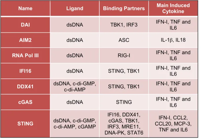
Up to now, more than ten DNA receptors or sensor have been identified (Table 1). In the recent years, significant attention has been drawn to cytosolic DNA receptors and the mechanisms by which they recognize DNA and activate the downstream signaling. One of the key findings is the discovery of a transmembrane protein, STING (also known as MITA, MPYS, ERIS and TMEM173) (Ishikawa and Barber, 2008;
the function and molecular mechanism of STING and several other important cytosolic DNA receptors.
DAI
Toll-like receptor 9 was found to be the DNA receptor that recognizes endosomal DNA and hypomethylated DNA (Krieg et al., 1995). Later on, it was found that cytosolic dsDNA could trigger innate immune response, specifically IFN-I induction, independent of TLR9. DNA-dependent activator of IFN-regulatory factor (DAI) was previously
identified in tumor stromal tissue and named DLM-1 or Z-DNA binding protein 1 (ZBP1). Overexpression of DAI led to an increased in IFN-I production after B-DNA stimulation while shRNA-mediated knockdown of DAI led to decreased IFN-I and ISGs production leading to the conclusion that DAI is important for DNA recognition. In addition, the expression level of DAI was highly inducible by B-DNA or IFN-I stimulation.
Mechanistically, DAI binds to B form of DNA in the cell cytosol and also binds to IRF3 and TBK1. The recognition of B-DNA by DAI enhances DNA binding to IRF3 and TBK1, both of which activate the downstream IFN-I pathway (Takaoka et al., 2007). However, Dai-/- mice showed normal IFN-I production after B-DNA stimulation. This suggests that there might be other DNA receptors that DAI are redundant to (Lippmann et al., 2008). This finding then led to the researchers to look for other cytosolic DNA receptors.
AIM2
However, researchers also found that Nlrp3 deficient cells displayed a normal caspse-1 activity in response to transfected double stranded DNA from various source (Hornung et al., 2008; Muruve et al., 2008). This suggests that intracellular dsDNA-induced caspase-1 activation is NLRP3 independent, and other factors are responsible for sensing intracellular dsDNA and activation of caspase-1. Using both human and mouse cells, researcher found that absent in melanoma 2 (AIM2), pyrin and HIN
domain-containing protein (PYHIN) 8 family member, serves as a receptor to cytosolic DNA and activate both caspase-1 and NF-κB (Fernandes-Alnemri et al., 2009; Hornung et al.,
2009; Rathinam et al., 2010; Schroder et al., 2009). Knockdown and knockout of Aim2 inhibited caspase-1 activation after stimulation with both cytosolic dsDNA and dsDNA viruses (Fernandes-Alnemri et al., 2009; Hornung et al., 2009). Mechanistically, AIM2 binds to dsDNA through HIN200 domain and associates with the adaptor protein
apoptosis-associated speck-like protein containing a caspase activation and recruitment domain (ASC) through pyrin domain (Fernandes-Alnemri et al., 2009; Hornung et al., 2009). Taken together, AIM2 is a cytoplasimc DNA receptor that binds to dsDNA and forms AIM2-containing inflammasome to activate caspase-1. However, AIM2 is not responsible for cytosolic dsDNA-induced IFN-I activation (Fernandes-Alnemri et al., 2009; Hornung et al., 2009; Rathinam et al., 2010; Schroder et al., 2009).
RNA POL III
mystery in the field was that when AT-rich DNA was introduced into the cytosol, RIG-I deficient cells showed decreased IFN-I production compared to WT cells. Later on, RNA polymerase III was identified as the missing link. RNA polymerase III utilizes AT-rich dsDNA as a template to transcribe dsRNA with 5’-ppp. 5’-ppp dsRNA then serves as RIG-I ligand and activates RIG-I-dependent pathways. Researchers also tested a panel of stimuli and found that RNA polymerase III mediates herpes simplex virus type 1, Epstein Barr virus (EBV) and Legionella pneumophila induced IFN-I production (Chiu et al., 2009; Choi et al., 2009). RNA polymerase III was previously known to localize in both nucleus and cytoplasm (Jaehning and Roeder, 1977), however, its function in the cytoplasm remained unknown until it was found to detect dsDNA and transcribe
cytosolic DNA into RIG-I ligand.
IFI16
complex upon stimulation with dsDNA (Unterholzner et al., 2010). Later on, it was revealed that IFI16 forms inflammasome with ASC and caspase-1 in the nucleus and peri-nuclear region upon KSHV infection (Kerur et al., 2011). However, further studies are needed to elucidate how IFN16 distinguishes self- and foreign-DNA in the nucleus and whether KSHV-induced IFN-I pathway is also dependent on IFI16.
DDX41
Several members of DExD/H-box helicase superfamily have been identified to be cytosolic nucleic acid sensors in myrloid dendritic cells as well as plasmacytoid dentritic cells, including DDX1-DDX21-DHX36 complex, DHX33, DHX36 and DHX9 (Kim et al., 2010; Mitoma et al., 2013; Zhang et al., 2011a; Zhang et al., 2011c). Yong-Jun Liu’s group screened all 59 members of the DExD/H-box helicase superfamily by using small interfering RNA (siRNA). This led them to the identification of another cytocolic DNA senser, DDX41 (Figure 3) (Cavlar et al., 2012; Zhang et al., 2011b). The results showed that overexpression of DDX41 and STING enhanced IFN-b promoter activity.
Conversely, reduction of DDX41 by shRNA resulted in a dampened IFN-I and proinflammatory cytokine production in response to dsDNA transfection and HSV-1 infection. Mechanistically, DDX41 associates with both dsDNA and STING in the cytosol and causing activation of the downstream signaling pathways, including IRF3
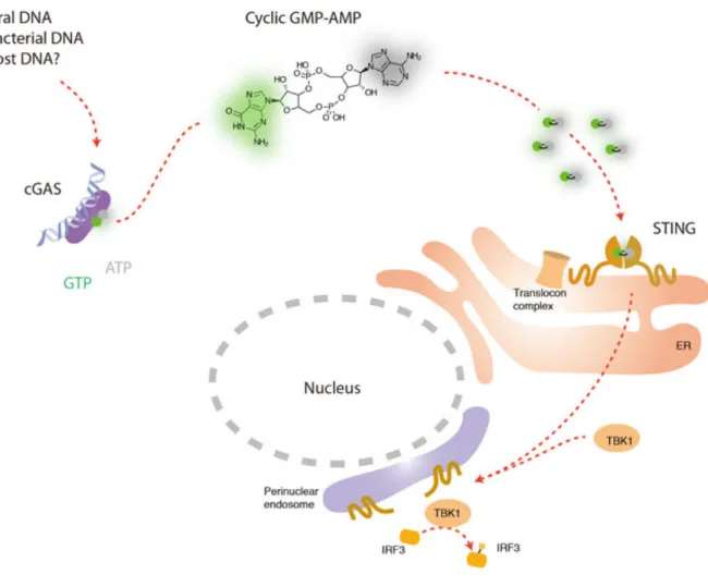
cGAS
Although it is now well accepted that STING is a central adaptor protein for cytosolic DNA sensing and that all cytosolic dsDNAs activate STING and the
downstream pathways in a sequence-independent manner, it is yet to be elucidated how DNA sensors activate STING. Chen’s group hypothesized that cytoplasmic dsDNA directly bound to DNA receptor, which then directly or indirectly activated the adaptor protein, STING. They carried out a well-designed in vitro experimental system using two cell lines, one as a screening cell line to screen for the candidate DNA receptor(s), the other as a reporter cell line to indicate the activation of STING-dependent pathways. These results showed that the potential molecule(s) induced by dsDNA stimulation and activated STING are not DNA, RNA or protein and is not heat-sensitive. Further
investigation showed that the STING-activating molecule is cyclic GMP-AMP (cGAMP) and is synthesized by cGAMP synthase (cGAS), a nucleotidyltransferase (NTase) and male abnormal 21 (Mab21) domain containing protein (Figure 4) (Ablasser and Hornung, 2013). Collectively, the studies suggested that cGAS directly bound to cytosolic dsDNA, which then led to the synthesis of a second messenger, cGAMP. cGAMP then directly bound to and activated the adaptor protein STING and STING-dependent pathways (Sun et al., 2013; Wu et al., 2013). The conclusions were later on confirmed in gene knockout cells and mice (Li et al., 2013).
convert GMP and AMP into cGAMP, which will then activate STING and induced IFN-I and proinflammatory cytokine production (Gao et al., 2013).
STING
STING, also known as mediator of interferon regulatory factor 3 activation (MITA)
(Zhong et al., 2008), plasma membrane tetraspanner (MPYS) (Jin et al., 2008) or endoplasmic reticulum interferon stimulator (ERIS) (Sun et al., 2009) and
transmembrane protein 173 (TMEM173), was first discovered by an expression cloning
technique using cDNA library (Ishikawa and Barber, 2008; Zhong et al., 2008). The
readout of the screening process was IFN-β promoter activity induced by the
transfected expression construct in 293T cells. Studies showed that STING is located to
ER, mitochondria outer membrane or mitochondria-associated ER membrane in
unstimulated cells. It is also ubiquitously expressed by all human tissues tested, and
more predominantly expressed in most macrophages, dendritic cells, T cells, B cells
and a variety of epithelial cells, endothelial cells and fibroblasts. Functionally, STING
was shown to be an adaptor protein that associates with different cytosolic DNA
sensors, such as IFI16, DDX41, cGAS (Figure 2-4). Upon activation, STING recruits a
downstream protein kinase, TBK1 and become phosphorylated by TBK1 and then
recruits transcription factor IRF3 to the interaction complex. STING has been shown to
GMP has been resolved by several groups and they showed that the CTD of STING
forms a homodimer and c-di-GMP binds to the pocket formed by this dimer (Huang et
al., 2012; Ouyang et al., 2012; Shang et al., 2012; Shu et al., 2012). The c-di-GMP
bound form of STING has a stronger association with TBK1 and resulted in an
enhanced IFN-I production (Barber, 2014; Gurtler and Bowie, 2013).
1.3 NLR proteins are negative regulators of innate immunity
The nucleotide-binding domain and leucine-rich-repeat-containing (NLR) protein family is discovered by genomic mining of proteins with similar structures as MHC class II transactivator (CIITA) (Ting et al., 2008a). NLR proteins have tripartite domain
architecture: N-terminal effector domain, central nucleotide binding domain and C-terminal leucine-rich-repeat domain. NLR family members have been further
categorized as several different sub-families according to their specific effector domains. NLRC proteins are one sub-family that contains one or more caspase recruitment
domains (CARD) (Harton et al., 2002; Inohara and Nunez, 2003; Ting et al., 2008a; Tschopp et al., 2003). Five NLR proteins belong to this sub-family, including NOD1, NOD2, NLRC3, NLRC4 and NLRC5. NLRP proteins have a pyrin domain (PYR) as effector domain. Other effector domains include transactivation domain (AD),
baculovirus inhibitor of apoptosis repeat domains (BIR) and uncharacterized domain (X) (Ting et al., 2008a).
The functional and mechanistic characterization of NLR proteins has been a major focus of research in the past decade. NLR proteins are pivotal in host proinflammatory responses to various stimuli. The NLR family includes about 22 members (human) that have distinct functions in various signaling pathways (Davis et al., 2011; Ting et al., 2010; Ting et al., 2008a). Functionally, NLR proteins can be classified into two categories: inflammasome-forming NLRs and noninflammasome NLRs (Ting et al., 2010). Inflammasome regulates activation of caspase-1 and is a big protein complex that is formed by multi-components, including NLRs, ASC and caspase-1. Activation of caspase-1 results in the proteolytic cleavage of IL-1β and IL-18 (Martinon et al., 2002). Electron microscopy (EM) has shown that inflammasome structures share striking structural similarities to the apoptosome (Faustin et al., 2007). NLRP1, 2, 3, 6, 12, NLRC4, NOD2 are all found to form inflammasome with caspase-1, with or without the adaptor ASC to facilitate IL-1β processing when ectopically overexpressed (Agostini et al., 2004; Grenier et al., 2002; Martinon et al., 2002; Poyet et al., 2001; Wang et al., 2002).
Although inflammasome-forming NLRs are thought to be crucial in infectious disease, metabolic disorders, tumorigenesis and autoimmune disorders, studies are converging to suggest that non-inflammasome NLRs are equally important (Ting et al., 2010). Studies have shown that NLRs function in the regulation of type I interferon production, NF-κB activity, MAPK activation, host antimicrobial response and sterile
inflammation. NOD1 and NOD2 represent the first NLRs that mediate
nonflammasome NLRs as negative regulators of innate immunity. We will focus on introducing the functions of several negative-regulator-NLRs in this section.
NLRX1
NLRX1 was the first NLR molecule shown to regulate intracellular RNA and RNA virus sensing pathway. Overexpression of NLRX1 dampened IFN-I and proinflammatory cytokine production in response to several RNA virus and RNA analogue poly(I:C). Knockdown of NLRX1 resulted in an enhanced production of IFN-I and proinflammatory cytokine (Moore et al., 2008). These findings were later confirmed by using
gene-deletion cells and mice (Allen et al., 2011; Xia et al., 2011). Mechanistically, NLRX1 localized to mitochondria membrane and associated with as MAVS to block MAVS-RIG-I association, which is required for the downstream signaling. NLRX1 has also been shown to directly bind to dsRNA by crystallography (Hong et al., 2012; Xiao and Ting, 2012). Studies performed in Nlrx1-/- mice also showed that NLRX1 is crucial in
regulating lung inflammation in response to Influenza A virus (IAV) infection or
intranasal delivery of LPS. Nlrx1-/- mice showed an enhanced production of IFN-β and
increased IFN-I and proinflammatory cytokine production and a decreased
autophagosome formation, this gave the host cell an advantage to fight against viral infection. Mechanistically, NLRX1 recruits TUFM, which in turn recruits ATG5-ATG12 and forms a protein complex to regulate autophagy response.
Most recently, the function of NLRX1 has again been expanded. The literature showed that Nlrx1-/- mice are more susceptible to experimental autoimmune
encephalomyelitis (EAE), a mouse model for multiple sclerosis (MS) (Eitas et al., 2014). Phenotypically, Nlrx1-/- mice showed a worsened clinical scores and increased tissue damage in central nervous system (CNS) during EAE, accompanied by increased inflammatory and cytokine responses (Eitas et al., 2014). This again affirms that NLRX1 reduces inflammatory responses.
NLRP6
NLRP6 was shown to suppress bacteria infection-induced NF-κB activation and
MAPK activation (Anand and Kanneganti, 2012; Anand et al., 2012). During Listeria monocytogenes and Salmonella typhimurium infection, Nlrp6 deficiency led to an elevated level of p65 activation and ERK phosphorylation. Nlrp6 deficient cells also showed faster bacteria clearance.
affected due to deficiency of Nlrp6. Further biochemical and molecular evidence are yet to be done.
NLRP12
NLRP12 was the first studied inhibitory NLR protein. It was found to inhibit both canonical (Williams et al., 2005) and noncanonical NF-κB pathway (Lich et al., 2007).
The inhibitory function of NLRP12 in the noncanonical pathway is through its association of NIK. It has been shown that upon binding to NIK, NLRP12 can de-stabilize NIK and thus inhibit the downstream signaling. However, when looking at the activation of canonical NF-κB activation, a modest effect was observed when NLRP12
expression was knocked down with shRNA, although it was confirmed later in gene deletion cells that NLRP12 inhibits canonical NF-κB activation (Zaki et al., 2011).
The function of NLRP12 in regulating NF-κB activation led researchers to the
studies of the role of NLRP12 in inflammation-associated diseases. In a colitis and colitis-associated colon cancer model, Nlrp12-/- animals showed an increased number of polyps and increased disease susceptibility (Allen et al., 2012; Zaki et al., 2011).
Mechanistically, canonical and noncanonical NF-κB was hyper-activated in Nlrp12
-/-mice and resulted in an elevated production of the downstream chemokines and proinflammatory cytokines, including Cxcl12, Cxcl13, TNF and IL6, which are
canonical and noncanonical NF-κB activation, inflammation and
inflammation-associated tumorigenesis.
It has also been proposed that NLRP12 forms an inflammasome under certain stimuli, such as attenuated Yersinia infection (Vladimer et al., 2012). It was shown that NLRP12 recognizes acylated lipid A in in vitro assays. In addition, Nlrp12−/− mice
showed a dampened IL-18 production and were more susceptible to an attenuated strain of Yersinia pestis infection. In humans, there is an association between
inflammasome activation and NLRP12 polymorphisms during periodic fever syndromes (Jeru et al., 2008). Most recently, it was shown that NLRP12 negatively regulates
Salmonella enterica serovar Typhimurium-induced innate immune responses (Zaki et al., 2014). This effect is independent of inflammasome activation. In this study, it was found that Nlrp12−/− mice showed reduced bacteria burden in both the liver and spleen after
salmonella infection, but increased inflammatory cytokine and antibacterial peptide production. A further investigation showed NLRP12 inhibits canonical NF-κB and ERK
activation, as well as NO production.
1.4 The intersection of innate immunity and DNA damage-repair response Brief overview of DNA damage response
chemicals (Ciccia and Elledge, 2010; Jackson and Bartek, 2009). Though the majority of DNA damages are challenges to genomic integrity, there are also programmed-DNA lesions, such as developmentally-regulated genome rearrangements in lymphocytes and germ cells (Jackson and Bartek, 2009; Longhese et al., 2009; Tsai and Lieber, 2010).
DNA damage could be detrimental to cells if dysregulated, leading to gene mutation, aberration of chromosomal structures and loss of genetic information. Thus, eukaryotic cells have evolved a signal transduction network during DNA damage, named DNA damage response (DDR), in order to preserve genome integrity and avoid the adverse effects caused by DNA damage (Hoeijmakers, 2001; Tsai and Lieber, 2010).
DDR signaling pathway are tightly regulated temporally and spatially to sense DNA damage as soon as it occurs, then amplify the signal by recruiting the responsible factors and then activate the downstream effector proteins to repair damage and
appropriately determine cell fate (Ciccia and Elledge, 2010; Jackson and Bartek, 2009). The signal transduction of DDR is through protein-protein interaction and cascades of protein post-translational modifications, which allow the signaling to be fast and reversibly-regulated (Polo and Jackson, 2011).
The major players in DDR signaling pathway (Figure 5) is a group of
recombination (Lee and Kim, 2002; Smith and Jackson, 1999). ATM senses the double strand DNA breaks (DSB), the most toxic form of DNA damage to cells. ATM directly phosphorylates the relevant transcription factors, such as the tumor suppressor p53 (Banin et al., 1998; Canman et al., 1998). There is also evidence that DNA damage can activate the transcription factor NF-κB in an ATM-dependent manner (Piret et al., 1999).
However, the mechanism remains to be elucidated.
DNA damage response-associated diseases
The DNA-damage-and-repair response is an ancient and conserved way of protecting our genomic stability and integrity (Kastan, 2008). The consequence of DNA damage if left unrepaired is diverse (Bohr, 2002). Acute and short-term DNA-damage results in cell cycle-arrest or even cell death. Accumulating and long-term DNA damage leads to gene mutation and oncogenesis, as well as other genetic diseases (Greenberg et al., 2006; O'Driscoll, 2012).
These four different pathways are partly overlapping and can cover almost all kinds of DNA damage (Maisonobe et al., 2013).
Homologous recombination and end-joining pathways are developed to deal with double strand breaks (DSBs), which is the most toxic and problematic form of DNA damage (Carr and Lambert, 2013). Homologous recombination usually takes place in S and G2 phase of DNA replication, when DNA is already replicated and has a second copy to align the breaks. On the other hand, end joining is less accurate and usually happens during G1 phase of cell cycle when the sister chromatin is not available.
BER and NER are in charge of repairing damages on a single strand of DNA (Seo and Jung, 2004). BER is responsible for repairing small chemical alteration of bases. This kind of lesions often cause miscoding and lead to gene mutation though they sometimes do not even impede transcription or replication. Thus, BER is
particularly crucial for preventing gene mutagenesis (Parsons and Dianov, 2013). NER targets a wide class of helix-distorting lesions, which usually interfere with transcription and replication (Melis et al., 2013). The other interesting fact worth mentioning is that BER-targeted lesions are usually endogenously originated, though not exclusively so, whereas NER-targeted lesions are usually caused by exogenous insults.
As we mentioned above, DNA damage can be very toxic if left unattended. One of the clearest associations of DNA damage with diseases is cancer (Wallace et al., 2012).
Genome instability is a fundamental feature of cancer. Cancer can be thought of as a disease of our genes and is driven by genome instability and fuelled by a variety of DNA damage and replication errors (O'Driscoll, 2012). An example that illustrates the intimate relationship between cancer and DNA damage is that most carcinogens work by causing DNA damage and generating gene mutations. In addition, inherited DNA repair defects often serve as predisposing factors for cancer, such as xeroderma pigmentosum (XP) (Niedernhofer et al., 2011; Schwertman et al., 2013), a syndrome associated with inborn defects in NER (Sepe et al., 2013). Patients with XP exhibit more than 1000-fold of incidence of sun-induced skin cancer compare to patients without the disease. Mutations in mice that target almost all of the factors in NER signaling pathway showed a pronounced cancer predisposition (de Boer and
Hoeijmakers, 1999). Another prime example is the inherited mutations in BRCA1 and BRCA2, which are associated with a strong predisposition to breast cancer (Lord and Ashworth, 2013).
Oncogene activation or tumor suppressor inactivation can both lead to abnormal cell proliferation, which generates DNA-replication-associated genomic stress, thus activating DDR, more specifically, ATM/ATR-dependent pathways to induce cell death or senescence (Bartkova et al., 2005; Gorgoulis et al., 2005).
Thus, DDR is activated to create a barrier to protect against tumorigenesis and malignancy. However, defects in this DDR barrier can also lead to higher frequency of cancer incidence.
DDR factors are required during immune system development, specifically during V(D)J recombination to generate a big repertoire of immune cell receptors. Ataxia
telangiectasia (AT) patients and Nijmegen breakage syndrome (NBS) patients have mutation or defects in ATM and NBS1, respectively (Marechal and Zou, 2013). One of the most challenging symptoms is that those patients are highly prone to sometimes-fatal infections, and this is largely due to immune deficiency. Furthermore, class-switch recombination is affected in AT patients.
Aging
There is an emerging interest in understanding the mechanism of aging as a means to increase longevity and mitigate aging-associated diseases. One of the theories for the cause of aging is the accumulation of DNA lesions (Schumacher et al., 2008). The links between DNA damage and aging are briefly summarized as followings. Firstly, patients with inherited defects in DDR often suffer from a premature or
accelerated aging symptom, such as Werner’s syndrome, which is caused by mutation in WRN gene; Hutchinson-Gilford progeria syndrome (HGPS), which has defects in Lamin-A (responsible for DSB repair signaling pathway) (Musich and Zou, 2011). Researchers also generated several mouse models with defects in DDR factors, some of the animals display a dramatic accelerated or premature ageing pheonotype,
thought to contribute to aging by exhaustion of stem cell in the process of tissue regeneration. DNA damage was thought to contribute to aging by inducing cell senescence and apoptosis, as well as causing stem cell exhaustion (Insinga et al., 2014).
Interplay of DNA damage and innate immune responses
Evolutionarily, DNA damage response should arose earlier than immune response given the fact that cells must protect itself from errors in replication or environmental stimulus that could break genomic integrity for basic cell survival. DNA damage responses have been studied intensively in the last 50 years, and much has been elucidated. By contrast the molecular mechanisms by which innate immune responses respond to and recognize nucleic acids has a history that is less than a decade. However, there are accumulating evidence showing the crosstalk between these two distinct and interwoven pathways. Several work has shown that factors in DNA damage response pathway play a role in innate immune signaling and vice versa. Understanding the interplay of DNA damage responses and innate immune responses potentially have translational implications and could help develop new treatment for immune disorders. Next, we will review several the factors that are involved in both pathways.
IFI16
2010). It was then shown that IFI16 is also involved in autoimmune disorders (Costa et al., 2011). Specifically, antibodies against IFI16 were found in the sera of SLE patients. In assays performed in cell culture, IFI16 translocates to the cytoplasm from nucleus after UV-B irradiation. Although the detailed mechanisms are lacking, this finding provides evidence that innate immune sensors can be involved in cellular response against UV-B irradiation.
DNA-PK
On the contrary to IFI16, DNA-PK was first found to be crucial in DDR. It was one of several major players in DSB signaling pathway (Anderson, 1993; Anderson and Lees-Miller, 1992; Sirbu and Cortez, 2013). DNA-PK can be activated and recruited by Ku70 and 80, which recognize DSB (Sirbu and Cortez, 2013). Previous work also showed that DNA-PK is able to activate NF-κB through activation of MEK-ERK-IKK
signaling pathway after DNA damage (Sabatel et al., 2011). Recently, DNA-PK was characterized as a cytosolic dsDNA sensor that directly binds to bacterial or viral dsDNA and activate transcription factor NF-κB and IRF3 which then can trigger IFN-I production
(Ferguson et al., 2012). There are also studies showing that DNA-PK can interact with transcription factor Aire, which then upregulates the expression level of TLR1, TLR3 and TLR8 (Kong et al., 2011).
Collectively, DNA-PK is crucial in both innate immune response and DNA
cellular response to the stimuli. It is also worth looking at how different pathways downstream of DNA-PK affect each other.
MRE11
MRE11 was part of the MRE11-RAD50-NBS1 (MRN) complex that is responsible for DSB repair signaling pathway. MRN complex was shown to bind to DSB directly in vitro and in vivo and was thought to be important for the initiation of DSB repair process and ATM recruitment and activation (Lavin, 2007; Lee and Paull, 2007). Later on, MRE11 was shown to directly bind to cytosolic dsDNA and activate IFN-I response in a STING-dependent manner. MRE11 can also regulate STING trafficking upon binding to cytosolic dsDNA, which is crucial for downstream signaling (Kondo et al., 2013). These findings again reinforce point that there is crosstalk between DNA damage response and innate immune response.
AIM2
Absent in melanoma 2 (AIM2) was named based on the finding that the expression of AIM2 is extremely low in melanoma. However, there is no detailed mechanistic study to show how AIM2 expression is regulated in melanoma or the significance of the low expression of AIM2. Functional characterization of AIM2 showed that AIM2 forms inflammasome with ASC and caspase-1 in the cytosol to process pro-IL1b and pro-IL18 to mature form in response to intracellular dsDNA stimulation (Fernandes-Alnemri et al., 2009; Hornung et al., 2009; Rathinam et al., 2010).
in a DSS/AOM animal model. Mechanistically, AIM2 associate with DNA-PK and AKT to downregulate tumorigenesis in an inflammasome-independent manner. It will be
Table 1: Cytosolic DNA sensors
Name Ligand Binding Partners Main Induced Cytokine
DAI dsDNA TBK1, IRF3 IFN-I, TNF and IL6
AIM2 dsDNA ASC IL-1β, IL18
RNA Pol III dsDNA RIG-I IFN-I, TNF and
IL6
IFI16 dsDNA STING, TBK1 IFN-I, TNF and IL6
DDX41 dsDNA, c-di-GMP, c-di-AMP STING, TBK1 IFN-I, TNF and IL6
cGAS dsDNA STING IFN-I, TNF and
IL6
STING dsDNA, c-di-GMP, c-di-AMP, cGAMP
IFI16, DDX41, cGAS, TBK1, IRF3, MRE11, DNA-PK, STAT6
IFN-I, CCL2, CCL20, MCP-3,
Figure 1. Canonical and noncanonical NF-κB signaling pathway
Figure 2. Nucleic acid induced IRF3/7 activation
Figure 3. IFI16 and DDX41 downstream signaling
Figure 4. cGAS downstream signaling
Figure 5. DNA damage response pathway
REFERENCES
Ablasser, A., and Hornung, V. (2013). DNA sensing unchained. Cell research 23, 585-587.
Agostini, L., Martinon, F., Burns, K., McDermott, M.F., Hawkins, P.N., and Tschopp, J. (2004). NALP3 forms an IL-1beta-processing inflammasome with increased activity in Muckle-Wells autoinflammatory disorder. Immunity 20, 319-325.
Allen, I.C., Moore, C.B., Schneider, M., Lei, Y., Davis, B.K., Scull, M.A., Gris, D., Roney, K.E., Zimmermann, A.G., Bowzard, J.B., et al. (2011). NLRX1 protein attenuates
inflammatory responses to infection by interfering with the RIG-I-MAVS and TRAF6-NF-kappaB signaling pathways. Immunity 34, 854-865.
Allen, I.C., Wilson, J.E., Schneider, M., Lich, J.D., Roberts, R.A., Arthur, J.C., Woodford, R.M., Davis, B.K., Uronis, J.M., Herfarth, H.H., et al. (2012). NLRP12 suppresses colon inflammation and tumorigenesis through the negative regulation of noncanonical NF-kappaB signaling. Immunity 36, 742-754.
Anand, P.K., and Kanneganti, T.D. (2012). Targeting NLRP6 to enhance immunity against bacterial infections. Future microbiology 7, 1239-1242.
Anand, P.K., Malireddi, R.K., Lukens, J.R., Vogel, P., Bertin, J., Lamkanfi, M., and Kanneganti, T.D. (2012). NLRP6 negatively regulates innate immunity and host defence against bacterial pathogens. Nature 488, 389-393.
Anderson, C.W. (1993). DNA damage and the DNA-activated protein kinase. Trends in biochemical sciences 18, 433-437.
Anderson, C.W., and Lees-Miller, S.P. (1992). The nuclear serine/threonine protein kinase DNA-PK. Critical reviews in eukaryotic gene expression 2, 283-314.
Atianand, M.K., and Fitzgerald, K.A. (2013). Molecular basis of DNA recognition in the immune system. Journal of immunology 190, 1911-1918.
Banin, S., Moyal, L., Shieh, S., Taya, Y., Anderson, C.W., Chessa, L., Smorodinsky, N.I., Prives, C., Reiss, Y., Shiloh, Y., and Ziv, Y. (1998). Enhanced phosphorylation of p53 by ATM in response to DNA damage. Science 281, 1674-1677.
Barber, G.N. (2014). STING-dependent cytosolic DNA sensing pathways. Trends in immunology 35, 88-93.
Barnes, B.J., Moore, P.A., and Pitha, P.M. (2001). Virus-specific activation of a novel interferon regulatory factor, IRF-5, results in the induction of distinct interferon alpha genes. The Journal of biological chemistry 276, 23382-23390.
Bartkova, J., Horejsi, Z., Koed, K., Kramer, A., Tort, F., Zieger, K., Guldberg, P., Sehested, M., Nesland, J.M., Lukas, C., et al. (2005). DNA damage response as a candidate anti-cancer barrier in early human tumorigenesis. Nature 434, 864-870.
Blasius, A.L., and Beutler, B. (2010). Intracellular toll-like receptors. Immunity 32, 305-315.
Bohr, V.A. (2002). Repair of oxidative DNA damage in nuclear and mitochondrial DNA, and some changes with aging in mammalian cells. Free radical biology & medicine 32, 804-812.
Bulek, K., Liu, C., Swaidani, S., Wang, L., Page, R.C., Gulen, M.F., Herjan, T., Abbadi, A., Qian, W., Sun, D., et al. (2011). The inducible kinase IKKi is required for IL-17-dependent signaling associated with neutrophilia and pulmonary inflammation. Nature immunology 12, 844-852.
Burdette, D.L., Monroe, K.M., Sotelo-Troha, K., Iwig, J.S., Eckert, B., Hyodo, M., Hayakawa, Y., and Vance, R.E. (2011). STING is a direct innate immune sensor of cyclic di-GMP. Nature 478, 515-518.
Canman, C.E., Lim, D.S., Cimprich, K.A., Taya, Y., Tamai, K., Sakaguchi, K., Appella, E., Kastan, M.B., and Siliciano, J.D. (1998). Activation of the ATM kinase by ionizing radiation and phosphorylation of p53. Science 281, 1677-1679.
Carr, A.M., and Lambert, S. (2013). Replication stress-induced genome instability: the dark side of replication maintenance by homologous recombination. Journal of
Cavlar, T., Ablasser, A., and Hornung, V. (2012). Induction of type I IFNs by intracellular DNA-sensing pathways. Immunology and cell biology 90, 474-482.
Chiu, Y.H., Macmillan, J.B., and Chen, Z.J. (2009). RNA polymerase III detects
cytosolic DNA and induces type I interferons through the RIG-I pathway. Cell 138, 576-591.
Choi, M.K., Wang, Z., Ban, T., Yanai, H., Lu, Y., Koshiba, R., Nakaima, Y., Hangai, S., Savitsky, D., Nakasato, M., et al. (2009). A selective contribution of the RIG-I-like receptor pathway to type I interferon responses activated by cytosolic DNA.
Proceedings of the National Academy of Sciences of the United States of America 106, 17870-17875.
Ciccia, A., and Elledge, S.J. (2010). The DNA damage response: making it safe to play with knives. Molecular cell 40, 179-204.
Claudio, E., Brown, K., Park, S., Wang, H., and Siebenlist, U. (2002). BAFF-induced NEMO-independent processing of NF-kappa B2 in maturing B cells. Nature immunology 3, 958-965.
Costa, S., Borgogna, C., Mondini, M., De Andrea, M., Meroni, P.L., Berti, E., Gariglio, M., and Landolfo, S. (2011). Redistribution of the nuclear protein IFI16 into the
cytoplasm of ultraviolet B-exposed keratinocytes as a mechanism of autoantigen processing. The British journal of dermatology 164, 282-290.
Darnay, B.G., Ni, J., Moore, P.A., and Aggarwal, B.B. (1999). Activation of NF-kappaB by RANK requires tumor necrosis factor receptor-associated factor (TRAF) 6 and NF-kappaB-inducing kinase. Identification of a novel TRAF6 interaction motif. The Journal of biological chemistry 274, 7724-7731.
Darnell, J.E., Jr., Kerr, I.M., and Stark, G.R. (1994). Jak-STAT pathways and
transcriptional activation in response to IFNs and other extracellular signaling proteins. Science 264, 1415-1421.
Davis, B.K., Wen, H., and Ting, J.P. (2011). The inflammasome NLRs in immunity, inflammation, and associated diseases. Annual review of immunology 29, 707-735.
Delhase, M., Hayakawa, M., Chen, Y., and Karin, M. (1999). Positive and negative regulation of IkappaB kinase activity through IKKbeta subunit phosphorylation. Science 284, 309-313.
Ea, C.K., Deng, L., Xia, Z.P., Pineda, G., and Chen, Z.J. (2006). Activation of IKK by TNFalpha requires site-specific ubiquitination of RIP1 and polyubiquitin binding by NEMO. Molecular cell 22, 245-257.
Eitas, T.K., Chou, W.C., Wen, H., Gris, D., Robbins, G.R., Brickey, J., Oyama, Y., and Ting, J.P. (2014). The Nucleotide-binding Leucine-rich Repeat (NLR) Family Member NLRX1 Mediates Protection against Experimental Autoimmune Encephalomyelitis and Represses Macrophage/Microglia-induced Inflammation. The Journal of biological chemistry 289, 4173-4179.
Elinav, E., Strowig, T., Kau, A.L., Henao-Mejia, J., Thaiss, C.A., Booth, C.J., Peaper, D.R., Bertin, J., Eisenbarth, S.C., Gordon, J.I., and Flavell, R.A. (2011). NLRP6
inflammasome regulates colonic microbial ecology and risk for colitis. Cell 145, 745-757.
Faustin, B., Lartigue, L., Bruey, J.M., Luciano, F., Sergienko, E., Bailly-Maitre, B., Volkmann, N., Hanein, D., Rouiller, I., and Reed, J.C. (2007). Reconstituted NALP1 inflammasome reveals two-step mechanism of caspase-1 activation. Molecular cell 25, 713-724.
Ferguson, B.J., Mansur, D.S., Peters, N.E., Ren, H., and Smith, G.L. (2012). DNA-PK is a DNA sensor for IRF-3-dependent innate immunity. eLife 1, e00047.
Fernandes-Alnemri, T., Yu, J.W., Datta, P., Wu, J., and Alnemri, E.S. (2009). AIM2 activates the inflammasome and cell death in response to cytoplasmic DNA. Nature 458, 509-513.
Gao, D., Wu, J., Wu, Y.T., Du, F., Aroh, C., Yan, N., Sun, L., and Chen, Z.J. (2013). Cyclic GMP-AMP synthase is an innate immune sensor of HIV and other retroviruses. Science 341, 903-906.
Garceau, N., Kosaka, Y., Masters, S., Hambor, J., Shinkura, R., Honjo, T., and Noelle, R.J. (2000). Lineage-restricted function of nuclear factor kappaB-inducing kinase (NIK) in transducing signals via CD40. The Journal of experimental medicine 191, 381-386.
Ghosh, S., May, M.J., and Kopp, E.B. (1998). NF-kappa B and Rel proteins:
evolutionarily conserved mediators of immune responses. Annual review of immunology 16, 225-260.
Gorgoulis, V.G., Vassiliou, L.V., Karakaidos, P., Zacharatos, P., Kotsinas, A., Liloglou, T., Venere, M., Ditullio, R.A., Jr., Kastrinakis, N.G., Levy, B., et al. (2005). Activation of the DNA damage checkpoint and genomic instability in human precancerous lesions. Nature 434, 907-913.
Greenberg, R.A., Sobhian, B., Pathania, S., Cantor, S.B., Nakatani, Y., and Livingston, D.M. (2006). Multifactorial contributions to an acute DNA damage response by
BRCA1/BARD1-containing complexes. Genes & development 20, 34-46.
Grenier, J.M., Wang, L., Manji, G.A., Huang, W.J., Al-Garawi, A., Kelly, R., Carlson, A., Merriam, S., Lora, J.M., Briskin, M., et al. (2002). Functional screening of five PYPAF family members identifies PYPAF5 as a novel regulator of NF-kappaB and caspase-1. FEBS letters 530, 73-78.
Gurtler, C., and Bowie, A.G. (2013). Innate immune detection of microbial nucleic acids. Trends in microbiology 21, 413-420.
Hacker, H., and Karin, M. (2006). Regulation and function of IKK and IKK-related kinases. Science's STKE : signal transduction knowledge environment 2006, re13.
Harton, J.A., Linhoff, M.W., Zhang, J., and Ting, J.P. (2002). Cutting edge: CATERPILLER: a large family of mammalian genes containing CARD, pyrin,
nucleotide-binding, and leucine-rich repeat domains. Journal of immunology 169, 4088-4093.
He, J.Q., Zarnegar, B., Oganesyan, G., Saha, S.K., Yamazaki, S., Doyle, S.E.,
Dempsey, P.W., and Cheng, G. (2006). Rescue of TRAF3-null mice by p100 NF-kappa B deficiency. The Journal of experimental medicine 203, 2413-2418.
Hemmi, H., Takeuchi, O., Sato, S., Yamamoto, M., Kaisho, T., Sanjo, H., Kawai, T., Hoshino, K., Takeda, K., and Akira, S. (2004). The roles of two IkappaB kinase-related kinases in lipopolysaccharide and double stranded RNA signaling and viral infection. The Journal of experimental medicine 199, 1641-1650.
factor. Journal of interferon & cytokine research : the official journal of the International Society for Interferon and Cytokine Research 19, 1-13.
Hoeijmakers, J.H. (2001). Genome maintenance mechanisms for preventing cancer. Nature 411, 366-374.
Hoffmann, A., and Baltimore, D. (2006). Circuitry of nuclear factor kappaB signaling. Immunological reviews 210, 171-186.
Hong, M., Yoon, S.I., and Wilson, I.A. (2012). Structure and functional characterization of the RNA-binding element of the NLRX1 innate immune modulator. Immunity 36, 337-347.
Hornung, V., Ablasser, A., Charrel-Dennis, M., Bauernfeind, F., Horvath, G., Caffrey, D.R., Latz, E., and Fitzgerald, K.A. (2009). AIM2 recognizes cytosolic dsDNA and forms a caspase-1-activating inflammasome with ASC. Nature 458, 514-518.
Hornung, V., Bauernfeind, F., Halle, A., Samstad, E.O., Kono, H., Rock, K.L., Fitzgerald, K.A., and Latz, E. (2008). Silica crystals and aluminum salts activate the NALP3
inflammasome through phagosomal destabilization. Nature immunology 9, 847-856.
Huang, Y.H., Liu, X.Y., Du, X.X., Jiang, Z.F., and Su, X.D. (2012). The structural basis for the sensing and binding of cyclic di-GMP by STING. Nature structural & molecular biology 19, 728-730.
Inohara, N., and Nunez, G. (2003). NODs: intracellular proteins involved in inflammation and apoptosis. Nature reviews. Immunology 3, 371-382.
Insinga, A., Cicalese, A., and Pelicci, P.G. (2014). DNA damage response in adult stem cells. Blood cells, molecules & diseases 52, 147-151.
Isaacs, A., Cox, R.A., and Rotem, Z. (1963). Foreign nucleic acids as the stimulus to make interferon. Lancet 2, 113-116.
Ishii, K.J., Coban, C., Kato, H., Takahashi, K., Torii, Y., Takeshita, F., Ludwig, H., Sutter, G., Suzuki, K., Hemmi, H., et al. (2006). A Toll-like receptor-independent antiviral
Ishikawa, H., and Barber, G.N. (2008). STING is an endoplasmic reticulum adaptor that facilitates innate immune signalling. Nature 455, 674-678.
Ishikawa, H., Ma, Z., and Barber, G.N. (2009). STING regulates intracellular DNA-mediated, type I interferon-dependent innate immunity. Nature 461, 788-792.
Iyama, T., and Wilson, D.M., 3rd (2013). DNA repair mechanisms in dividing and non-dividing cells. DNA repair 12, 620-636.
Jackson, S.P., and Bartek, J. (2009). The DNA-damage response in human biology and disease. Nature 461, 1071-1078.
Jaehning, J.A., and Roeder, R.G. (1977). Transcription of specific adenovirus genes in isolated nuclei by exogenous RNA polymerases. The Journal of biological chemistry 252, 8753-8761.
Janeway CA Jr, T.P., Walport M, Shlomchik M (2001). Immumobiology, 5 edn (New York: Garland).
Janeway, C.A., Jr. (1993). How the immune system recognizes invaders. Scientific American 269, 72-79.
Jeru, I., Duquesnoy, P., Fernandes-Alnemri, T., Cochet, E., Yu, J.W., Lackmy-Port-Lis, M., Grimprel, E., Landman-Parker, J., Hentgen, V., Marlin, S., et al. (2008). Mutations in NALP12 cause hereditary periodic fever syndromes. Proceedings of the National
Academy of Sciences of the United States of America 105, 1614-1619.
Jin, L., Waterman, P.M., Jonscher, K.R., Short, C.M., Reisdorph, N.A., and Cambier, J.C. (2008). MPYS, a novel membrane tetraspanner, is associated with major
histocompatibility complex class II and mediates transduction of apoptotic signals. Molecular and cellular biology 28, 5014-5026.
Kastan, M.B. (2008). DNA damage responses: mechanisms and roles in human disease: 2007 G.H.A. Clowes Memorial Award Lecture. Molecular cancer research : MCR 6, 517-524.
Kaufmann, S.H. (2007). The contribution of immunology to the rational design of novel antibacterial vaccines. Nature reviews. Microbiology 5, 491-504.
Kawai, T., and Akira, S. (2010). The role of pattern-recognition receptors in innate immunity: update on Toll-like receptors. Nature immunology 11, 373-384.
Kerur, N., Veettil, M.V., Sharma-Walia, N., Bottero, V., Sadagopan, S., Otageri, P., and Chandran, B. (2011). IFI16 acts as a nuclear pathogen sensor to induce the
inflammasome in response to Kaposi Sarcoma-associated herpesvirus infection. Cell host & microbe 9, 363-375.
Kim, T., Pazhoor, S., Bao, M., Zhang, Z., Hanabuchi, S., Facchinetti, V., Bover, L., Plumas, J., Chaperot, L., Qin, J., and Liu, Y.J. (2010). Aspartate-glutamate-alanine-histidine box motif (DEAH)/RNA helicase A helicases sense microbial DNA in human plasmacytoid dendritic cells. Proceedings of the National Academy of Sciences of the United States of America 107, 15181-15186.
Kondo, T., Kobayashi, J., Saitoh, T., Maruyama, K., Ishii, K.J., Barber, G.N., Komatsu, K., Akira, S., and Kawai, T. (2013). DNA damage sensor MRE11 recognizes cytosolic double-stranded DNA and induces type I interferon by regulating STING trafficking. Proceedings of the National Academy of Sciences of the United States of America 110, 2969-2974.
Kong, X., Shen, Y., Jiang, N., Fei, X., and Mi, J. (2011). Emerging roles of DNA-PK besides DNA repair. Cellular signalling 23, 1273-1280.
Krieg, A.M., Yi, A.K., Matson, S., Waldschmidt, T.J., Bishop, G.A., Teasdale, R.,
Koretzky, G.A., and Klinman, D.M. (1995). CpG motifs in bacterial DNA trigger direct B-cell activation. Nature 374, 546-549.
Lavin, M.F. (2007). ATM and the Mre11 complex combine to recognize and signal DNA double-strand breaks. Oncogene 26, 7749-7758.
Lee, S.H., and Kim, C.H. (2002). DNA-dependent protein kinase complex: a
multifunctional protein in DNA repair and damage checkpoint. Molecules and cells 13, 159-166.
Lei, Y., Wen, H., Yu, Y., Taxman, D.J., Zhang, L., Widman, D.G., Swanson, K.V., Wen, K.W., Damania, B., Moore, C.B., et al. (2012). The mitochondrial proteins NLRX1 and TUFM form a complex that regulates type I interferon and autophagy. Immunity 36, 933-946.
Li, Q., and Verma, I.M. (2002). NF-kappaB regulation in the immune system. Nature reviews. Immunology 2, 725-734.
Li, X.D., Wu, J., Gao, D., Wang, H., Sun, L., and Chen, Z.J. (2013). Pivotal roles of cGAS-cGAMP signaling in antiviral defense and immune adjuvant effects. Science 341, 1390-1394.
Liao, G., Zhang, M., Harhaj, E.W., and Sun, S.C. (2004). Regulation of the NF-kappaB-inducing kinase by tumor necrosis factor receptor-associated factor 3-induced
degradation. The Journal of biological chemistry 279, 26243-26250.
Lich, J.D., Williams, K.L., Moore, C.B., Arthur, J.C., Davis, B.K., Taxman, D.J., and Ting, J.P. (2007). Monarch-1 suppresses non-canonical NF-kappaB activation and
p52-dependent chemokine expression in monocytes. Journal of immunology 178, 1256-1260.
Lin, R., Heylbroeck, C., Pitha, P.M., and Hiscott, J. (1998). Virus-dependent phosphorylation of the IRF-3 transcription factor regulates nuclear translocation,
transactivation potential, and proteasome-mediated degradation. Molecular and cellular biology 18, 2986-2996.
Lindahl, T., and Barnes, D.E. (2000). Repair of endogenous DNA damage. Cold Spring Harbor symposia on quantitative biology 65, 127-133.
Lippmann, J., Rothenburg, S., Deigendesch, N., Eitel, J., Meixenberger, K., van Laak, V., Slevogt, H., N'Guessan P, D., Hippenstiel, S., Chakraborty, T., et al. (2008). IFNbeta responses induced by intracellular bacteria or cytosolic DNA in different human cells do not require ZBP1 (DLM-1/DAI). Cellular microbiology 10, 2579-2588.
Longhese, M.P., Bonetti, D., Guerini, I., Manfrini, N., and Clerici, M. (2009). DNA double-strand breaks in meiosis: checking their formation, processing and repair. DNA repair 8, 1127-1138.
Lord, C.J., and Ashworth, A. (2013). Mechanisms of resistance to therapies targeting BRCA-mutant cancers. Nature medicine 19, 1381-1388.
Lund, J., Sato, A., Akira, S., Medzhitov, R., and Iwasaki, A. (2003). Toll-like receptor 9-mediated recognition of Herpes simplex virus-2 by plasmacytoid dendritic cells. The Journal of experimental medicine 198, 513-520.
Maisonobe, M., Giglia-Mari, G., and Eckert, D. (2013). DNA repair: a changing geography? (1964-2008). DNA repair 12, 466-471.
Mamane, Y., Heylbroeck, C., Genin, P., Algarte, M., Servant, M.J., LePage, C., DeLuca, C., Kwon, H., Lin, R., and Hiscott, J. (1999). Interferon regulatory factors: the next
generation. Gene 237, 1-14.
Marechal, A., and Zou, L. (2013). DNA damage sensing by the ATM and ATR kinases. Cold Spring Harbor perspectives in biology 5.
Martinon, F., Burns, K., and Tschopp, J. (2002). The inflammasome: a molecular platform triggering activation of inflammatory caspases and processing of proIL-beta. Molecular cell 10, 417-426.
Medzhitov, R. (2007). Recognition of microorganisms and activation of the immune response. Nature 449, 819-826.
Melis, J.P., van Steeg, H., and Luijten, M. (2013). Oxidative DNA damage and nucleotide excision repair. Antioxidants & redox signaling 18, 2409-2419.
Mitoma, H., Hanabuchi, S., Kim, T., Bao, M., Zhang, Z., Sugimoto, N., and Liu, Y.J. (2013). The DHX33 RNA helicase senses cytosolic RNA and activates the NLRP3 inflammasome. Immunity 39, 123-135.