Effect of bracing or surgical treatments on
balance control in idiopathic scoliosis: three case
studies
Jean-Philippe Pialasse,
DC, MSc, PhD(c) a,bMartin Simoneau,
PhD a,ba Faculté de médecine, Département de kinésiologie, Université Laval, 2300 Rue de la Terrasse, Québec, Québec, Canada G1V 0A6 b Centre de recherche du CHU de Québec, Québec, Québec, Canada
Corresponding Author:
Jean-Philippe Pialasse, PhD(c), 1(418)656-2131 extension 14784, [email protected] Université Laval, 2300 Rue de la Terrasse, Québec, Québec, Canada G1V 0A6
All participants gave their informed consent according to the Laval University Biomedical Ethics Committee.
This project has been partly supported by Fondation Cotrel de l’Institut de France. JPP has been awarded a scholarship from Fond de Recherche
du Québec en Santé, and has been supported by Fondation de Recherche Chiropratique du Québec and the European Chiropractic Union.
©JCCA 2014
Scoliosis is the most frequent spinal deformity among adolescents. In 80 % of cases, it is defined as idiopathic as no individual cause has been identified. However, several factors linked to Adolescent Idiopathic Scoliosis (AIS) have been identified and are under investigation. One of these factors is neurological dysfunction. Increase in body sway has been observed either during or following sensory manipulation in AIS patients. It is believed that impairment in sensory processing could be related to scoliosis onset. Impairment in sensory processing could induce a body schema distortion. The aim of this case series was to evaluate if conventional orthopaedic treatments could improve balance control thus implying a better body representation. Although, no strong conclusion can be drawn from a case series, results suggest that alteration in body representation should be investigated in future studies.
(JCCA 2014;58(2):131-140)
k e y w o r d s: scoliosis, adolescent, sensory
impairment, chiropractic
La scoliose est la déformation de la colonne vertébrale la plus fréquente chez les adolescents. Dans 80 % des cas, on la définit comme idiopathique, puisqu’on n’a jamais déterminé de cause unique. Toutefois, plusieurs facteurs liés à la scoliose idiopathique de l’adolescent (SIA) ont été déterminés, et font actuellement l’objet d’études. L’un de ces facteurs est la dysfonction neurologique. Une augmentation du déséquilibre corporel a été observée durant ou après la manipulation sensorielle chez les patients atteints de SIA. On croit qu’un trouble du traitement sensoriel pourrait être lié à l’apparition de la scoliose. Un trouble du traitement sensoriel pourrait entraîner une distorsion du schéma postural. Le but de cette série d’études de cas était d’évaluer si les traitements orthopédiques classiques pouvaient améliorer le contrôle de l’équilibre, et ainsi améliorer la posture du corps. Même s’il est impossible de tirer des conclusions solides d’une série d’études de cas, les résultats suggèrent néanmoins que les modifications de la posture du corps devraient faire l’objet d’études ultérieures.
(JCCA 2014;58(2):131-140)
m o t s c l é s : scoliose, adolescent, trouble sensoriel,
Introduction
Scoliosis is the most common spinal deformity among adolescents.1 It can be congenital or have an early onset
between birth and 3 years of age (infantile), develop be-tween 2 and 10 (juvenile), or it even develops during adulthood as a degenerative scoliosis. Scoliosis takes place mostly during adolescence, the prevalence is ap-proximately 2-3% in children ages 10 to 16 years, and is more frequent in females.2,3 Scoliosis is characterized or
classically defined as a lateral deviation of the spine, but in fact, it is a three-dimensional (3D) deformation indu-cing geometric and morphologic changes in trunk and rib cage.4
Etiology
Harrington5 has suggested that over 50 pathologies
gen-erate a secondary scoliosis. Among these pathologies, various neuromuscular diseases such as anterior polio-myelitis with trunk paralysis, multiple sclerosis, but also malformations such as congenital hemi-vertebra cause secondary scoliosis. Nonetheless, 80% of scoliosis is still considered as idiopathic.5 It is unlikely, however, that the
etiopathogenesis of idiopathic scoliosis results from a unique factor. In contrast, it is believed that various factors are involved and interact with various genetic predispos-ing factors.6,7 The current trend in scoliosis research is to
detect biomarkers that could predict either spine deforma-tion onset or progression risk.6 The common factors that
are being investigated could be aggregated into 6 groups: genetic, neurological, hormonal and metabolic, skeletal growth, biomechanical, environmental.8 During the last
decades, various studies have investigated whether AIS patients had perceptual or sensorimotor impairments. It has been reported that AIS patients have deficits in sen-sorimotor adaptation and balance control and perceptual impairments.9
Vestibular system and scoliosis
An efficient control of upright balance implies the detec-tion of instability (i.e., its direcdetec-tion and amplitude) and the selection of appropriate motor commands to restore stability.10,11 Therefore, these processes require accurate
sensory systems, optimal sensory processing and senso-rimotor transformation. Altering the quality of sensory information allows studying the ability of the brain to re-weight the sensory signal and select the appropriate
mo-tor commands to ascertain proper balance control. Results from studies assessing balance control have demonstrated that AIS patients have poorer balance control than con-trols and manipulating the availability of visual infor-mation or the quality of lower limb sensory inforinfor-mation increased their disequilibrium.12-15 The role of ankle
pro-prioception, for controlling balance, has been studied in AIS patients by co-vibrating the tendon of the ankle joint, which altered the sensory information, and led to greater instability of AIS patients than controls.16 Furthermore,
following a brief period of sensory deprivation it has been shown that reintegration of ankle proprioception, whether vision was available or not, led to larger variability of the CP velocity in AIS patients whereas the age-matched con-trols reduced their CP velocity variability.17
Another sensory system that is worth investigating as a potential factor for scoliosis onset is the vestibular apparatus.18-20 For instance, the vestibular nuclei occupy
a prominent position in the brainstem. Since the lateral vestibulospinal tract controls axial muscles21, it is thought
that alteration in the brainstem or the cortical network involved in sensorimotor transformation, during body growth (i.e., preadolescent and adolescent period) may translate into abnormal trunk muscles activation caus-ing permanent spinal deformities.19,22 It has been reported
that AIS patients, when asked to judge the amplitude of the whole body rotation, underestimated the amplitude of the angular displacement to a greater extent than con-trols.18 However, in this last study, the vestibulo-ocular
reflex (VOR) gain (defined as eye speed divided by head speed) of the AIS patients was similar to controls. These latest results promote the suggestion that it is the cortical mechanisms performing the sensory processing and sen-sorimotor transformation rather than the brainstem that is malfunctioning in AIS patients.23-25
One way to assess sensorimotor transformation capabil-ity is to manipulate sensory information and quantify its effect on motor control. For instance, the role of vestibu-lar information on upright balance control can be evalu-ated using bipolar binaural galvanic vestibular stimulation (GVS).22,26-28 With the head in neutral position, GVS evokes
body sway mainly along the frontal plane and the direction is toward the side of the anode.29 By changing the polarity
been observed in AIS patients; compared to controls AIS patients demonstrated larger body sway either during or immediately after GVS cessation.30
It has been suggested that scoliosis could be related to a delay in the development or a distortion of the body schema.9,31 Although attractive, this suggestion should
be further investigated. Body schema refers to specific neural cortical networks holding an updated map of the body shape, dimension and posture. In other words, at the cortical level, the processing of the various sensory sig-nals forms a sensory map of the body.32 As an example,
when using a tool to elongate the hand the brain needs to take into account the change in the body dynamics to ascertain proper movements.33 In such a case, the body
schema is updated; the participants perceive their arm as
being longer.34 Proprioception and vision are crucial for
body schema updating, however, it has been recently sug-gested that vestibular information also contributes to body schema updating.33,35-37 For instance, it has been
demon-strated that vestibular stimulation enhances
somatosen-sory input and even modulates visual processing.36,38
Furthermore, it has been reported that patients with ves-tibular disorders might encounter distortions of their body schema.37 Consequently, dysfunction in the mechanisms
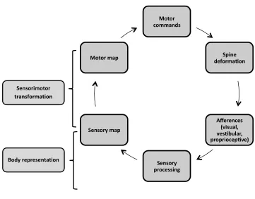
processing sensory information can cause asymmetry or a change in the amplitude of the vestibulomotor commands and alters the body schema. During rapid spine growth, this condition would lead to spine deformation and asym-metrical trunk proprioception promoting the updating of a distorted body schema (Fig. 1).31,34,39
Motor commands
Spine deformaFon
Afferences (visual, vesFbular, propriocepFve)
Sensory processing Sensory map
Motor map
Body representation Sensorimotor transformation
Figure 1:
Theoretical model of the association between a distorted body representation and the development of spine deformation. Alteration in the processing of sensory information could create a deformation of the body representation. Consenquently, the motor commands from the
sensorimotor transformation process would be altered (e.g., asymmetrical). During a critical period of the development, this would create spine deformation. As a result, torso proprioception
The present study is part of a research programme as-sessing the vestibulomotor control of balance in AIS. The objective of the present study was to establish an experi-mental framework for testing whether spine deformation could be related to a distortion of the body schema. Since the body schema is continuously updated through sensory signals, it is possible that surgical intervention that dras-tically reduces spine deformation or bracing that creates proprioceptive rehabilitation, through torso propriocep-tive cues, lead to a recalibration of the body schema. If this is the case, improvement in balance control either during or after sensory manipulation should be observed follow-ing spine surgery or long-term bracfollow-ing. If this hypothesis is supported, it would indicate that the weight of pro-prioceptive information from the torso is larger than the weight of vestibular information (participants are tested in absence of vision) in the updating of body schema. An alternate hypothesis is that balance control improvement is caused by a decrease in the biomechanical forces acting on the spine due to a lessening of the spinal curvature. It has been demonstrated, however, that reintegration of sensory information altered balance in AIS patients which favours the first hypothesis.17 In contrast, if body sway
does not decrease following spine surgery or long-term bracing, it would suggest that the cortical mechanisms involved in sensorimotor transformation are impaired. In this case, although straightening the spine or bracing would improve torso proprioceptive cues, it would not be sufficient to recalibrate effectively the body schema.
Methods
Three participants were involved in this study. All of them gave their written informed consent according to Laval University biomedical ethics committee. Ves-tibular stimulations were delivered using a DS5 bipolar constant current stimulator (Digitimer Ltd, Garden City, UK). The skin behind the ears over the mastoid process was prepared bilaterally using electrode skin prep pad (Dynarex, Orangeburg NY, USA) before placing the PALS Platinum 3.2 cm electrodes (Axelgaard Manufac-turing Co Ltd, Failbrook CA, USA). The electrodes were secured using 3M Transpore Tape 1527-1(3M). Par-ticipants performed the same tasks; they stood upright with their eyes closed and their feet 2 cm apart and with each foot standing on a force platform. Balance control was assessed using two force platforms (AMTI-model
Case 1: Effect of spine surgery on balance control
This case concerns a 17-year-old male. He was 14 years old when he first saw his orthopedic surgeon. The assess-ment of his balance control was performed when he was 15 year old. There were 3 other known cases of scoliosis in his family: his 2-year younger sister (mild scoliosis, Cobb angle = 20°), his mother (unknown Cobb angle), and his mother’s sister (she probably had a severe spine deformation since she had had corrective spinal surgery). At the initial balance control assessment (T0), his Risser sign was 1 (i.e., index of osseous maturity based on iliac crest ossification, ranging from 0 to 5) and he had a 52° right thoracic curve and a 34° left lumbar curve. At the age of 16, he underwent surgery. Pre-surgery neurological routine examination did not report any findings. Motor conductance was normal in both lower limbs, sensory conductance was difficult to obtain on the right side but lumbar spine MRI was normal. The surgery consisted of reducing the curves and vertebrae rotations using transpe-dicular screws from the third thoracic to third lumbar vertebrae and two Harrington rods. Following the sur-gery (T1), 18-months later, he had an 18° right thoracic curve and a 14° left lumbar curve. His Risser sign was 5. Because spine deformation and surgical instrumenta-tion necessarily constrained trunk mobility, the partici-pant’s trunk maximal voluntary range of motion along the frontal plane was quantified using the sensors located on the 5th lumbar vertebra (L5), and on the 7th cervical
ver-tebra (C7). Right and left maximal voluntary trunk flex-ions were 30°and 38° before surgery (T0) and 23° and 27° following surgery (T1). Maximal torso deviations, due to vestibular stimulation, were smaller than his voluntary range of motion: 4° and 6° at T0 and 2° and 2° at T1 for right and left movements, respectively.
Before spine surgery, his balance instability was much larger than controls during and after vestibular stimula-tion; the vertical force RMS values were 2.4 times greater than controls during GVS ([0-2]) and 4.9 times immedi-ately following GVS (i.e., sensory reintegration epoch, [2-3]) (Fig. 2). Following spine surgery (T1), however, his balance control slightly improved. For instance, his verti-cal force RMS values were both 1.3 times greater than controls for the GVS and sensory reintegration epochs, respectively. It is worth noting that, following spine sur-gery, his vertical force RMS values diverged slightly from controls during the GVS epoch mainly because the verti-cal force slightly increased toward the end of the interval whereas it leveled out for controls. Overall, for this AIS patient, it seems that the spine surgery improved his bal-ance control.
Case 2: Effect of bracing on balance control
Case 2 is a 15-year-old girl and the sister of case 1. Her balance control assessments were performed the same day as her brother. At that time (T0), she was 13 when a 16° right thoracic curve and a 13° left lumbar curve Table 1:
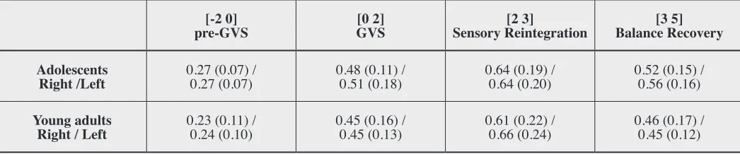
Root mean square (RMS) values of the vertical force before (pre), during or after galvanic vestibular stimulation (GVS). These data are from a group of healthy adolescent (n=16) and a group of healthy young adult (n=15). Data are the means
(standard deviation) of 15 trials per side. [-2 0]
pre-GVS GVS[0 2] Sensory Reintegration[2 3] Balance Recovery[3 5]
Adolescents
Right /Left 0.27 (0.07) / 0.27 (0.07) 0.48 (0.11) / 0.51 (0.18) 0.64 (0.19) / 0.64 (0.20) 0.52 (0.15) / 0.56 (0.16)
Young adults
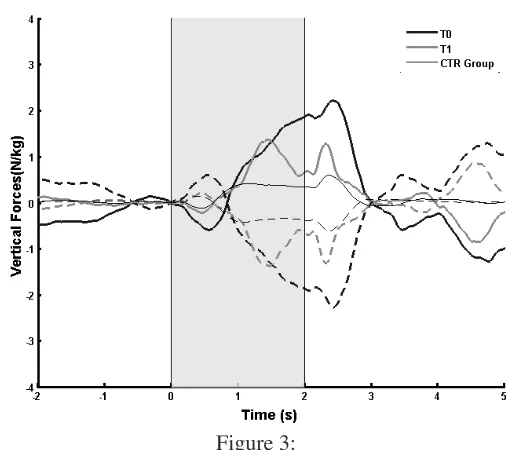
were detected. At the time, her Risser sign was 2. Be-fore the first balance control evaluation, the patient had been wearing a Providence brace for 2 months and was still wearing it 18-months later (i.e., at T1). Bracing did not change much her spine deformation; she had a 17° right thoracic curve and a 23° left lumbar curve and her Risser sign was 4. At initial evaluation (T0), during the vestibular stimulation, her balance control was impaired compared to controls; the vertical force RMS value was 2.4 times larger (Fig. 3). Furthermore, her vertical force RMS value was 3 times larger than controls immediately following GVS (i.e., sensory reintegration interval, [2 3]) and she could not recover her balance to the same extent as the controls (balance recovery interval, [3 5]). Eighteen months later (T1), during GVS, her vertical force RMS
value was 2.6 larger than control. Although it seems that the amplitude of her vertical force slightly decreased; her balance control was still impaired compared to controls. Immediately following the cessation of GVS (i.e., sen-sory reintegration interval), her vertical force RMS value was 2.6 times greater than controls. Finally, it is worth noting that compared to controls, she had trouble recover-ing her balance; the amplitude of her vertical forces did not reach a steady state. Overall, the present results sug-gest that long-term torso proprioceptive cue provided by the brace partly improved (but still larger than controls) balance control while her lumbar deformation increased by 10°. This latest result suggests that the amplitude of the spine deformation is not necessary related to balance control impairment.
Figure 2:
Case 1 mean vertical forces from 2 seconds before GVS onset to 3 seconds after GVS cessation. GVS onset starts at 0-s and lasts 2-s (shaded area). Regular lines present data for the right stimulation whereas the dashed
lines depict data for the left stimulation. The thin lines represent mean data for age-matched controls (CTR
group) and thick lines illustrate the data of the AIS patients before (T0: thick gray lines) and after spine
surgery (T1: thick light gray lines).
Figure 3:
Case 2 mean vertical forces from 2 seconds before GVS onset to 3 seconds after GVS cessation. GVS onset starts at 0-s and lasts 2-s (shaded area). Regular lines present data for the right stimulation whereas the dashed
lines depict data for the left stimulation. The thin lines represent mean data for age-matched controls (CTR
group) and thick lines illustrate the data of the AIS patients before (T0: thick gray lines) and 18-months
Case 3: Effect of spine surgery in adult on balance control
This participant is a 20-year-old woman. There are two other known cases of scoliosis in her family: her grand-mother and her older sister underwent spine surgery. Her scoliosis has been diagnosed when she was 11. Between the diagnosis and the surgery, she had been braced. A first surgery was performed when she was 14 and a second surgery when she was 16. Before the first surgery, she had a 70° right thoracic curve and a 55° left lumbar curve. The last assessment of her spine deformation revealed that she still had a 35° right thoracic curve and a 30° left lumbar curve. The balance control assessment was realized fol-lowing both spine surgeries. The analysis of the vertical force time-series during GVS revealed that her balance
control was worse than controls; her vertical force RMS value was 2.3 larger than controls (Fig. 4). Furthermore, immediately following vestibular stimulation (i.e., sen-sory reintegration epoch [2 3]), her balance control was still worse than controls; her vertical force RMS value was 4.5 times larger. Across time (i.e., balance recovery epoch, [3 5]), however, her vertical forces drastically de-creased but her RMS value was still 1.9 larger than con-trols. Overall, it is concluded that despite the absence of a complete reduction in her spine deformation, compared to controls, the cortical mechanisms performing sensorimo-tor transformation are impaired.
Discussion
Visual, proprioceptive and vestibular information contrib-ute to the perception of the body shape, dimension and relative limb position with respect to each other (body representation). Since it has long been reported that AIS patients have sensory processing impairments16,17,19,40-43, it
is plausible to suggest that AIS patients could have a dis-torted body representation. The aim of this study was to present an experimental framework to evaluate this sug-gestion. It was hypothesized that reducing spine deforma-tion, through conventional treatment, should allow recali-brating body schema. As a result, reduction in spine de-formation should translate into balance control improve-ment either during or following sensory manipulation.
Bracing or surgery effect
Results have demonstrated that for cases 1 and 2, either the spine surgery or bracing slightly improved balance control. For both cases, however, balance control was still impaired during or following vestibular stimulation. For these patients, altering the asymmetry in torso tion through spine surgery or providing torso propriocep-tive cue via bracing partly improved balance control. The cortical mechanisms that update the body schema likely weight differently the sensory signals.17,44 Consequently,
for some patients, straightening the spine or wearing a brace could partly reduce body representation distortion. For these individuals, alteration in the sensorimotor trans-formation of vestibular intrans-formation would not be com-pletely eliminated by the torso proprioception. In conclu-sion, it is speculated that for these two cases, improvement in balance control during sensory deprivation or sensory reintegration implies a better body representation.
Figure 4:
Case 3 mean vertical forces from 2 seconds before GVS onset to 3 seconds after GVS cessation. GVS onset starts at 0-s and lasts 2-s (shaded area). Regular lines present data for the right stimulation whereas the dashed lines depict data for the left stimulation. The thin lines represent mean data for age-matched controls (CTR group) and thick lines illustrate
For case 3, the reduction in spine deformation, through two surgeries, did not reduce her balance sway to the same extent as controls either during or immediately follow-ing sensory manipulation. Nonetheless, it is worth men-tioning that she still had a spine deformation post-surgery (i.e., 35° right thoracic curve and a 30° left lumbar curve). Therefore, one may suggest that balance control impair-ment was related to biomechanical factor. The increase in vertical force immediately following vestibular stimula-tion rule out this suggesstimula-tion as performing sensory reinte-gration led to balance control impairment. As a result, it seems that asymmetrical torso proprioceptive information (i.e., distorted body representation) led to suboptimal sen-sorimotor transformation and inefficient balance control.
Treatment of AIS
The recommendation from the Scoliosis Research Soci-ety (SRS) indicates that for curves between 25° and 40° patients should be braced.45-47 For these curve severities,
surgical treatment is not necessary as long as the curve remains below 45° even if it progresses despite bracing. Surgical treatment is recommended for patients that are still growing with curve greater than 45°, or if the curve is larger than 45° and continues to progress even if growth has stopped. The purpose of surgical intervention is two-fold: i) to prevent curve progression and ii) to reduce spine deformation. On the other hand, bracing only slows curve progression. Therefore, to be efficient, bracing must be prescribed as soon as possible. Bracing is considered an effective treatment with 72% of success ( i.e., the curve
did not worsen) compared to 42% after observation.48
Furthermore, there is a significant positive association be-tween hours of bracing and treatment success; 12.9 daily hours of bracing entails a success rate of 90 %.48
Limitations and research recommendation
Undoubtedly, scoliosis onset or progression involves multiple factors. Alteration in the processing of sensory information or in the mechanisms performing sensorimo-tor transformation could be related to a genetic defect, for example. Therefore, alterations in sensorimotor trans-formation, for example due to a distortion in body rep-resentation, might be related to scoliosis onset or progres-sion in some patients. This case series propose a tentative experimental framework to explore whether a potential link between body representation and scoliosis exists.
This study has various limitations. Obviously, to better test the experimental framework and draw any conclu-sion, more AIS patients need to be tested before and after spine surgery to thoroughly verify whether reduction in spine deformation translate into a better body represen-tation. Because of its complex aetiology, it is proposed that grouping AIS patients based on the severity of the spine deformation could mix patients with various causes (e.g., genetic, neurological dysfunction, hormonal). Con-sequently, an approach based on detecting the prevalence of a biomarker (e.g., vestibular impairment) should be used.49
The motor response evoked by GVS is reliable in healthy individuals and individuals with vestibular path-ology over weeks (personal communication with the au-thors).50 Although in the present study balance control
was studied after several months, we are confident that this period did not affect our results since the motor re-sponses evoked by GVS are unaffected up to 60 years old.
Conclusion
Overall, the present results suggest that reducing spine de-formation does not necessary translate in balance control improvement. The three cases demonstrated different be-haviour following conventional treatment. For instance, spine surgery improved to a great extent balance control in case 1 either during or following sensory manipulation. In contrast, bracing had a slight effect for case 2 while her lumbar deformation increased by 10°. For case 3, reduc-tion in spine deformareduc-tion through surgeries did not trans-late in balance control similar to controls. The absence of clear-cut results supports the idea that AIS is a multi-factorial pathology. Consequently, studying the effects of conventional treatment on balance control while manipu-lating sensory information (e.g., through GVS) could give some insights into the physiopathology of AIS patients with balance control impairment.
Acknowledgements
This project has been supported by Fondation Cotrel de
l’Institut de France. JPP has been awarded a scholarship from Fonds de Recherche du Québec en Santé, and has
been supported by Fondation de Recherche
References
1. Ueno M, Takaso M, Nakazawa T, et al. A 5-year
epidemiological study on the prevalence rate of idiopathic scoliosis in Tokyo: school screening of more than 250,000 children. J Orthop Sci. 2011;16(1):1-6.
2. Weinstein SL, Zavala DC, Ponseti IV. Idiopathic scoliosis: long-term follow-up and prognosis in untreated patients. J Bone Joint Surg Am. 1981;63(5):702-712.
3. Miller NH. Genetics of familial idiopathic scoliosis. Clin Orthop Relat Res. 2007;462:6-10.
4. Stokes IA. Three-dimensional terminology of spinal deformity. A report presented to the Scoliosis Research Society by the Scoliosis Research Society Working Group on 3-D terminology of spinal deformity. Spine. 1994;19(2):236-248.
5. Harrington PR. The etiology of idiopathic scoliosis. Clin Orthop Relat Res. 1977;(126):17-25.
6. Burwell RG, Dangerfield PH. Whither the etiopathogenesis (and scoliogeny) of adolescent idiopathic scoliosis? Stud Health Technol Inform. 2012;176:3-19.
7. Lowe TG, Edgar M, Margulies JY, et al. Etiology of idiopathic scoliosis: current trends in research. J Bone Joint Surg Am. 2000;82-A(8):1157-1168.
8. Wang WJ, Yeung HY, Chu WC, et al. Top theories for the etiopathogenesis of adolescent idiopathic scoliosis. J Pediatr Orthop. 2011;31(1 Suppl):S14-27.
9. Herman R, Mixon J, Fisher A, Maulucci R, Stuyck J. Idiopathic scoliosis and the central nervous system: a motor control problem. The Harrington lecture, 1983. Scoliosis Research Society. Spine. 1985;10(1):1-14. 10. Slobounov S, Wu T, Hallett M. Neural basis subserving
the detection of postural instability: an fMRI study. Motor Control. 2006;10(1):69-89.
11. Mergner T, Maurer C, Peterka RJ. A multisensory posture control model of human upright stance. Prog Brain Res. 2003;142:189-201.
12. Byl NN, Holland S, Jurek A, Hu SS. Postural imbalance and vibratory sensitivity in patients with idiopathic scoliosis: implications for treatment. J Orthop Sports Phys Ther. 1997;26(2):60-68.
13. Byl NN, Gray JM. Complex balance reactions in different sensory conditions: adolescents with and without
idiopathic scoliosis. J Orthop Res. 1993;11(2):215-227. 14. Haumont T, Gauchard GC, Lascombes P, Perrin PP.
Postural instability in early-stage idiopathic scoliosis in adolescent girls. Spine. 2011;36(13).
15. Silferi V, Rougier P, Labelle H, Allard P. [Postural control in idiopathic scoliosis: comparison between healthy and scoliotic subjects]. Rev Chir Orthop Reparatrice Appar Mot.2004;90(3):215-225.
16. Simoneau M, Richer N, Mercier P, Allard P, Teasdale N. Sensory deprivation and balance control in idiopathic scoliosis adolescent. Exp Brain Res. 2006;170(4):576-582. 17. Simoneau M, Mercier P, Blouin J, Allard P, Teasdale N.
Altered sensory-weighting mechanisms is observed in adolescents with idiopathic scoliosis. BMC Neurosci. 2006;7:68.
18. Simoneau M, Lamothe V, Hutin E, Mercier P, Teasdale N, Blouin J. Evidence for cognitive vestibular integration impairment in idiopathic scoliosis patients. BMC Neurosci. 2009;10:102.
19. Manzoni D, Miele F. Vestibular mechanisms involved in idiopathic scoliosis. Arch Ital Biol. 2002;140(1):67-80. 20. Wiener-Vacher SR, Mazda K. Asymmetric otolith
vestibulo-ocular responses in children with idiopathic scoliosis. J Pediatr. 1998;132(6):1028-1032.
21. Kandel E, Schwartz J, Jessell T. Principles of Neural Science, Fourth Edition. McGraw-Hill Companies,Incorporated; 2000.
22. Pialasse JP, Laurendeau S, Descarreaux M, Blouin J, Simoneau M. Is abnormal vestibulomotor responses related to idiopathic scoliosis onset or severity? Med Hypotheses. 2013;80(3):234-236.
23. Departments of Neurology R. John Leigh Professor NOBECWRUUHVAMCCO, Departments of Neurology David S. Zee Professor OOHNSN, Ocular Motor-Visual Testing Lab Johns Hopkins University rector BM. The Neurology of Eye Movements : Text and CD-ROM: Text and CD-ROM. Oxford University Press, USA; 1999. 24. Furman JM, Cass SP, Whitney SL. Vestibular Disorders: A
Case-study Approach to Diagnosis and Treatment. Oxford University Press; 2010.
25. Goldberg JM, Wilson VJ. The Vestibular System: A Sixth Sense. OUP USA; 2012.
26. Pialasse JP, Descarreaux M, Mercier P, Blouin J, Simoneau M. Sensorimotor integration in adolescent idiopathic scoliosis patients. In: Grivas TB, ed. Scoliosis: Intech; 2012:49-70.
27. Ardic FN, Latt LD, Redfern MS. Paraspinal muscle response to electrical vestibular stimulation. Acta Otolaryngol. 2000;120(1):39-46.
28. Marsden JF, Castellote J, Day BL. Bipedal distribution of human vestibular-evoked postural responses during asymmetrical standing. J Physiol. 2002;542(Pt 1):323-331. 29. Day BL, Severac Cauquil A, Bartolomei L, Pastor MA,
Lyon IN. Human body-segment tilts induced by galvanic stimulation: a vestibularly driven balance protection mechanism. J Physiol.1997;500 ( Pt 3):661-672.
30. Pialasse JP, Simoneau M, Descarreaux M. Kinematic and Kinetic Response to Galvanic Vestibular Stimulation in Adolescent Idiopathic Scoliosis: Preliminary Results. Paper presented at: Abstracts of ACC conference proceedings; 2012/04/01, 2012; Las Vegas.
32. Wang X, Merzenich MM, Sameshima K, Jenkins WM. Remodelling of hand representation in adult cortex determined by timing of tactile stimulation. Nature. 1995;378(6552):71-75.
33. Maravita A, Iriki A. Tools for the body (schema). Trends Cogn Sci. 2004;8(2):79-86.
34. Cardinali L, Frassinetti F, Brozzoli C, Urquizar C, Roy AC, Farne A. Tool-use induces morphological updating of the body schema. Curr Biol.
2009;19(12):R478-479.
35. Lopez C, Halje P, Blanke O. Body ownership and embodiment: vestibular and multisensory mechanisms. Neurophysiol Clin. 2008;38(3):149-161.
36. Ferre ER, Bottini G, Haggard P. Vestibular modulation of somatosensory perception. Eur J Neurosci.
2011;34(8):1337-1344.
37. Lopez C, Schreyer HM, Preuss N, Mast FW. Vestibular stimulation modifies the body schema. Neuropsychologia. 2012;50(8):1830-1837.
38. Ferre ER, Bottini G, Haggard P. Vestibular inputs modulate somatosensory cortical processing. Brain structure & function. 2012;217(4):859-864.
39. Dominici N, Daprati E, Nico D, Cappellini G,
Ivanenko YP, Lacquaniti F. Changes in the limb kinematics and walking-distance estimation after shank elongation: evidence for a locomotor body schema? J Neurophysiol. 2009;101(3):1419-1429.
40. Guo X, Chau WW, Hui-Chan CW, Cheung CS,
Tsang WW, Cheng JC. Balance control in adolescents with idiopathic scoliosis and disturbed somatosensory function. Spine. 2006;31(14):E437-440.
41. McInnes E, Hill DL, Raso VJ, Chetner B, Greenhill BJ, Moreau MJ. Vibratory response in adolescents who
have idiopathic scoliosis. J Bone Joint Surg Am. 1991;73(8):1208-1212.
42. Sahlstrand T, Ortengren R, Nachemson A. Postural equilibrium in adolescent idiopathic scoliosis. Acta Orthop Scand. 1978;49(4):354-365.
43. Wyatt MP, Barrack RL, Mubarak SJ, Whitecloud TS, Burke SW. Vibratory response in idiopathic scoliosis. J Bone Joint Surg Br. 1986;68(5):714-718.
44. So CW, Bent LR. Increased vestibular contribution to posture control in individuals with chronic headache. J Vestib Res. 2009;19(1-2):49-58.
45. Reamy BV, Slakey JB. Adolescent idiopathic scoliosis: review and current concepts. Am Fam Physician. 2001;64(1):111-116.
46. Richards BS, Bernstein RM, D’Amato CR, Thompson GH. Standardization of criteria for adolescent idiopathic scoliosis brace studies: SRS Committee on Bracing and Nonoperative Management. Spine. 2005;30(18):2068-2075; discussion 2076-2067.
47. Negrini S, Aulisa AG, Aulisa L, et al. 2011 SOSORT guidelines: Orthopaedic and Rehabilitation treatment of idiopathic scoliosis during growth. Scoliosis. 2012;7(1):3. 48. Weinstein SL, Dolan LA, Wright JG, Dobbs MB. Effects
of bracing in adolescents with Idiopathic Scoliosis. N Engl J Med. 2013;369(16):1512-21.
49. Vindras P, Desmurget M, Baraduc P. When one size does not fit all: a simple statistical method to deal with across-individual variations of effects. PLoS One. 2012;7(6):e39059.